. 2024 Jan 17;20(1):e1011907.
doi: 10.1371/journal.ppat.1011907. eCollection 2024 Jan.

Kaposi's sarcoma herpesvirus latency-associated nuclear antigen broadly regulates viral gene expression and is essential for lytic infection

Affiliations

Kaposi's sarcoma herpesvirus latency-associated nuclear antigen broadly regulates viral gene expression and is essential for lytic infection

Shijun Li et al. PLoS Pathog. .

Abstract

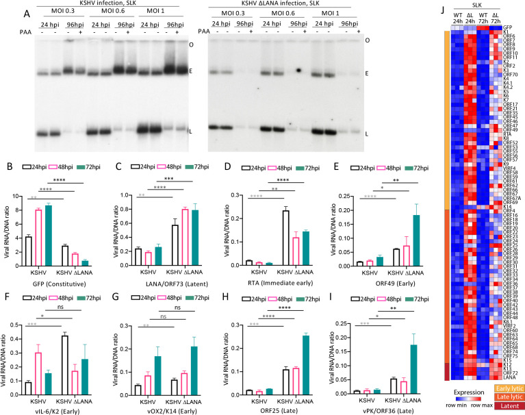
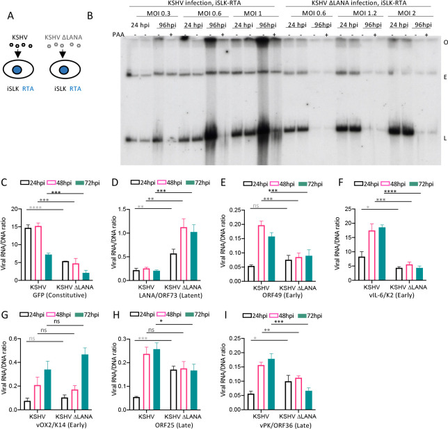
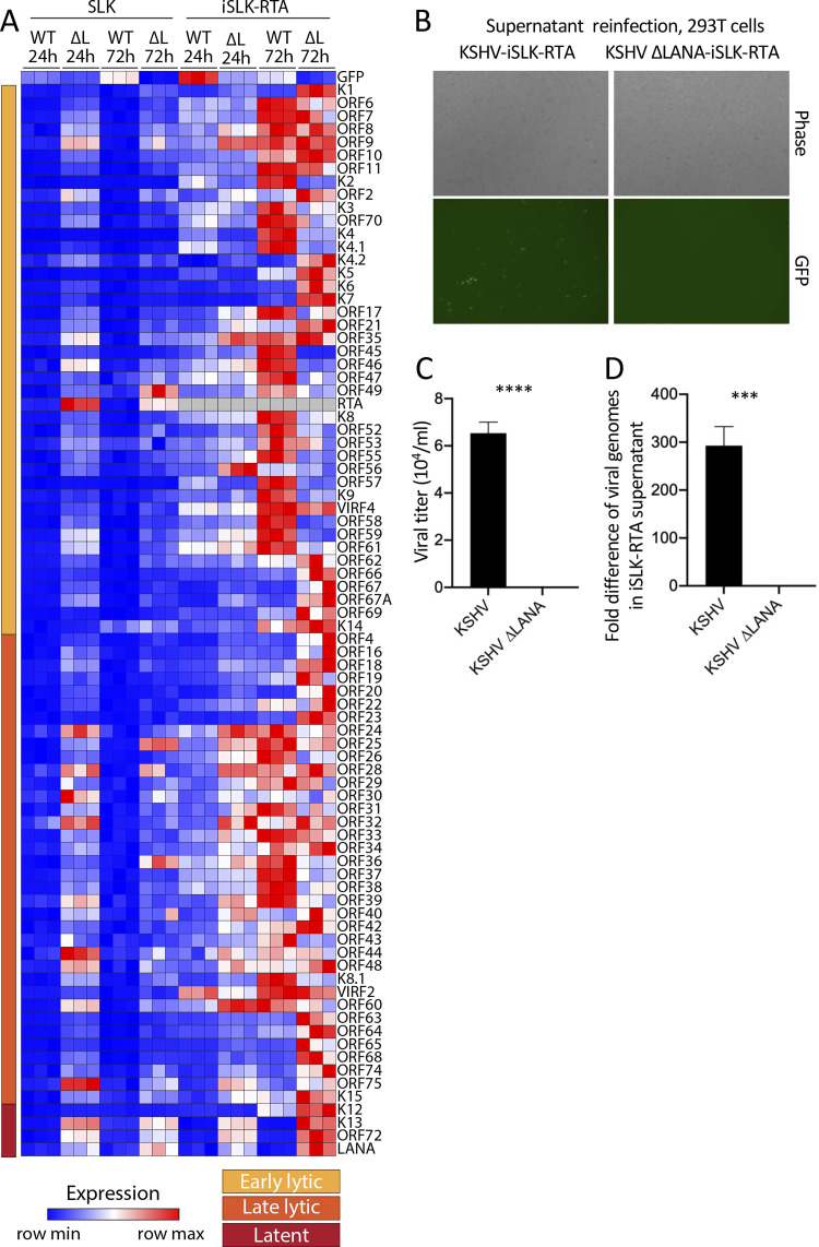
Kaposi's sarcoma herpesvirus (KSHV) is a leading cause of malignancy in AIDS and current therapies are limited. Like all herpesviruses, KSHV infection can be latent or lytic. KSHV latency-associated nuclear antigen (LANA) is essential for viral genome persistence during latent infection. LANA also maintains latency by antagonizing expression and function of the KSHV lytic switch protein, RTA. Here, we find LANA null KSHV is not capable of lytic replication, indicating a requirement for LANA. While LANA promoted both lytic and latent gene expression in cells partially permissive for lytic infection, it repressed expression in non-permissive cells. Importantly, forced RTA expression in non-permissive cells led to induction of lytic infection and LANA switched to promote, rather than repress, most lytic viral gene expression. When basal viral gene expression levels were high, LANA promoted expression, but repressed expression at low basal levels unless RTA expression was forcibly induced. LANA's effects were broad, but virus gene specific, extending to an engineered, recombinant viral GFP under control of host EF1α promoter, but not to host EF1α. Together, these results demonstrate that, in addition to its essential role in genome maintenance, LANA broadly regulates viral gene expression, and is required for high levels of lytic gene expression during lytic infection. Strategies that target LANA are expected to abolish KSHV infection.


Conflict of interest statement

The authors have declared that they have no competing interests.

Figures

Fig 1
Fig 1. KSHV LANA is required for spontaneous lytic infection in 293 cells.
A. Gardella gel of KSHV infected 293 cells at 24 hpi, with or without PAA incubation. B. Gardella gel of KSHV infected 293 cells at the indicated MOI at 24 or 96 hpi with or without PAA. C. Gardella gel following KSHVΔLANA infection at 24 or 96 hpi with or without PAA. Data representative of 2 experiments. A-C. Each lane pair contains independent infections. O, gel origin; E, episomal band; L, linear viral DNA. Linear band signal quantification using Image J is shown below.
Fig 2
Fig 2. LANA promotes KSHV gene expression in 293 cells.
A-H Ratios of viral RNA to DNA following KSHV or KSHVΔLANA infection of 293 cells for the indicated viral genes at 24, 48 or 72 hpi. ns, p>0.05; *, p<0.05; **, p<0.01; ***, p<0.001;****, p<0.0001 by unpaired t test. I. Immunoblot of the indicated gene products following KSHV or KSHVΔLANA infection of 293 cells at 24 or 48 hpi at indicated MOI’s. The multiple LANA bands, which are more prominent at 48 hpi, are due to alternative initiation of translation and an alternative poly A signal.[71,72] GFP is expressed from the EF1α promoter in the recombinant viral genome. J. Gardella gel is shown at left. Phase microscopy (right, top) or detection of GFP expression by fluorescence microscopy (right, bottom) following infection of KSHV or KSHVΔLANA at 24 hpi. Cells were detected using the 4x objective on an EVOS M7000 imaging system (ThermoFisher). K. Heatmap of z score values showing relative viral gene expression for all KSHV genes following KSHV (WT) or KSHVΔLANA (ΔL) at 24 or 72 hpi in 293 cells. Three independent biological replicates are shown for each time point. Early lytic, late lytic, or latent classes of genes are indicated at left.
Fig 3
Fig 3. LANA expressed in trans promotes KSHVΔLANA broad viral gene transcription following infection in 293FRT cells.
A. Immunoblot of LANA in 293FRT/LANA or BCBL1 PEL cells. 5x10⁴ cells were loaded per lane. (BCBL1 PEL cells have less cytoplasm than do 293FRT cells, accounting for less tubulin in the BCBL1 lane.). B-I. Ratios of viral RNA to DNA following KSHVΔLANA infection of 293FRT/LANA or 293FRT cells for the indicated viral genes at 24, 48 or 72 hpi. **, p<0.01; ***, p<0.001; ****, p<0.0001 by unpaired t test. J. Heatmap of z score values showing relative viral gene expression for all KSHV genes following KSHVΔLANA infection of 293FRT or 293FRT LANA stably expressing cells at 24 or 72 hpi. Three independent biological replicates are shown for each time point. Early lytic, late lytic, or latent classes of genes are indicated at left. Gray color indicates absent values.
Fig 4
Fig 4. KSHV does not undergo spontaneous lytic infection following infection of NOK cells and LANA inhibits most viral gene expression.
A. Gardella gel at 24 or 96 hpi at the indicated MOI following KSHV or KSHVΔLANA infection of NOK cells. Each lane pair contains independent infections. O, gel origin; E, episomal DNA; L, linear viral genomic DNA. B-I. Ratios of viral RNA to DNA following KSHV or KSHVΔLANA infection of NOK cells for the indicated viral genes at 24, 48 or 72 hpi. ns, p>0.05; *, p<0.05, **, p<0.01; ***, p<0.001;****, p<0.0001 by unpaired t test. J. Heatmap of z score values showing relative viral gene expression for all KSHV genes following KSHV (WT) or KSHVΔLANA (ΔL) at 24 or 72 hpi in NOK cells. Three independent biological replicates are shown for each time point. Early lytic, late lytic, or latent classes of genes are indicated at left.
Fig 5
Fig 5. KSHV does not undergo spontaneous lytic infection following infection of SLK cells and LANA inhibits most viral gene expression.
A. Gardella gel at 24 or 96 hpi at the indicated MOI following KSHV or KSHVΔLANA infection of SLK cells. Each lane pair contains independent infections. O, gel origin; E, episomal DNA; L, linear viral genomic DNA. B-I. Ratios of viral RNA to DNA following KSHV or KSHVΔLANA infection of SLK cells for the indicated viral genes at 24, 48 or 72 hpi. ns, p>0.05; *, p<0.05, **, p<0.01; ***, p<0.001;****, p<0.0001 by unpaired t test. J. Heatmap of z score values showing relative viral gene expression for all KSHV genes following KSHV (WT) or KSHVΔLANA (ΔL) at 24 or 72 hpi in SLK cells. Three independent biological replicates are shown for each time point. Early lytic, late lytic, or latent classes of genes are indicated at left.
Fig 6
Fig 6. RTA expression in trans induces KSHV, but not KSHVΔLANA, lytic replication in SLK cells and leads to LANA promotion of broad lytic viral gene expression.
A. Schematic diagram of KSHV or KSHVΔLANA infection of iSLK RTA cells. B. Gardella gel at 24 or 96 hpi at the indicated MOI following KSHV infection of iSLK RTA cells that were doxycycline induced for RTA expression, with or without PAA. Each lane pair contains independent infections. O, gel origin; E, episomal DNA; L, linear viral genomic DNA. C-I. Ratios of viral RNA to DNA following KSHV or KSHVΔLANA infection of iSLK RTA cells for the indicated viral genes at 24, 48 or 72 hpi. ns, p>0.05; *, p<0.05, **, p<0.01; ***, p<0.001;****, p<0.0001 by unpaired t test.
Fig 7
Fig 7. RTA expression in trans leads to LANA promotion of lytic gene expression and virus production in SLK cells.
A. Heatmap of z score values showing relative viral gene expression for all KSHV genes following KSHV (WT) or KSHVΔLANA (ΔL) at 24 or 72 hpi in SLK cells (derived from same data as in Fig 5J), or iSLK cells that were induced for Rta expression. Three independent biological replicates are shown for each time point. Early lytic, late lytic, or latent classes of genes are indicated at left. Relative expression values were compared to the mean of all values for both SLK and iSLK for each gene, resulting in different z scores for SLK here compared to Fig 5J, which only included SLK. Gray color indicates absent values. B. Phase (top panels) or fluorescent microscopy (bottom panels) was performed to detect GFP expression from 293T cells 24 hours following incubation with supernatant from KSHV or KSHVΔLANA infected iSLK-RTA cells that had been induced for RTA expression. Cells were detected using the 4x objective on an EVOS M7000 imaging system (ThermoFisher). C. Viral titer of RTA induced, iSLK-RTA cell supernatant, 96 hpi following KSHV or KSHVΔLANA infection. GFP expression was detected by flow cytometry. D. Fold difference in KSHV genome copy number determined by qPCR of RTA induced, iSLK-RTA cell supernatant 96 hpi following KSHV or KSHVΔLANA infection.

Similar articles

Cited by

References

    1. Chang Y, Cesarman E, Pessin MS, Lee F, Culpepper J, Knowles DM, et al.. Identification of Herpesvirus-like DNA Sequences in AIDS-Associated Kaposi’s Sarcoma. Science. 1994;266:1865–9. doi: 10.1126/science.7997879 - DOI - PubMed
    1. Cesarman E, Chang Y, Moore PS, Said JW, Knowles DM. Kaposi’s Sarcoma-Associated Herpesvirus-Like DNA Sequences in AIDS-Related Body-Cavity-Based Lymphomas. NEJM. 1995;332:1186–91. doi: 10.1056/NEJM199505043321802 - DOI - PubMed
    1. Moore PS, Chang Y. Detection of herpesvirus-like DNA sequences in Kaposi’s sarcoma in patients with and without HIV infection [see comments]. The New England journal of medicine. 1995;332(18):1181–5. - PubMed
    1. Soulier J, Grollet L, Oksenhendler E, Cacoub P, Cazals-Hatem D, Babinet P, et al.. Kaposi’s Sarcoma-Associated Herpesvirus-Like DNA Sequences in Multicentric Castleman’s Disease. Blood. 1995;86(4):1276–80. - PubMed
    1. Decker LL, Shankar P, Khan G, Freeman RB, Dezube BJ, Lieberman J, et al.. The Kaposi sarcoma-associated herpesvirus (KSHV) is present as an intact latent genome in KS tissue but replicates in the peripheral blood mononuclear cells of KS patients. J Exp Med. 1996;184(1):283–8. doi: 10.1084/jem.184.1.283 - DOI - PMC - PubMed