. 2024 Jan 17;16(730):eade2886.
doi: 10.1126/scitranslmed.ade2886. Epub 2024 Jan 17.

Splicing neoantigen discovery with SNAF reveals shared targets for cancer immunotherapy

Affiliations

Splicing neoantigen discovery with SNAF reveals shared targets for cancer immunotherapy

Guangyuan Li et al. Sci Transl Med. .

Abstract

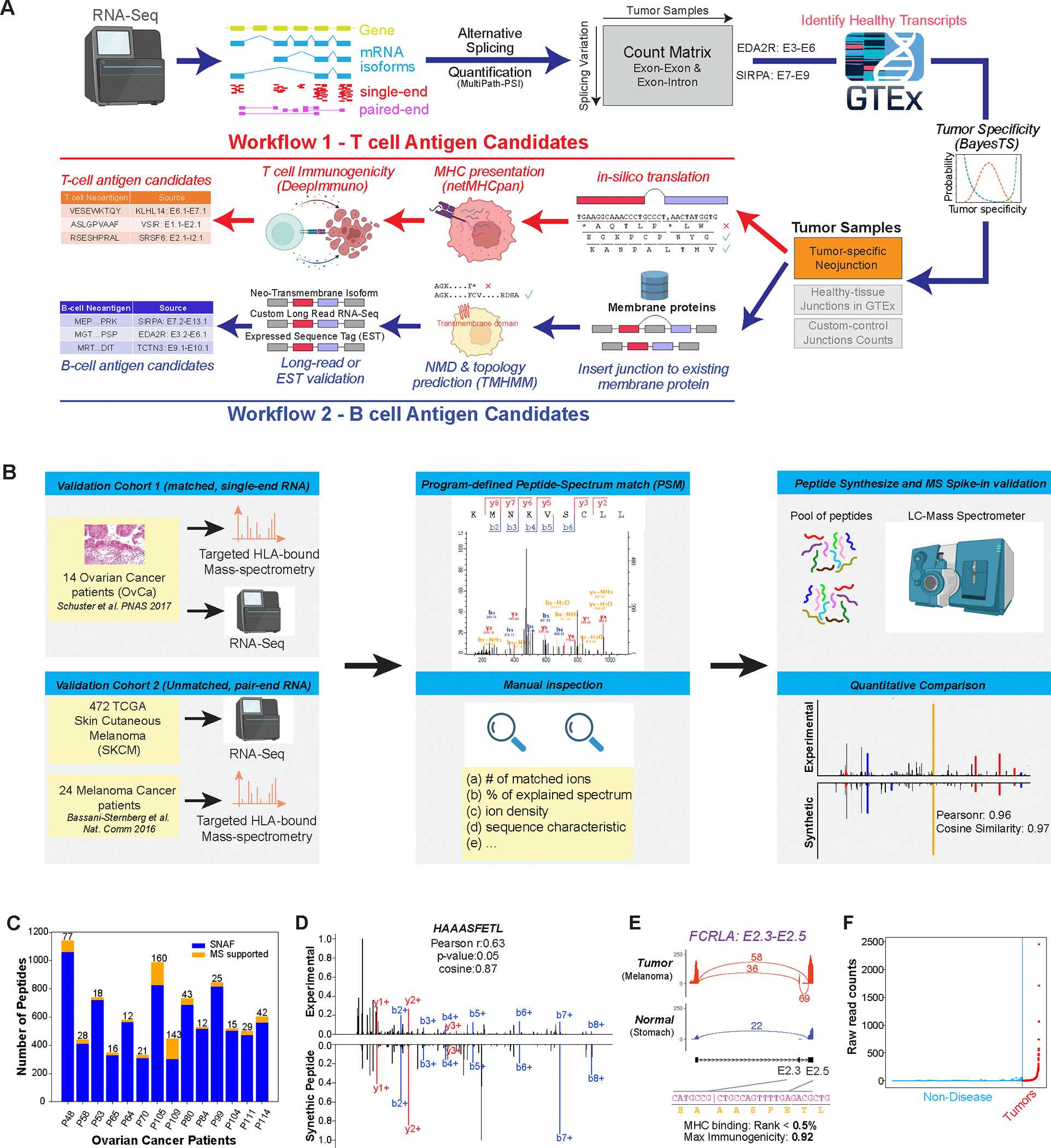
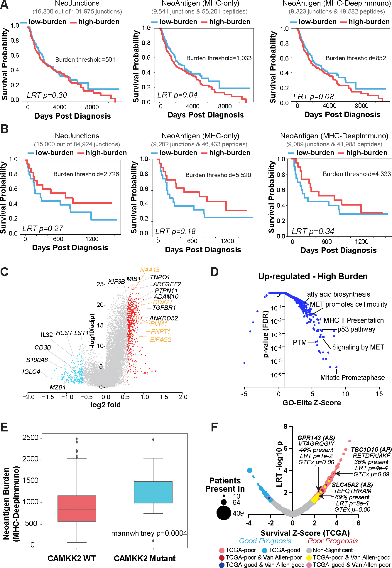
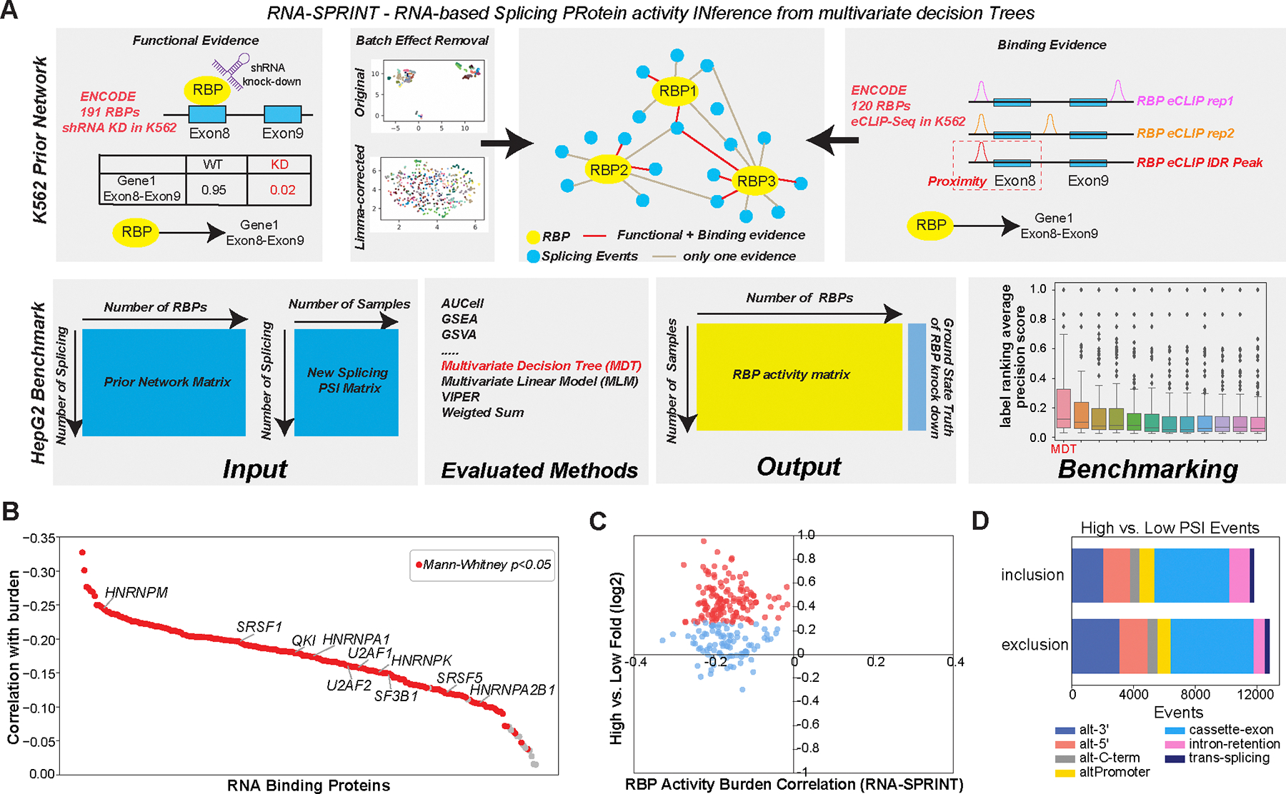
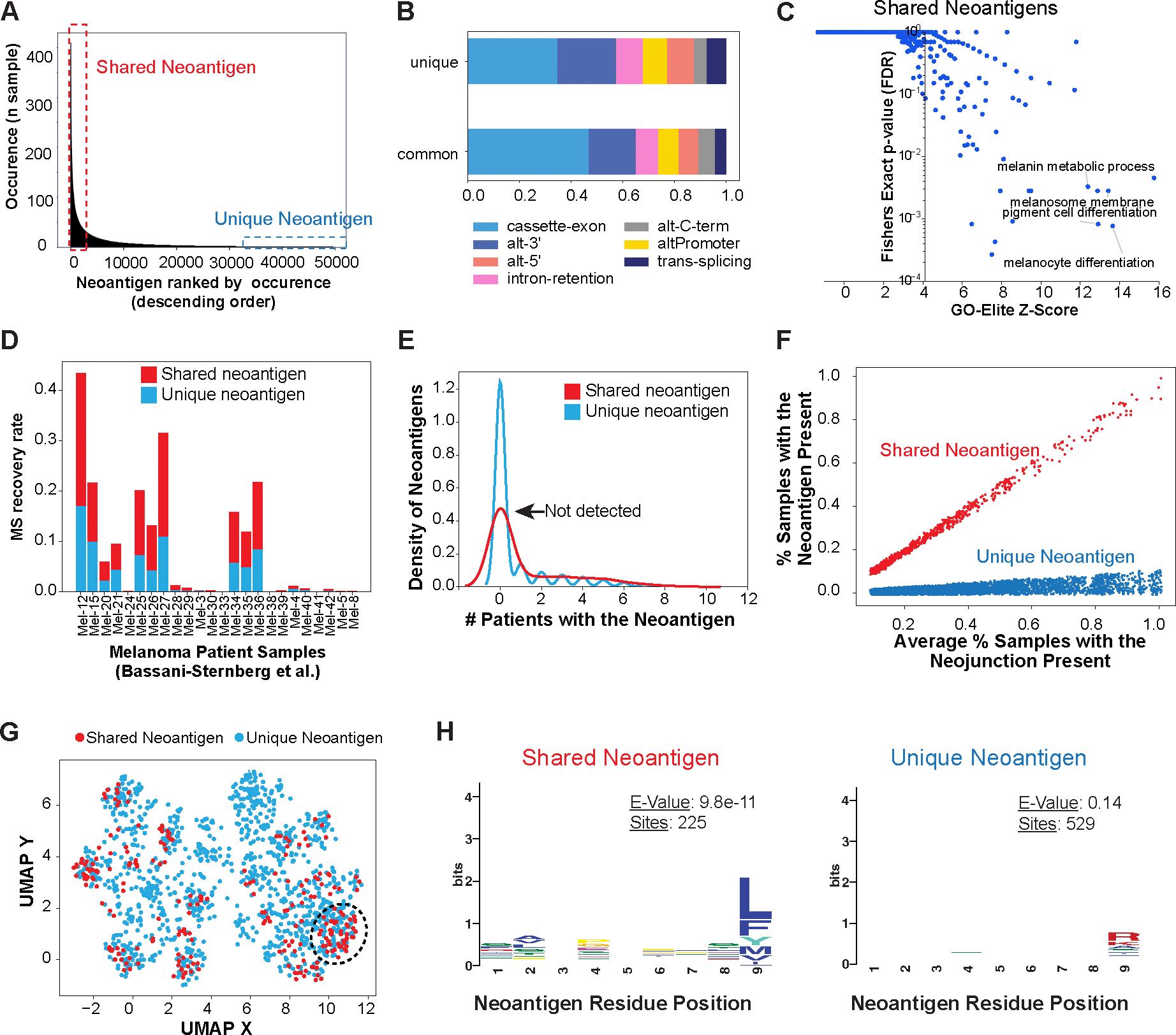
Immunotherapy has emerged as a crucial strategy to combat cancer by "reprogramming" a patient's own immune system. Although immunotherapy is typically reserved for patients with a high mutational burden, neoantigens produced from posttranscriptional regulation may provide an untapped reservoir of common immunogenic targets for new targeted therapies. To comprehensively define tumor-specific and likely immunogenic neoantigens from patient RNA-Seq, we developed Splicing Neo Antigen Finder (SNAF), an easy-to-use and open-source computational workflow to predict splicing-derived immunogenic MHC-bound peptides (T cell antigen) and unannotated transmembrane proteins with altered extracellular epitopes (B cell antigen). This workflow uses a highly accurate deep learning strategy for immunogenicity prediction (DeepImmuno) in conjunction with new algorithms to rank the tumor specificity of neoantigens (BayesTS) and to predict regulators of mis-splicing (RNA-SPRINT). T cell antigens from SNAF were frequently evidenced as HLA-presented peptides from mass spectrometry (MS) and predict response to immunotherapy in melanoma. Splicing neoantigen burden was attributed to coordinated splicing factor dysregulation. Shared splicing neoantigens were found in up to 90% of patients with melanoma, correlated to overall survival in multiple cancer cohorts, induced T cell reactivity, and were characterized by distinct cells of origin and amino acid preferences. In addition to T cell neoantigens, our B cell focused pipeline (SNAF-B) identified a new class of tumor-specific extracellular neoepitopes, which we termed ExNeoEpitopes. ExNeoEpitope full-length mRNA predictions were tumor specific and were validated using long-read isoform sequencing and in vitro transmembrane localization assays. Therefore, our systematic identification of splicing neoantigens revealed potential shared targets for therapy in heterogeneous cancers.


Conflict of interest statement

COMPETING INTERESTS

The authors declare no competing interests.

Figures

Figure 1.
Figure 1.. Automated discovery and confirmation of immunogenic and transmembrane splicing neoantigens with SNAF.
A) Outline of the two parallel workflows in the software SNAF to predict splicing neoantigens. SNAF begins with the identification and quantification of alternative splice junctions (exon-exon and exon-intron) from RNA-Seq BAM files and filters these against normal tissue reference RNA-Seq profiles (BayesTS). Retained tumor-specific splice junctions (neojunctions) are evaluated for T-cell (SNAF-T) and B-cell (SNAF-B) antigen production. SNAF-T performs in-silico translation of each junction, MHC binding affinity prediction (netMHCpan or MHCflurry) and identifies high-confidence immunogenic neoantigens through deep learning (DeepImmuno). SNAF-B predicts full-length protein coding isoforms that produce cancer-specific extracellular neo-epitopes (ExNeoEpitopes), considering existing transcript annotations and full-length isoform sequencing for targeted antibodies. B) Validation workflow for Ovarian cancer and Melanoma immunopeptidomics with either matched or unmatched RNA-Seq. MaxQuant is applied to find Peptide-Spectrum Match (PSM), followed by quantitative and expert MS2 spectra prioritization. HPLC-MS/MS confirmation is performed on synthesized nominated neoantigens. C) Number of SNAF-T predicted neoantigens and those confirmed by immunopeptidomics across 14 of patients. D) Mirror plot of the immunopeptidomics and spike-in MS spectrum for HAAASFETL. The lines indicate mass-to-charge ratios for distinct types of fragmented ions (red/blue). E) SashimiPlot visualization of HAAASFETL, derived from an exon-exon junction in the gene FCRLA, along with the junction/peptide sequence, binding affinity and immunogenicity prediction. F) Raw read counts of the FCRLA neojunction between normal controls (blue) and TCGA melanoma cohort (red).
Figure 2.
Figure 2.. Splicing-neoantigen burden predicts response to therapy in Melanoma.
A,B) Kaplan-Meier (KM) survival plots of Melanoma patient samples stratified into low and high neoantigen burden, considering overall survival for each sequential step in SNAF for two cohorts (A) TCGA (n=472), and (B) Van Allen (n=39). These steps are: 1) tumor-specific neojunctions (left column), MHC-bound neoantigens (middle column) and immunogenic neoantigens (right column). Van Allen cohort RNA-Seq samples were subject to immune checkpoint inhibitors whereas TCGA were not. The number of neojunctions or Neoantigen peptides are shown at the top of each plot. C) Volcano plot of genes differentially expressed in patients with high versus low immunogenic splicing neoantigen burden in TCGA-SKCM, with a fold>1.5 and eBayes t-test P<0.05 (FDR corrected). Red = genes that are up-regulated in the high burden group; blue = down-regulated genes in the high burden; orange = representative RNA binding proteins. D) Gene-set enrichment with GO-Elite of ToppFun pathway gene-sets (AltAnalyze) for genes up-regulated in patients with high splicing versus low neoantigen burden(panel C). E) Immunogenic splicing neoantigen burden between patients in the TCGA Melanoma cohort with or without mutations in CAMKK2. Mann Whitney two-sided test. F) Bubble-plot of survival associated splicing neoantigens from SNAF in TCGA-SKCM. Dot size corresponds to the number of patients with melanoma that the splicing neoantigen is detected in (10–470) and are colored according to their survival significance in the TCGA-SKCM and Van Allen cohorts (LRT P<0.05 and z ≥ 1). AS = alternative splicing.
Figure 3.
Figure 3.. Regulatory networks mediating splicing neoantigen burden in Melanoma.
A) Schematic overview of the software RNA-SPRINT and associated benchmarking steps. The workflow involves construction of an RNA Binding Protein (RBP) prior network to predict splicing regulatory interactions. Evaluation of the method is overviewed, consisting of RNA-SPRINT benchmarking relative to 12 transcription factor (TF) activity methods in HepG2 cell line RBP knockdown RNA-Seq datasets. B) The correlation of inferred RBP activity with splicing neoantigen burden for all TCGA patients with melanoma. C) Comparison of RBP activity-burden correlations with RBP differential gene expression, for high versus low burden (TCGA SKCM). Red = upregulated genes (fold>1.2 and eBayes t-test p<0.05, FDR corrected) in high burden. D) Type of splicing events observed with exon/intron inclusion or exclusion comparing high versus low burden.
Figure 4.
Figure 4.. Shared splicing neoantigens are frequently detected by MS and are defined by their sequence composition in Melanoma.
A) Identification of common (shared) and unique immunogenic splicing neoantigens in the TCGA Melanoma cohort, based on their frequency of occurrence among patients. B) Frequency of splicing-event types for shared and unique splicing neoantigen junctions in TCGA. C) Gene-set enrichment with GO-Elite of the Gene Ontology and pathways of shared neoantigens (present in >15% of patients with melanoma). D) MS recovery rate in an independent melanoma immunopeptidome dataset (Bassani-Sternberg et al.) between shared and unique neoantigens considered in the query database. E) Kernel density estimate plot comparing the observed occurrence in an independent immunopeptidomics MS experimental cohort, for all detected shared (>15% of patients with melanoma) versus unique splicing-neoantigens. F) Re-defined shared and unique neoantigens in TCGA by normalizing the occurrence of their parental splice junction, leveraging their respective observed amino acid bias. G) UMAP of splicing neoantigens based on their amino acid physiological properties in TCGA, highlighting neoantigens that cluster based on shared amino acid physicochemical features. H) Distinct enriched amino acid motifs (MEME), comparing shared versus unique neoantigens.
Figure 5.
Figure 5.. Shared splicing neoantigens bind HLA and induce T-cell reactivity.
(A) Histograms and (B) graph show HLA-A*02-PE staining on HLA-A*02 containing TAP deficient T2 cells without peptide (no pep), loaded with FLU and HCMV control peptides and RLLGTEFQT (RLL) and FQTTRRAMTL (FQT) peptide neoantigens. MFI = median fluorescence intensity. PE = Phycoerythrin conjugated antibodies. C) Dot plots and (D) graph show the percentage of Interferon gamma-positive (IFNγ+) CD8+ T-cells in response to 5 melanoma shared splicing antigens compared to negative (unstimulated, no pep) or positive (PMA/I, FLU, HMCV) controls. CD8+ T cells were primed using peptide loaded monocyte derived dendritic cells and thereafter tested against 721.221 cells selectively expressing the indicated HLA allele with and without peptide loading. Bars indicate median of 2–3 donors and lines interquartile range.
Figure 6.
Figure 6.. Splicing-neoantigen cell of origin is dependent on its mechanism of regulation.
A,B) Venn diagrams comparing the number of parental neojunctions for TCGA SKCM splicing neoantigens unique to a single-patient (A) or shared in >15% of patients (B) to the specific cell-types they derive from in independent melanoma tumor biopsies by single-cell RNA-Seq analysis. Neojunctions are defined as tumor or immune if they are >2 fold enriched in either cell-population (absolute number of reads in all patients and cells for each lineage). C) Neojunction expression in individual cell populations for select shared splicing neoantigens. Each dot denotes the combined neojunction read counts in a single patient (n=19) with melanoma, separately per cell annotated cell population.
Figure 7.
Figure 7.. SNAF-B finds full-length mRNAs and stable-proteoforms for targeted therapies.
A) Overview of the SNAF-B prediction workflow to define ExNeoEpitopes. The workflow begins with bulk RNA-Seq datasets and optional long-read sequencing data integration to produce results with multiple levels of in silico evidence. B) Comparison of a SNAF-B predicted full-length isoform in the transmembrane protein SIRPA to documented mRNA isoforms and those predicted from PacBio long-read IsoSeq of melanoma cell lines. C) SashimiPlot of alternative 3’ splice site selection in Melanoma and Brain RNA-Seq for SIRPA. D) Specificity of the indicated SIRPA ExNeoEpitope for TCGA melanoma samples versus an integrated healthy controls tissue database (GTEx + TCGA). E) Alphafold2 3D modeling of the reference isoform and the long-read verified ExNeoEpitope. Arrow denotes the deleted region in the alternative isoform. F,G) Co-localization of the SIRPA reference (F) or Melanoma-specific (G) splice isoform by confocal microscopy with a cell surface stain (phalloidin). The arrow indicates the cross-section used to quantify fluorophore spatial coincidence.

Similar articles

Cited by

References

    1. Hanahan D, Weinberg RA, Hallmarks of cancer: the next generation. Cell 144, 646–674 (2011). - PubMed
    1. Tran E, Robbins PF, Rosenberg SA, “Final common pathway” of human cancer immunotherapy: targeting random somatic mutations. Nat. Immunol. 18, 255–262 (2017). - PMC - PubMed
    1. Ott PA, Hu Z, Keskin DB, Shukla SA, Sun J, Bozym DJ, Zhang W, Luoma A, Giobbie-Hurder A, Peter L, Chen C, Olive O, Carter TA, Li S, Lieb DJ, Eisenhaure T, Gjini E, Stevens J, Lane WJ, Javeri I, Nellaiappan K, Salazar AM, Daley H, Seaman M, Buchbinder EI, Yoon CH, Harden M, Lennon N, Gabriel S, Rodig SJ, Barouch DH, Aster JC, Getz G, Wucherpfennig K, Neuberg D, Ritz J, Lander ES, Fritsch EF, Hacohen N, Wu CJ, An immunogenic personal neoantigen vaccine for patients with melanoma. Nature 547, 217–221 (2017). - PMC - PubMed
    1. Hu Z, Leet DE, Allesøe RL, Oliveira G, Li S, Luoma AM, Liu J, Forman J, Huang T, Iorgulescu JB, Holden R, Sarkizova S, Gohil SH, Redd RA, Sun J, Elagina L, Giobbie-Hurder A, Zhang W, Peter L, Ciantra Z, Rodig S, Olive O, Shetty K, Pyrdol J, Uduman M, Lee PC, Bachireddy P, Buchbinder EI, Yoon CH, Neuberg D, Pentelute BL, Hacohen N, Livak KJ, Shukla SA, Olsen LR, Barouch DH, Wucherpfennig KW, Fritsch EF, Keskin DB, Wu CJ, Ott PA, Personal neoantigen vaccines induce persistent memory T cell responses and epitope spreading in patients with melanoma. Nat. Med. 27, 515–525 (2021). - PMC - PubMed
    1. Bauman J, Burris H, Clarke J, Patel M, Cho D, Gutierrez M, Julian R, Scott A, Cohen P, Frederick J, Robert-Tissot C, Zhou H, Mody K, Keating K, Meehan R, Gainor J, 798 Safety, tolerability, and immunogenicity of mRNA-4157 in combination with pembrolizumab in subjects with unresectable solid tumors (KEYNOTE-603): an update. J Immunother Cancer 8 (2020), doi:10.1136/jitc-2020-SITC2020.0798. - DOI

Publication types