. 2024 Jan 19;10(3):eadk7957.
doi: 10.1126/sciadv.adk7957. Epub 2024 Jan 17.

Four-dimensional computational ultrasound imaging of brain hemodynamics

Affiliations

Four-dimensional computational ultrasound imaging of brain hemodynamics

Michael D Brown et al. Sci Adv. .

Abstract

Four-dimensional ultrasound imaging of complex biological systems such as the brain is technically challenging because of the spatiotemporal sampling requirements. We present computational ultrasound imaging (cUSi), an imaging method that uses complex ultrasound fields that can be generated with simple hardware and a physical wave prediction model to alleviate the sampling constraints. cUSi allows for high-resolution four-dimensional imaging of brain hemodynamics in awake and anesthetized mice.


Figures
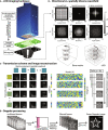
Fig. 1.
Fig. 1.. cUSi of blood flow with a matrix probe.
(A) Rendering of the cUSi imaging hardware. We modified a matrix probe populated with acoustically large, sensitive elements by attaching a plastic encoding mask that scrambles the transmit and receive wavefields. We coupled the mask to a waveguide that confines the transmitted fields and provides the necessary imaging offset. (B) Scrambling the transmitted wavefield provides a broader sampling of k-space and allows us to trade lateral resolution and side-lobe intensity. We illustrate this with the two renderings, which compare the imaging performance of the bare (without mask) and cUSi (with mask) probe on a numerical spiral phantom occupying an 8 mm × 8 mm × 8 mm volume. We simulated data using the experimentally measured forward fields (i.e., using y = Ax) and used a matched filter to reconstruct. (C) We drove the probe using a Hadamard-encoded synthetic aperture scheme, signals from different transmissions are separately reconstructed using a model-based approach then coherently summed to form each 3D volume. The system response is calibrated with a one-time measurement and then correlated with each set of measurements to recover an image. (D) To generate Doppler images, we continuously transmit to acquire data that are used to reconstruct separate volumes at a rate of ∼400 Hz. The data are spatiotemporally filtered, and the power associated with blood flow in each voxel over time is evaluated to form the PDI.
Fig. 2.
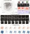
Fig. 2.. cUSis of hemodynamics in the anesthetized mouse brain.
(A) 3D rendering of the reconstructed PDI of the anesthetized mouse brain. Image was formed by compounding a filtered dataset composed of 8041 volumes using stable data acquired over 60 s. (B) Axial, coronal, and sagittal maximum amplitude projections through the reconstructed power (top) and color (bottom) Doppler volumes. (C) Subprojections through the PDI rendered in (A). Each slice was formed from a maximum amplitude projection through a set of planes with a thickness of 480 μm (corresponding to 12 planes in the reconstructed volume). The approximate position of each projection in the brain is denoted on the image. For the coronal slices, the positions are indicated relative to the bregma. (D) 3D rendering of vessels (bottom) and flow direction (top) in a fixed 2.48 mm by 2.48 mm by 2.4 mm region of the cortex formed via summation of an increasing number of frames. The time indicated denotes the acquisition time for the number of frames in the stable data ensemble that were used to form the image.

References

    1. Tanter M., Fink M., Ultrafast imaging in biomedical ultrasound. IEEE Trans. Ultrason. Ferroelectr. Freq. Control 61, 102–119 (2014). - PubMed
    1. Couade M., The advent of ultrafast ultrasound in vascular imaging: A review. J. Vasc. Diagn. Interv. 4, 9–22 (2016).
    1. Sebag F., Vaillant-Lombard J., Berbis J., Griset V., Henry J. F., Petit P., Oliver C., Shear wave elastography: A new ultrasound imaging mode for the differential diagnosis of benign and malignant thyroid nodules. J. Clin. Endocrinol. Metabol. 95, 5281–5288 (2010). - PubMed
    1. Vappou J., Luo J., Konofagou E. E., Pulse wave imaging for noninvasive and quantitative measurement of arterial stiffness in vivo. Am. J. Hypertens. 23, 393–398 (2010). - PMC - PubMed
    1. Errico C., Pierre J., Pezet S., Desailly Y., Lenkei Z., Couture O., Tanter M., Ultrafast ultrasound localization microscopy for deep super-resolution vascular imaging. Nature 527, 499–502 (2015). - PubMed

Publication types