. 2024 Feb 19;37(2):234-247.
doi: 10.1021/acs.chemrestox.3c00255. Epub 2024 Jan 17.

Tissue Organoid Cultures Metabolize Dietary Carcinogens Proficiently and Are Effective Models for DNA Adduct Formation

Affiliations

Tissue Organoid Cultures Metabolize Dietary Carcinogens Proficiently and Are Effective Models for DNA Adduct Formation

Angela L Caipa Garcia et al. Chem Res Toxicol. .

Abstract

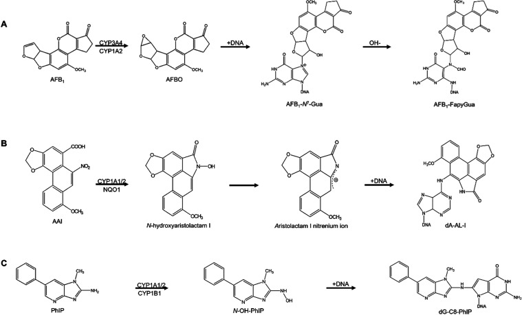
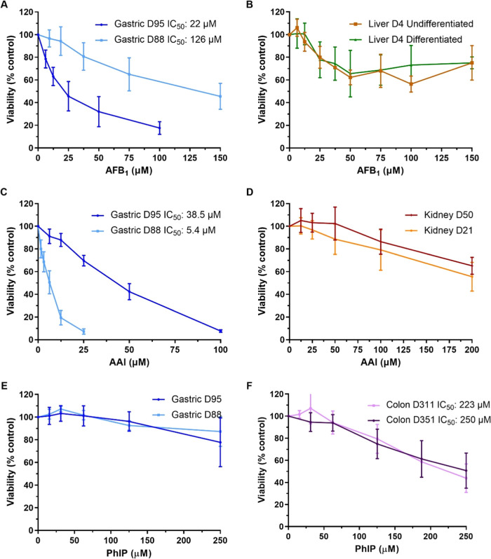
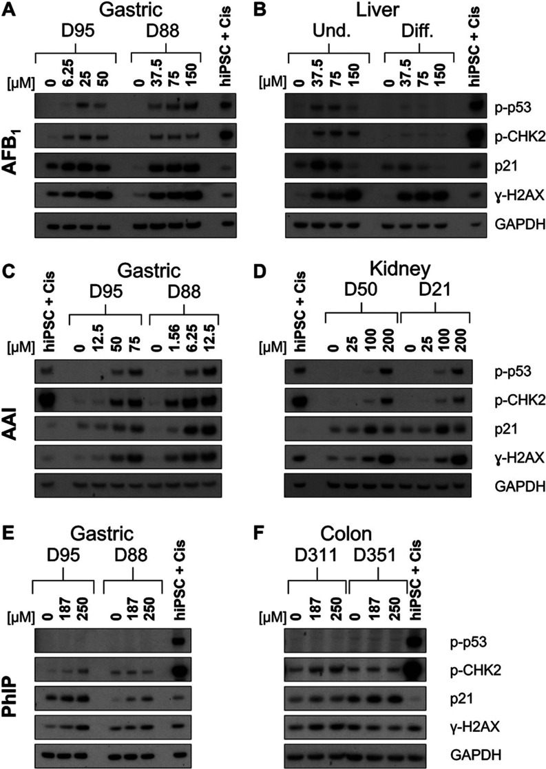
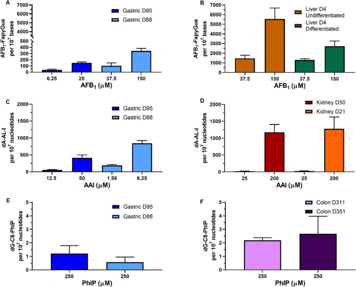
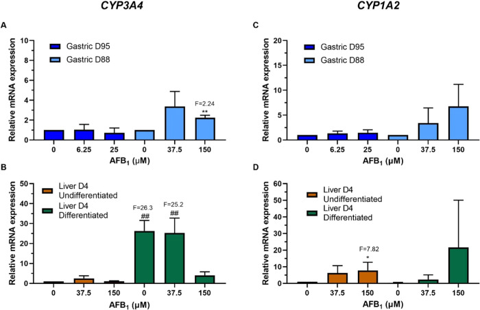
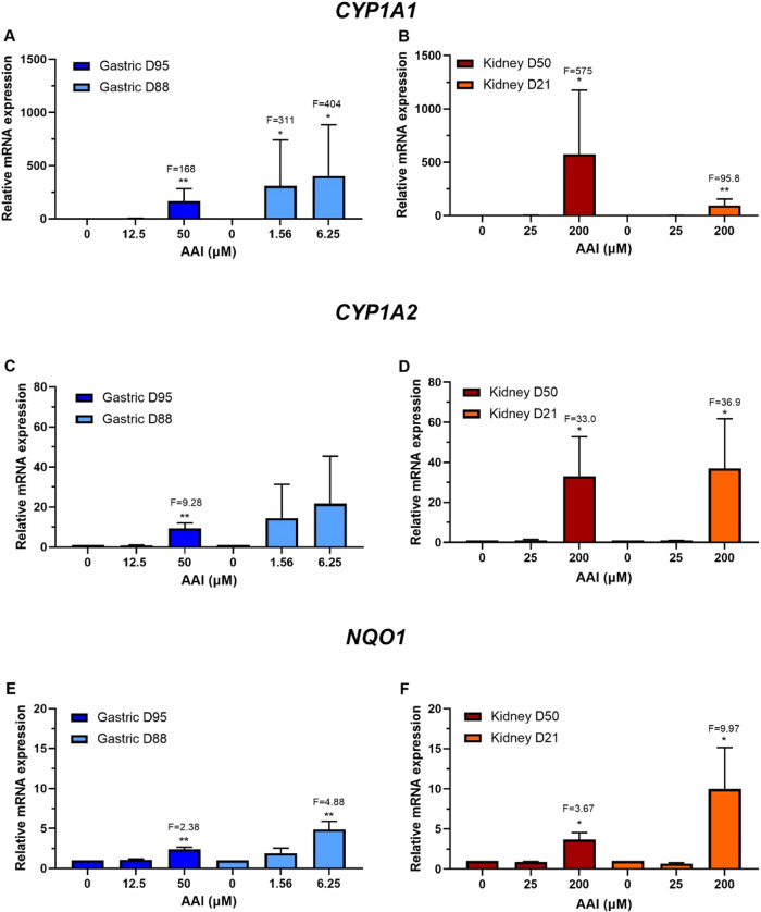
Human tissue three-dimensional (3D) organoid cultures have the potential to reproduce in vitro the physiological properties and cellular architecture of the organs from which they are derived. The ability of organoid cultures derived from human stomach, liver, kidney, and colon to metabolically activate three dietary carcinogens, aflatoxin B1 (AFB1), aristolochic acid I (AAI), and 2-amino-1-methyl-6-phenylimidazo[4,5-b]pyridine (PhIP), was investigated. In each case, the response of a target tissue (liver for AFB1; kidney for AAI; colon for PhIP) was compared with that of a nontarget tissue (gastric). After treatment cell viabilities were measured, DNA damage response (DDR) was determined by Western blotting for p-p53, p21, p-CHK2, and γ-H2AX, and DNA adduct formation was quantified by mass spectrometry. Induction of the key xenobiotic-metabolizing enzymes (XMEs) CYP1A1, CYP1A2, CYP3A4, and NQO1 was assessed by qRT-PCR. We found that organoids from different tissues can activate AAI, AFB1, and PhIP. In some cases, this metabolic potential varied between tissues and between different cultures of the same tissue. Similarly, variations in the levels of expression of XMEs were observed. At comparable levels of cytotoxicity, organoids derived from tissues that are considered targets for these carcinogens had higher levels of adduct formation than a nontarget tissue.


Conflict of interest statement

The authors declare no competing financial interest.

Figures

Figure 1
Figure 1
Pathways to the activation of the carcinogens in this study. (A) Aflatoxin B1 (AFB1); (B) aristolochic acid I (AAI); (C) 2-amino-1-methyl-6-phenylimidazo[4,5-b]pyridine (PhIP).
Figure 2
Figure 2
Cell viability in human tissue organoids treated with AFB1, AAI, and PhIP. Organoids from normal human stomach (A, C, and E; D95 and D88), liver (B; D4 undifferentiated and differentiated), kidney (D; D50 and D21), and colon (F; D351 and D311) tissues were treated with various concentrations of AFB1 (A, B), AAI (C, D), and PhIP (E, F) for 48 h. Vehicle controls DMSO (A, B, E, and F) or water (C, D) were included. Cell viability (% control) was measured using the CellTiter-Glo assay. Results are shown as mean ± SD (n ≥ 3).
Figure 3
Figure 3
DDR in normal human tissue organoids treated with AFB1, AAI, and PhIP. Organoids from gastric (D95 and D88; A, C, and E), liver (D4 undifferentiated and differentiated; B), kidney (D50 and D21; D), and colon (D351 and D311; F) tissues were treated with the indicated concentrations of AFB1 (A, B), AAI (C, D), and PhIP (E, F) for 48 h, and lysates were analyzed by Western blotting. Various DDR proteins (p-p53, p-CHK2, p21, and γ-H2AX) were detected, and GAPDH was used as a loading control. iPSC + Cis (hiPSC treated with 3.125 μM cisplatin) was used as the positive control. Representative blots are shown (n = 2).
Figure 4
Figure 4
DNA adduct levels in human tissue organoids after treatment with AFB1, AAI, and PhIP. Gastric (D95 and D88; A, C, and E), liver undifferentiated and differentiated (D4; B), kidney (D50 and D21; D), and colon (D311 and D351; F) organoids were treated with the indicated concentrations of AFB1, AAI, and PhIP for 48 h. Vehicle controls (DMSO or water) were included (not shown). AFB1–FapyGua adduct was quantified using LC-MS/MS (A, B). dA-AL-I (C, D) and dG-C8-PhIP (E, F) adduct formation was quantified by using UPLC-ESI/MS3. Results are shown as mean ± SD (n ≥ 3).
Figure 5
Figure 5
Relative gene expression of XMEs in human tissue organoids after AFB1 treatment. RT-qPCR and the 2–ΔΔCT method were used to determine CYP3A4 and CYP1A2 expression in gastric (D95 and D88; A, C) and liver undifferentiated and differentiated (D4; B, D) organoids treated with the indicated AFB1 concentrations for 48 h. Values were normalized to mRNA expression of the housekeeping gene GAPDH and are relative to the vehicle control (0.5% DMSO); for liver organoids, the values are relative to the undifferentiated control. Results are shown as mean ± SD (n ≥ 3). Statistical analysis was performed by log 2 transforming the data and a one-sample t-test with Bonferroni correction against the control mean of 0: *p < 0.05; **p < 0.01 compared to untreated control; ##p < 0.01 compared to undifferentiated liver control.
Figure 6
Figure 6
Relative gene expression of XMEs in human tissue organoids after AAI treatment. RT-qPCR and the 2–ΔΔCT method were used to determine CYP1A1, CYP1A2, and NQO1 expression in gastric (D95 and D88; A, C, and E) and kidney (D50 and D21; B, D, and F) organoids treated with the indicated AAI concentrations for 48 h. Values were normalized to mRNA expression of the housekeeping gene GAPDH and are relative to the vehicle control (0.1–1% water). Results are shown as mean ± SD (n ≥ 3). Statistical analysis was performed by log2 transforming the data and a one-sample t-test with Bonferroni correction against the control mean of 0 (*p < 0.05; **p < 0.01).

Similar articles

Cited by

References

    1. Garcia A. L. C.; Arlt V. M.; Phillips D. H. Organoids for Toxicology and Genetic Toxicology: Applications with Drugs and Prospects for Environmental Carcinogenesis. Mutagenesis 2022, 37 (2), 143–154. 10.1093/mutage/geab023. - DOI - PMC - PubMed
    1. Lancaster M. A.; Huch M. Disease Modelling in Human Organoids. Dis. Model Mech. 2019, 12, dmm03934710.1242/dmm.039347. - DOI - PMC - PubMed
    1. Huch M.; Gehart H.; van Boxtel R.; Hamer K.; Blokzijl F.; Verstegen M. M. A.; Ellis E.; van Wenum M.; Fuchs S. A.; de Ligt J.; van de Wetering M.; Sasaki N.; Boers S. J.; Kemperman H.; de Jonge J.; Ijzermans J. N. M.; Nieuwenhuis E. E. S.; Hoekstra R.; Strom S.; Vries R. R. G.; van der Laan L. J. W.; Cuppen E.; Clevers H. Long-Term Culture of Genome-Stable Bipotent Stem Cells from Adult Human Liver. Cell 2015, 160 (1–2), 299–312. 10.1016/j.cell.2014.11.050. - DOI - PMC - PubMed
    1. Garcia A. L. C.; Kucab J. E.; Al-serori H.; Beck R. S. S.; Fischer F.; Hufnagel M.; Hartwig A.; Floeder A.; Balbo S.; Francies H.; Garnett M.; Huch M.; Drost J.; Zilbauer M.; Arlt V. M.; Phillips D. H. Metabolic Activation of Benzo[a]Pyrene by Human Tissue Organoid Cultures. Int. J. Mol. Sci. 2023, 24, 60610.3390/ijms24010606. - DOI - PMC - PubMed
    1. Liu Y.; Wu F. Global Burden of Aflatoxin-Induced Hepatocellular Carcinoma: A Risk Assessment. Environ. Health Perspect. 2010, 118 (6), 818–824. 10.1289/ehp.0901388. - DOI - PMC - PubMed

Publication types