

Pulmonary veno-occlusive disease: illustrative cases and literature review
- PMID: 38232988
- PMCID: PMC10792437
- DOI: 10.1183/16000617.0156-2023
Pulmonary veno-occlusive disease: illustrative cases and literature review
Abstract
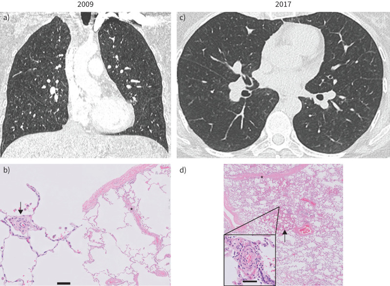
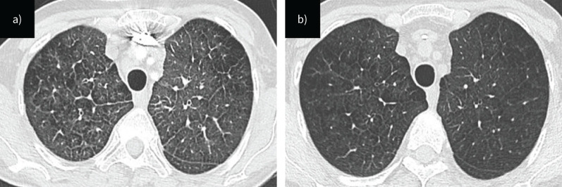
Pulmonary veno-occlusive disease (PVOD), also known as "pulmonary arterial hypertension (PAH) with overt features of venous/capillary involvement", is a rare cause of PAH characterised by substantial small pulmonary vein and capillary involvement, leading to increased pulmonary vascular resistance and right ventricular failure. Environmental risk factors have been associated with the development of PVOD, such as occupational exposure to organic solvents and chemotherapy, notably mitomycin. PVOD may also be associated with a mutation in the EIF2AK4 gene in heritable forms of disease. Distinguishing PVOD from PAH is critical for guiding appropriate management. Chest computed tomography typically displays interlobular septal thickening, ground-glass opacities and mediastinal lymphadenopathy. Life-threatening pulmonary oedema is a complication of pulmonary vasodilator therapy that can occur with any class of PAH drugs in PVOD. Early referral to a lung transplant centre is essential due to the poor response to therapy when compared with other forms of PAH. Histopathological analysis of lung explants reveals microvascular remodelling with typical fibrous veno-occlusive lesions. This review covers the main features distinguishing PVOD from PAH and two clinical cases that illustrate the challenges of PVOD management.
Copyright ©The authors 2024.
Conflict of interest statement
Conflict of interest: B. Lechartier reports travel support from Janssen and advisory board participation with MSD, outside the submitted work. A. Boucly reports grants from Acceleron, Janssen and MSD; lecture honoraria from Janssen, MSD, AOP Orphan and Ferrer; and travel support from Janssen, MSD and Ferrer, outside the submitted work. D. Montani reports grants from Acceleron, Janssen and MSD; consulting fees from Acceleron, Merck MSD, Janssen and Ferrer; and lecture honoraria from Bayer, Janssen, Boehringer, Chiesi, GSK, Ferrer and Merck MSD, outside the submitted work. All other authors have nothing to disclose.
Figures
Comment in
References
Publication types
MeSH terms
Substances
LinkOut - more resources
Full Text Sources
Medical