. 2024 Mar 4;11(3):ENEURO.0268-23.2023.
doi: 10.1523/ENEURO.0268-23.2023. Print 2024 Mar.

Hypercapnia Causes Injury of the Cerebral Cortex and Cognitive Deficits in Newborn Piglets

Affiliations

Hypercapnia Causes Injury of the Cerebral Cortex and Cognitive Deficits in Newborn Piglets

Karen Fritz et al. eNeuro. .

Abstract

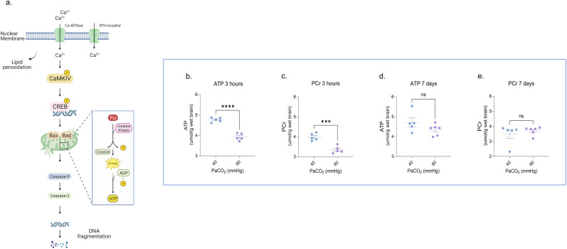
In critically ill newborns, exposure to hypercapnia (HC) is common and often accepted in neonatal intensive care units to prevent severe lung injury. However, as a "safe" range of arterial partial pressure of carbon dioxide levels in neonates has not been established, the potential impact of HC on the neurodevelopmental outcomes in these newborns remains a matter of concern. Here, in a newborn Yorkshire piglet model of either sex, we show that acute exposure to HC induced persistent cortical neuronal injury, associated cognitive and learning deficits, and long-term suppression of cortical electroencephalogram frequencies. HC induced a transient energy failure in cortical neurons, a persistent dysregulation of calcium-dependent proapoptotic signaling in the cerebral cortex, and activation of the apoptotic cascade, leading to nuclear deoxyribonucleic acid fragmentation. While neither 1 h of HC nor the rapid normalization of HC was associated with changes in cortical bioenergetics, rapid resuscitation resulted in a delayed onset of synaptosomal membrane lipid peroxidation, suggesting a dissociation between energy failure and the occurrence of synaptosomal lipid peroxidation. Even short durations of HC triggered biochemical responses at the subcellular level of the cortical neurons resulting in altered cortical activity and impaired neurobehavior. The deleterious effects of HC on the developing brain should be carefully considered as crucial elements of clinical decisions in the neonatal intensive care unit.

Keywords: cortex; hypercapnia; hypercarbia; neonatal; permissive; piglet.


Figures

Figure 1.
Figure 1.
Neonatal HC induces persistent abnormal neuronal cortical activity and behavioral deficits. a, Severe (PaCO2 80 mmHg) HC piglets exhibited markedly inferior task scores and (b) experienced a prolonged time to complete the task compared with their NC counterparts. c–j, Bipolar-distance EEGs showed that a 3 h exposure to severe HC significantly increased EEG frequencies in all channels, which normalized after 1 h of spontaneous recovery to room air. After a 7 d recovery period, the cortical activity globally decreased in the HC piglets compared with the baseline in all channels. k, Depiction of peroxidation of cellular and subcellular neuronal membranes. l, Measurement of indirect lipid peroxidation products from synaptosomes showed an increase in FC after 3 h of severe HC, m, which remained elevated even 7 d after the initial exposure. The above noninstrumented chamber piglets were exposed to 3 h of severe (CO2, 80 mmHg) HC with and without 7 d of NC recovery after HC. Each group was compared with a group of sham piglets with a similar timeline of events except for the EEG studies where each HC piglet served as their control by using their initial NC period as the control. Extended Data Figure 1-1 demonstrates the experimental animal protocols for the piglets. Extended Data Figure 1-2 shows the representation of the milk-drinking training system for evaluating behavioral and cognitive function. Extended Data Figure 1-3 demonstrates the effect of 1 h of severe HC (PaCO2 80 mmHg) on bipolar-distance EEGs. HC, hypercapnia; EEG, electroencephalogram; FC, fluorescent compounds; PaCO2, partial pressure of carbon dioxide; NC, normocapnia. Statistical analysis was performed using one-way ANOVA for multiple groups and two-tailed t tests for two groups by Prism statistical software, and the graph displays the mean ± SEM values; *p < 0.05, **p < 0.01, ***p < 0.001, ****p < 0.0001; n = 5–7/group. Part of this figure was created with BioRender.
Figure 2.
Figure 2.
Neonatal HC–induced neuronal energy failure is transient. a, Schematic representation illustrating the impact of peroxidation of neuronal membranes on energy demand. b,c, Severe HC for 3 h transiently decreased tissue ATP and PCr levels in cortical neurons. d,e, However, ATP and PCr levels in cortical neurons returned to normal after 7 d of recovery following the HC insult. The above noninstrumented chamber piglets were exposed to 3 h of severe (PaCO2 80 mmHg) HC with and without 7 d of NC recovery post HC. Each group was compared with a group of NC sham piglets with a similar timeline of events. HC, hypercapnia; ATP, adenosine triphosphate; PCr, phosphocreatine; PaCO2, partial pressure of carbon dioxide; NC, normocapnia. Statistical analysis was performed using one-way ANOVA for multiple groups and two-tailed t tests for two groups by Prism statistical software, and the graph displays the mean ± SEM values; *p < 0.05, **p < 0.01, ***p < 0.001, ****p < 0.0001; n = 5–6/group. Part of this figure was created with BioRender.
Figure 3.
Figure 3.
Neonatal HC induces persistent dysregulation of calcium-dependent proapoptotic signaling in the cerebral cortex. a, Schematic representation of the HC-induced activation of Ca2+-dependent pathways and its consequential impact on the expression of apoptotic proteins. HC increased both (b,c) intranuclear Ca2+ influx and (d,e) CaMK IV activity after 3 h of exposure which persisted 7 d after the insult. f,g, Three hours of HC also increased ratios of Bax/Bcl-2 and of Bad/Bcl-xl in the nucleus, cytosol, and mitochondria, which persisted at 7 d of recovery after HC, especially in the cytosol (Bax) and nucleus (Bad). The above noninstrumented chamber piglets were exposed to 3 h of severe (PaCO2 80 mmHg) HC with and without 7 d of NC recovery post HC. Each group was compared with a group of NC sham piglets with a similar timeline of events. Extended Data Figure 3-1 illustrates the impact of HC on nuclear Ca2+ signaling and Extended Data Figure 3-2 demonstrates the expression of pro- and antiapoptotic proteins following HC exposure. Representative Western blots of Bax, Bad, Bcl-2, and Bcl-xl expression are shown in Extended Data Figure 3-3. Ca2+, calcium; HC, hypercapnia; CaMK IV, calmodulin-dependent protein kinase IV; NC, normocapnia, PaCO2, partial pressure of carbon dioxide. Statistical analysis was performed using one-way ANOVA for multiple groups and two-tailed t tests for two groups by Prism statistical software, and the graph displays the mean ± SEM values; *p < 0.05, **p < 0.01, ***p < 0.001, ****p < 0.0001; n = 5–7/group. Part of this figure was created with BioRender.
Figure 4.
Figure 4.
Neonatal HC causes neuronal apoptosis and DNA fragmentation. a, The schematic representation illustrates the cell death process where activation of proapoptotic signals leads to the activation of caspases, which, in turn, results in nuclear DNA fragmentation and prolonged cell death. b–e, Three hours of HC increased levels of caspase-9 and caspase-3 expression, which returned to baseline after 7 d of recovery under NC conditions; (f) however, DNA fragmentation was elevated at 7 d after HC compared with NC controls. The above noninstrumented chamber piglets were exposed to 3 h of severe (PaCO2 80 mmHg) HC with and without 7 d of NC recovery post HC. Each group was compared with a group of NC sham piglets with a similar timeline of events. Extended Data Figure 4-1 illustrates the effect of HC on caspase-9 and caspase-3 expression and activity and on nuclear DNA fragmentation. Representative Western blots of caspase-9 and caspase-3 expression are shown in Extended Data Figures 4-3 and 4-4 and of DNA fragmentation in Extended Data Figure 4-2. DNA, deoxyribonucleic acid; HC, hypercapnia; NC, normocapnia; PaCO2, partial pressure of carbon dioxide. Statistical analysis was performed using one-way ANOVA for multiple groups and two-tailed t tests for two groups by Prism statistical software, and the graph displays the mean ± SEM values; *p < 0.05, **p < 0.01, ***p < 0.001, ****p < 0.0001; n = 5/group. Part of this figure was created with BioRender.
Figure 5.
Figure 5.
Rapid resuscitation to normocapnia does not prevent HC-induced cortical injury. Rapid normalization of PaCO2 following 1 h of HC in newborn piglets (a–d) showed no effect on ATP and PCr levels either after 1 h of HC or following the rapid resuscitation of HC. However, following rapid resuscitation after HC, there was a delayed elevation of (e–h) synaptosomal lipid peroxidation products, FC and CDs, (i–l) intranuclear Ca2+ influx, and caspase-3 activity. Each of the above groups of ventilated piglets was exposed to either 1 h of moderate (PaCO2, 65 mmHg) or severe (PaCO2, 80 mmHg) HC or 1 h of HC followed by 1 h of rapid resuscitation. Each group was compared with a group of NC sham piglets with similar instrumentation and timeline of events. PaCO2, partial pressure of carbon dioxide; HC, hypercapnia; ATP, adenosine triphosphate; PCr, phosphocreatine; FC, fluorescent compounds; CD, conjugated dienes; Ca2+, calcium; NC, normocapnia. Statistical analysis was performed using one-way ANOVA for multiple groups and two-tailed t tests for two groups by Prism statistical software, and the graph displays the mean ± SEM values; *p < 0.05, **p < 0.01, ***p < 0.001, ****p < 0.0001; n = 4–10/group.

Similar articles

Cited by

References

    1. Akhter W, Ashraf QM, Zanelli SA, Mishra OP, Delivoria-Papadopoulos M (2001) Effect of graded hypoxia on cerebral cortical genomic DNA fragmentation in newborn piglets. Biol Neonate 79:187–193. 10.1159/000047089 - DOI - PubMed
    1. Austoker J, Cox D, Mathias AP (1972) Fractionation of nuclei from brain by zonal centrifugation and a study of the ribonucleic acid polymerase activity in the various classes of nuclei. Biochem J 129:1139–1155. 10.1042/bj1291139 - DOI - PMC - PubMed
    1. Ayala A, Munoz MF, Arguelles S (2014) Lipid peroxidation: production, metabolism, and signaling mechanisms of malondialdehyde and 4-hydroxy-2-nonenal. Oxid Med Cell Longev 2014:360438. 10.1155/2014/360438 - DOI - PMC - PubMed
    1. Booth RF, Clark JB (1978) A rapid method for the preparation of relatively pure metabolically competent synaptosomes from rat brain. Biochem J 176:365–370. 10.1042/bj1760365 - DOI - PMC - PubMed
    1. Buonocore G, Liberatori S, Bini L, Mishra OP, Delivoria-Papadopoulos M, Pallini V, Bracci R (1999) Hypoxic response of synaptosomal proteins in term guinea pig fetuses. J Neurochem 73:2139–2148. 10.1046/j.1471-4159.1999.02139.x - DOI - PubMed

LinkOut - more resources