. 2024 Jan 17;10(1):34.
doi: 10.1038/s41420-023-01794-3.

NMDARs activation regulates endothelial ferroptosis via the PP2A-AMPK-HMGB1 axis

Affiliations

NMDARs activation regulates endothelial ferroptosis via the PP2A-AMPK-HMGB1 axis

Wei-Min Han et al. Cell Death Discov. .

Abstract

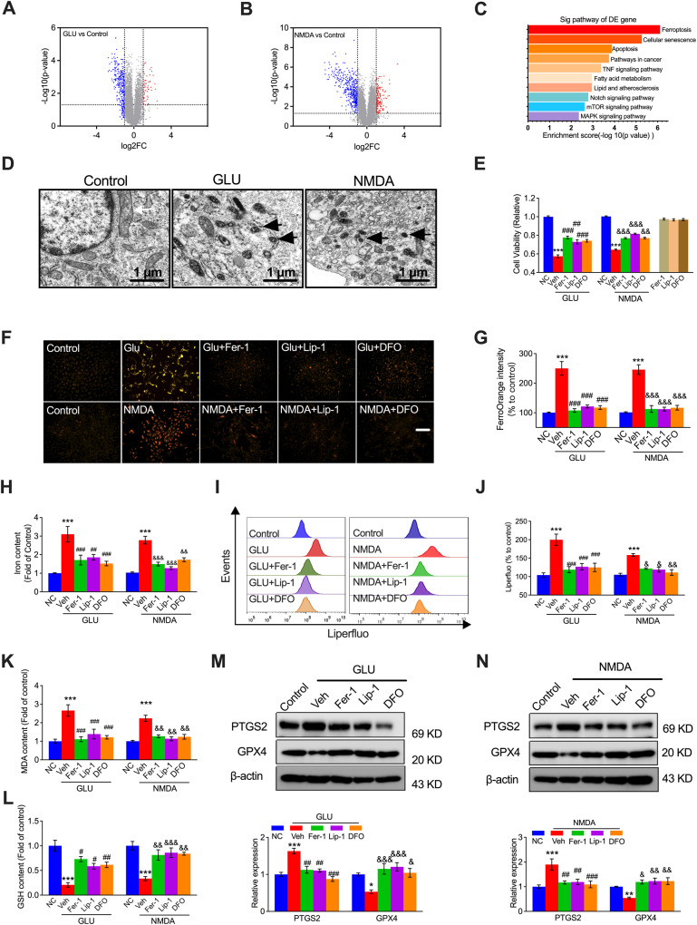
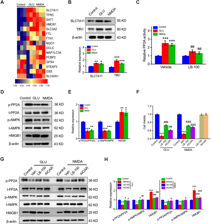
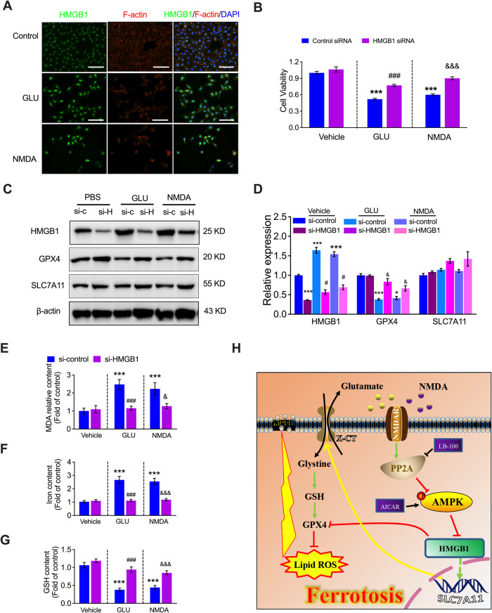
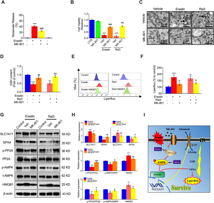
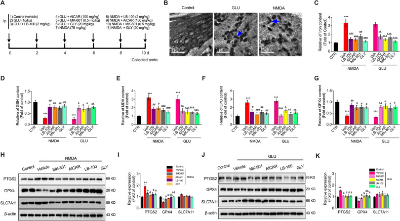
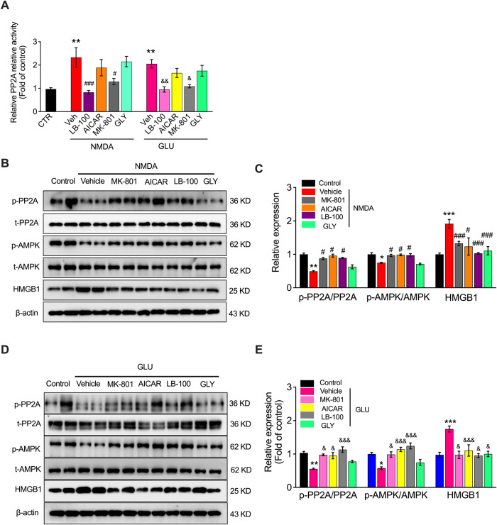
N-methyl-D-aspartate receptors (NMDARs) are ligand-gated, voltage-dependent channels of the ionotropic glutamate receptor family. The present study explored whether NMDAR activation induced ferroptosis in vascular endothelial cells and its complicated mechanisms in vivo and in vitro. Various detection approaches were used to determine the ferroptosis-related cellular iron content, lipid reactive oxygen species (LOS), siRNA molecules, RNA-sequence, MDA, GSH, and western blotting. The AMPK activator Acadesine (AICAR), HMGB1 inhibitor glycyrrhizin (GLY), PP2A inhibitor LB-100, and NMDAR inhibitor MK801 were used to investigate the involved in vivo and in vitro pathways. The activation of NMDAR with L-glutamic acid (GLU) or NMDA significantly promoted cellular ferroptosis, iron content, MDA, and the PTGS2 expression, while decreasing GPX4 expression and GSH concentration in human umbilical vein endothelial cells (HUVECs), which was reversed by ferroptosis inhibitors Ferrostatin-1(Fer-1), Liproxstatin-1 (Lip-1), or Deferoxamine (DFO). RNA-seq revealed that ferroptosis and SLC7A11 participate in NMDA or GLU-mediated NMDAR activation. The PP2A-AMPK-HMGB1 pathway was majorly associated with NMDAR activation-induced ferroptosis, validated using the PP2A inhibitor LB-100, AMPK activator AICAR, or HMGB1 siRNA. The role of NMDAR in ferroptosis was validated in HUVECs induced with the ferroptosis activator errasin or RSL3 and counteracted by the NMDAR inhibitor MK-801. The in vivo results showed that NMDA- or GLU-induced ferroptosis and LOS production was reversed by MK-801, LB-100, AICAR, MK-801, and GLY, confirming that the PP2A-AMPK-HMGB1 pathway is involved in NMDAR activation-induced vascular endothelium ferroptosis. In conclusion, the present study demonstrated a novel role of NMDAR in endothelial cell injury by regulating ferroptosis via the PP2A-AMPK-HMGB1 pathway.


Conflict of interest statement

The authors declare no competing interests.

Figures

Fig. 1
Fig. 1. Ferroptosis participates in NMDAR activation-mediated endothelial cell damage in vitro.
RNA sequencing was performed after in HUVECs treated with GLU (20 mM) or NMDA (1 mM) for 24 h, and the results of volcano plot (A, B) and KEGG analysis (C) were shown. D Ultrastructure of the mitochondria in HUVECs treated with GLU or NMDA, visualized by TEM. Black arrowheads: shrunken mitochondria (Scale bars, 1 μm). E MTT assay and (F) ferrous ion levels visualized by FerroOrange staining (Scale bars, 200 μm) and (G) corresponding quantification showing cell viability and ferrous ion levels in HUVECs pretreated with ferroptosis inhibitors (Fer-1: 10 μM, Lip-1: 2 μM, or DFO: 10 μM) for 1 h followed by GLU and NMDA treatment for 24 h. The relative levels of iron content (H), flow cytometry plots (I), and quantification of lipid ROS (LOS) production (J), MDA (K), and GSH (L) in HUVECs pretreated with Fer-1 (10 μM), Lip-1 (2 μM), or DFO (10 μM) for 1 h followed by GLU or NMDA treatment for 24 h. The relative levels of ferroptosis biomarkers PTGS2 and GPX4 shown by immunoprotein blots and quantification in the presence of GLU (M) or NMDA (N) and treatment with Fer-1 (10 μM), Lip-1 (2 μM), or DFO (10 μM). *P < 0.05, **P < 0.01 and ***P < 0.001 vs control; #P < 0.05, ##P < 0.01 and ###P < 0.001 vs GLU; &P < 0.05, &&P < 0.01 and &&&P < 0.001 vs NMDA.
Fig. 2
Fig. 2. Activation of SLC7A11 via the PP2A-AMPK axis contributes to NMDAR-activated ferroptosis in HUVECs.
A Differentially expressed genes closely associated with HUVEC ferroptosis in NMDA- or GLU-treated cells determined by RNA-seq. Blue: low expression levels; Red: high expression levels. BProtein blotting and quantitation of SLC7A11 and TfR1 expression in HUVECs in response to GLU or NMDA treatment. *P < 0.05, **P < 0.01 and ***P < 0.001 vs control. C The relative PP2A activity of HUVECs after pretreatment with LB-100 (0.5 μM) followed by NMDA or GLU treatment (n = 6–8). ***P < 0.001 vs control with vehicle; ##P < 0.01 < 0.001 vs control with LB-100. D Protein expression levels and (E) quantitation of SLC7A11 upstream, including PP2A, AMPK, and HMGB1, were analyzed by western blotting (n = 6). *P < 0.05 **P < 0.01, and ***P < 0.001 vs control. F MTT assay detected the cell viability of HUVECs treated with NMDA or GLU and/or LB-100 (0.5 μM) and AICAR (25 μM) for 24 h (n = 6). ***P < 0.001 vs control; ##P < 0.01 and ###P < 0.001 vs GLU; &&&P < 0.001 vs NMDA. G Protein expression levels and (H) quantitation of LB-100 and AICAR with the relative expression of p-PP2A, PP2A, p-AMPK, p-AMPK, and HMGB1 in HUVECs (n = 5). *P < 0.05, **P < 0.01 and ***P < 0.001 vs control; #P < 0.05, ##P < 0.01, and ###P < 0.001 vs GLU; &P < 0.05 and &&&P < 0.001 vs NMDA.
Fig. 3
Fig. 3. Knockdown of HMGB1 suppresses NMDAR activation-induced ferroptosis.
A Representative image of immunostaining of HMGB1 in the presence of GLU or NMDA in HUVECs. B Representative cell viability of the control siRNA or HMGB1 siRNA-transfected HUVECs in the presence of GLU or NMDA with MTT assay (n = 5). C Protein expression levels and (D) quantitation of HMGB1, GPX4, and SLC7A1 in the control siRNA or HMGB1 siRNA-transfected HUVECs in the presence of GLU or NMDA (n = 5). E MDA content, (F) iron content, and (G) GSH levels in the control siRNA or HMGB1 siRNA-transfected HUVECs in the presence of GLU or NMDA (n = 5). H Scheme of the mechanism of NMDAR inhibitor MK-801 in VEC ferroptosis. *P < 0.05, ***P < 0.001 vs control siRNA; ###P < 0.001 vs control siRNA + GLU, &P < 0.05 and &&&P < 0.001 vs control siRNA + NMDA.
Fig. 4
Fig. 4. Inhibition of NMDAR ameliorates Erastin- or RSL3-induced ferroptosis in vitro.
A Glutamate release in HUVECs treated with Erastin in the presence of the NMDAR inhibitor MK801. B MTT assay showing HUVECs treated with Erastin or RSL3 in the presence of MK-801. C Ultrastructure of mitochondria in HUVECs treated with Erastin or RSL3 in the presence of MK-801. Black arrowheads: shrunken mitochondria. Scale bars, 5 μm. D GSH content in HUVECs pretreated with ferroptosis inducers (Erastin, 10 μM; RSL3, μM) for 24 h and then treated with MK801. E Representative flow cytometry image and quantification (F) of LOS production in HUVECs pretreated with a ferroptosis inducer (Erastin, 10 μM; RSL3, μM) for 24 h and treated with MK801. G Immunoblots and quantification (H) of SLC7A11, GPX4, p-PP2A, PP2A, p-AMPK, AMPK, and HMGB1 in HUVECs pretreated with a ferroptosis inducer (Erastin, 10 μM; RSL3, μM) for 24 h and treated with MK801. I Scheme of the protective mechanism of inhibiting HMGB1 overexpression by MK-801 treatment against ferroptosis in VECs. **P < 0.01 and ***P < 0.001 vs control; #P < 0.05, ##P < 0.01 and ###P < 0.001 vs Erastin; &P < 0.05 and &&P < 0.01 vs RSL3.
Fig. 5
Fig. 5. NMDAR and PP2A-AMPK-HMGB1 pathway mediate VEC ferroptosis in vivo.
A Experimental design summarizing the procedure. B Ultrastructure of the mitochondria of the vascular tissue of mice treated with GLU (1 g/kg) or NMDA (75 mg/kg) for 10 days, visualized by TEM. Black arrowheads: shrunken mitochondria. Scale bars, 5 μm. The relative levels of (C) iron, (D) GSH, (E) MDA, (F) LPO, and (G) GPX4 content in the vascular tissue of the treated mice. H Western blot image and (I) quantification of PTGS2, GPX4, and SLC7A11 in NMDA and/or LB100-, AICAR-, MK-801-, and GLY-treated mice. ***P < 0.001 vs control; #P < 0.05, ##P < 0.01 and ###P < 0.001 vs NMDA, &P < 0.05, &&P < 0.01 and &&&P < 0.001 vs GLU. J Western blot image and (K) quantification of PTGS2, GPX4, and SLC7A11 in GLU and/or LB100-, AICAR-, MK-801-, and GLY-treated mice. *P < 0.05, **P < 0.01 and ***P < 0.001 vs control; #P < 0.05, ##P < 0.01 and ###P < 0.001 vs NMDA/GLU in PTGS2 expression; &P < 0.05, &&P < 0.01 and &&&P < 0.001 vs NMDA/GLU in GPX4 expression.
Fig. 6
Fig. 6. NMDAR activation-mediated VEC ferroptosis is regulated by the PP2A-AMPK-HMGB1 pathway in vivo.
A PP2A activity in vascular tissue after treatment with LB100, AICAR, MK-801, and GLY in the presence or absence of NMDA or GLU in vivo. B Western blot image and (C) quantification of p-PP2A, PP2A, p-AMPK, AMPK, and HMGB1 in GLU and/or LB100-, AICAR-, MK-801-, and GLY-treated mice. D Western blot image and (E) quantification of p-PP2A, PP2A, p-AMPK, AMPK, and HMGB1 in GLU and/or LB100-, AICAR-, MK-801-, and GLY-treated mice. *P < 0.05, **P < 0.01 and ***P < 0.001 vs control; #P < 0.05 and ###P < 0.001 vs NMDA; &P < 0.05, &&P < 0.01 and &&&P < 0.001 vs GLU.

References

    1. Wardlaw JM, Smith C, Dichgans M. Small vessel disease: mechanisms and clinical implications. Lancet Neurol. 2019;18:684–96. doi: 10.1016/S1474-4422(19)30079-1. - DOI - PubMed
    1. Sturtzel C. Endothelial cells. Adv Exp Med Biol. 2017;1003:71–91. doi: 10.1007/978-3-319-57613-8_4. - DOI - PubMed
    1. Fels J, Kusche-Vihrog K. Endothelial nanomechanics in the context of endothelial (Dys)function and inflammation. Antioxid Redox Signal. 2019;30:945–59. doi: 10.1089/ars.2017.7327. - DOI - PMC - PubMed
    1. Gimbrone MA, Jr., Garcia-Cardena G. Endothelial cell dysfunction and the pathobiology of atherosclerosis. Circ Res. 2016;118:620–36. doi: 10.1161/CIRCRESAHA.115.306301. - DOI - PMC - PubMed
    1. Yang Q, Xu J, Ma Q, Liu Z, Sudhahar V, Cao Y, et al. PRKAA1/AMPKalpha1-driven glycolysis in endothelial cells exposed to disturbed flow protects against atherosclerosis. Nat Commun. 2018;9:4667. doi: 10.1038/s41467-018-07132-x. - DOI - PMC - PubMed