. 2024 May;29(5):1322-1337.
doi: 10.1038/s41380-024-02412-z. Epub 2024 Jan 17.

The amygdala NT3-TrkC pathway underlies inter-individual differences in fear extinction and related synaptic plasticity

Affiliations

The amygdala NT3-TrkC pathway underlies inter-individual differences in fear extinction and related synaptic plasticity

Gianluca Masella et al. Mol Psychiatry. 2024 May.

Abstract

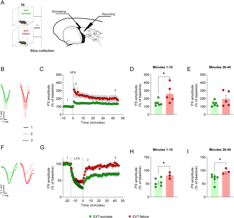
Fear-related pathologies are among the most prevalent psychiatric conditions, having inappropriate learned fear and resistance to extinction as cardinal features. Exposure therapy represents a promising therapeutic approach, the efficiency of which depends on inter-individual variation in fear extinction learning, which neurobiological basis is unknown. We characterized a model of extinction learning, whereby fear-conditioned mice were categorized as extinction (EXT)-success or EXT-failure, according to their inherent ability to extinguish fear. In the lateral amygdala, GluN2A-containing NMDAR are required for LTP and stabilization of fear memories, while GluN2B-containing NMDAR are required for LTD and fear extinction. EXT-success mice showed attenuated LTP, strong LTD and higher levels of synaptic GluN2B, while EXT-failure mice showed strong LTP, no LTD and higher levels of synaptic GluN2A. Neurotrophin 3 (NT3) infusion in the lateral amygdala was sufficient to rescue extinction deficits in EXT-failure mice. Mechanistically, activation of tropomyosin receptor kinase C (TrkC) with NT3 in EXT-failure slices attenuated lateral amygdala LTP, in a GluN2B-dependent manner. Conversely, blocking endogenous NT3-TrkC signaling with TrkC-Fc chimera in EXT-success slices strengthened lateral amygdala LTP. Our data support a key role for the NT3-TrkC system in inter-individual differences in fear extinction in rodents, through modulation of amygdalar NMDAR composition and synaptic plasticity.


Conflict of interest statement

The authors declare no competing interests.

Figures

Fig. 1
Fig. 1. Inter-individual variation in context fear extinction.
A Schematic representation of the Pavlovian contextual fear conditioning and extinction paradigm to which young adult (8 to 12 weeks old) C56BL/6J male mice were submitted throughout this study. B Quantification of the percentage of time spent freezing during fear extinction acquisition and extinction memory retrieval. Fear-conditioned mice were categorized as EXT-success (n = 5) or EXT-failure (n = 11) according to their extinction learning performance. A CTRL-no shock group was included that received no shocks (n = 9). Repeated measures two-way ANOVA with either Sidak or Tukey multiple comparisons test. * CTRL-no shock vs. EXT-failure; £ CTRL-no shock vs. EXT-success; # EXT-success vs. EXT-failure; * EXT-success, R/E1 vs. E6 and R/E1 vs. EM; * EXT-failure, R/E1 vs. E6. C Extinction learning performance, expressed as the percentage of freezing levels in E6 relative to freezing in R/E1. The dotted line marks the threshold of 30% reduction from R/E1 used to categorize mice as EXT-success or EXT-failure. *** two-tailed Student’s t test. D Extinction memory performance, expressed as the percentage of freezing levels in extinction retrieval relative to freezing in R/E1. *** Mann–Whitney U test. E Correlation of the freezing levels during the last trial of extinction acquisition (E6) with the freezing levels in extinction retrieval. Pearson r. F Correlation matrix of the total distance traveled, distance in open arms and time spent in the open arms in the EPM test with the percentage of time spent freezing during extinction acquisition trials and with ELP and EMP; statistics using Pearson r. CS conditioned stimulus, E1 to E6 extinction trials, EM extinction memory retrieval, ELP extinction learning performance, EMP extinction memory performance, EPM elevated plus maze, R fear retrieval, US unconditioned stimulus.; *,£,#p ≤ 0.05, **p ≤ 0.01, ***p ≤ 0.001.
Fig. 2
Fig. 2. EXT-success mice show attenuated LTP and increased LTD in LA synapses.
A Schematic representation of experimental conditions and localization of stimulating and recording electrodes in horizontal slices containing the LA. BE LTP was induced ex-vivo in the LA of EXT-success (n = 6) and EXT-failure (n = 5) brain slices with a HFS protocol (three 1 s duration trains of 100 Hz pulses, with 5 s inter-train interval; vertical dashed line). B Representative traces for EXT-success and EXT-failure slices at baseline (__), upon 10 min () and upon 45 min (---) after HFS. C Time course of LTP recorded for 45 min following LTP induction. PS amplitude was averaged for the (D) first 10 min and (E) last 10 min of recordings following LTP induction. FI LTD was induced ex-vivo in the LA of EXT-success (n = 6) and EXT-failure (n = 3) brain slices with a low-frequency stimulation protocol (900 stimuli at 1 Hz; between the two vertical lines). F Representative traces for EXT-success and EXT-failure slices at baseline (__), upon 10 min () and 45 min (---) of LTD induction with the LFS train. G Time course of LTD recorded for 45 min following induction. PS amplitude was averaged for the (H) first 10 min and (I) last 10 min of recordings. D, E, H, I Two-tailed Student’s t test and Mann–Whitney U test. BLA basolateral amygdala, Ce central amygdala, E6 extinction trial 6, HFS high-frequency stimulation, LA lateral amygdala, LFS low-frequency stimulation, PS population spikes. *p ≤ 0.05.
Fig. 3
Fig. 3. Differential surface density of GluN2A and GluN2B subunits of NMDA receptors in amygdala synaptoneurosomes from EXT-success and EXT-failure mice.
A Schematic representation of experimental conditions and biologic material used. B, E Representative images of synaptoneurosomes isolated from the amygdalae of EXT-success and EXT-failure mice, live stained for GluN2A and GluN2B subunits of NMDA receptors. Synaptoneurosomes were identified with co-staining against the postsynaptic marker PSD95 and the presynaptic marker VGlut1 and inspection of intact membranes by phase contrast. C, F Integrated density of GluN2A and GluN2B signal was quantified in synaptoneurosomes isolated from the amygdalae of EXT success (GluN2A n = 631, GluN2B n = 529) and EXT-failure (GluN2A n = 612; GluN2B n = 518) mice. D, G The percentage of GluN2A- and GluN2B-positive amygdala synaptoneurosomes was calculated for EXT-success and EXT-failure animals (n = 4 independent experiments). C, F Mann–Whitney U test, (D, G) two-tailed Student’s t test. E6 extinction trial 6, GluN2A subunit 2A of NMDA receptor, GluN2B subunit 2B of NMDA receptor, PSD95 postsynaptic density 95, VGluT1 vesicular glutamate transporter 1. Scale bar 0.5 µm. *p ≤ 0.05, ***p ≤ 0.001.
Fig. 4
Fig. 4. Amygdalar TrkC activation is associated with a successful fear extinction performance.
A Schematic representation of experimental conditions and dissected brain region. B Representative images of western blots for phosphorylated TrkC and total TrkC performed in brain extracts from the amygdala. The panel shows non-contiguous lanes from the same membrane. Quantification of (C) relative TrkC activation as measured by pTrkC/full-length TrkC ratio, (D) total pTrkC levels, (E) total full-length TrkC levels, (F) full-length TrkC/truncated TrkC ratio and (G) truncated TrkC levels. CTRL-no shock (n = 7), EXT-success (n = 7) and EXT-failure (n = 14) mice. β-actin was used as a loading control. CG One-way ANOVA with Tukey’s multiple comparisons test. CTRL control group, E6 extinction trial 6, pTrkC phosphorylated TrkC, TrkC tropomyosin receptor kinase C. *p ≤ 0.05.
Fig. 5
Fig. 5. NT3 infusion in the BLA rescues fear extinction deficits in EXT-failure mice.
Representative images of western blots for (A) pTrkC and (C) pTrkB performed using protein extracts from NT3-infused amygdalae versus contralateral not-infused amygdalae (n = 4 per condition). Quantification of total (B) pTrkC and (D) pTrkB levels. β-actin was used as a loading control. Two-tailed Student’s t test. E Schematic representation of experimental conditions and treatments. F Schematic representation of cannulas placement in the BLA. Circles represent the tip of the internal cannula. Mice with misplaced cannulas were excluded from the analysis (EXT-success n = 2, EXT-failure n = 1). G Quantification of the percentage of time spent freezing during extinction acquisition session. Fear-conditioned mice were categorized as EXT-success (n = 6) or EXT-failure (n = 12), according to their extinction learning performance. Repeated measures two-way ANOVA with Tukey multiple comparisons test. * EXT-success, R/E1 vs. E6. H Extinction learning performance, expressed as the percentage of freezing levels in E6 relative to freezing in R/E1. Mann–Whitney U test. I Extinction memory performance, expressed as the percentage of freezing levels in extinction memory retrieval relative to freezing in R/E1. The dotted line marks the threshold of 30% reduction from R/E1 used to categorize mice as EXT-success or EXT-failure. One-way ANOVA with Tukey multiple comparisons test. E1-E6, extinction trial 1 to 6; ELP, extinction learning performance; EMP, extinction memory performance; NT3, neurotrophin 3; pTrkC, phosphorylated tropomyosin receptor kinase C; pTrkB, phosphorylated tropomyosin receptor kinase B; R/E1, fear retrieval/extinction trial 1. *p ≤ 0.05, **p ≤ 0.01, ***p ≤ 0.001.
Fig. 6
Fig. 6. NT3 attenuates LTP at LA synapses in a GluN2B-dependent mechanism.
A Schematic representation of experimental conditions and localization of stimulating and recording electrode in horizontal brain slices containing the LA. A HFS protocol (three 1 s duration trains of 100 Hz pulses, with 5 s inter-train interval; dashed vertical line) was used to induce ex-vivo LTP in the LA of (BE) EXT-failure slices untreated (n = 6), treated with NT3 (n = 6, 50 ng/mL), treated with ifenprodil (n = 4, 3.25 µg/mL), treated with NT3+ifenprodil (n = 5, NT3 50 ng/mL and ifenprodil 3.25 µg/mL) and of (FI) EXT-success slices untreated (n = 6) and treated with TrkC-Fc chimera (n = 4, 0.5 µg/mL). Tested drugs were added 45 min before HFS and were maintained in the medium until the end of the experiment. B Representative traces for EXT-failure slices untreated and treated with NT3, ifenprodil and NT3+ifenprodil at baseline (__), upon 10 min () and 45 min (---) of HFS. C LTP time course recorded for 45 min following induction. Amplitude of PS was averaged for the (D) first 10 min and (E) last 10 min of recordings following LTP induction. Repeated measures two-way ANOVA with Tukey multiple comparisons test. F Representative traces for EXT-success slices untreated and treated with TrkC-Fc at baseline (__), upon 10 min () and 45 min (---) of HFS. G LTP time course recorded for 45 min following induction. PS amplitude was averaged for the (H) first 10 min and (I) last 10 min of recordings following LTP induction. Two-tailed Student’s t test. BLA basolateral amygdala, Ce central amygdala, E6 extinction trial 6, HFS high frequency stimulation, LA lateral amygdala, NT3 neurotrophin 3, PS population spikes. TrkC-Fc tropomyosin receptor kinase C–Fc chimera. *p ≤ 0.05.
Fig. 7
Fig. 7. Proposed theoretical model.
Contextual fear conditioning induces TrkC inactivation [32] (A) and synaptic accumulation of GluN2A-containing NMDAR in the amygdala [26] (B), which are responsible for LA LTP [25]. This leads to the induction of GluN2A-dependent LTP in the LA fear-specific microcircuit (C), a necessary step for the acquisition of conditioned fear [42]. Given that EXT-success and EXT-failure mice form equally strong fear memories, accumulation of GluN2A-containing NMDAR in synapses and LTP in the LA are not expected to differ in the two groups. Retrieval and concomitant extinction training initiates two parallel processes rendering fear memory labile and amenable for destabilization or reconsolidation on one side and activating the extinction microcircuit on the other side [28, 49]. At this critical timepoint, amygdalar TrkC activation, as observed in EXT-success mice (D), will promote the synaptic accumulation of GluN2B- in detriment of GluN2A-containing NMDAR (E), resulting in the attenuation of the fear microcircuit, weakening LA LTP (F), and activation of the extinction circuit inducing LTD, thereby promoting fear memory destabilization and extinction consolidation (G). In EXT-failure animals, insufficient amygdalar TrkC activation (H) will prevent the GluN2A to GluN2B switch at synapses (I) and thereby promoting the GluN2A-dependent memory stabilization and reconsolidation (J), maintaining levels of fear high.

Similar articles

Cited by

References

    1. Bandelow B, Michaelis S. Epidemiology of anxiety disorders in the 21st century. Dialogues Clin Neurosci. 2015;17:327–35. doi: 10.31887/DCNS.2015.17.3/bbandelow. - DOI - PMC - PubMed
    1. Michael T, Zetsche U, Margraf J. Epidemiology of anxiety disorders. Psychiatry. 2007;6:136–42. doi: 10.1016/j.mppsy.2007.01.007. - DOI
    1. Wicking M, Steiger F, Nees F, Diener SJ, Grimm O, Ruttorf M, et al. Deficient fear extinction memory in posttraumatic stress disorder. Neurobiol Learn Mem. 2016;136:116–26. doi: 10.1016/j.nlm.2016.09.016. - DOI - PubMed
    1. American Psychiatric Association AP. Diagnostic and Statistical Manual of Mental Disorders (DSM-5®). Washigton DC, United States: American Psychiatric Pub; 2013, 1414pp.
    1. Carpenter JK, Andrews LA, Witcraft SM, Powers MB, Smits JAJ, Hofmann SG. Cognitive behavioral therapy for anxiety and related disorders: a meta-analysis of randomized placebo-controlled trials. Depress Anxiety. 2018;35:502–14. doi: 10.1002/da.22728. - DOI - PMC - PubMed