. 2024 May;49(6):993-1006.
doi: 10.1038/s41386-024-01795-5. Epub 2024 Jan 17.

Dorsal peduncular cortex activity modulates affective behavior and fear extinction in mice

Affiliations

Dorsal peduncular cortex activity modulates affective behavior and fear extinction in mice

Justin J Botterill et al. Neuropsychopharmacology. 2024 May.

Abstract

The medial prefrontal cortex (mPFC) is critical to cognitive and emotional function and underlies many neuropsychiatric disorders, including mood, fear and anxiety disorders. In rodents, disruption of mPFC activity affects anxiety- and depression-like behavior, with specialized contributions from its subdivisions. The rodent mPFC is divided into the dorsomedial prefrontal cortex (dmPFC), spanning the anterior cingulate cortex (ACC) and dorsal prelimbic cortex (PL), and the ventromedial prefrontal cortex (vmPFC), which includes the ventral PL, infralimbic cortex (IL), and in some studies the dorsal peduncular cortex (DP) and dorsal tenia tecta (DTT). The DP/DTT have recently been implicated in the regulation of stress-induced sympathetic responses via projections to the hypothalamus. While many studies implicate the PL and IL in anxiety-, depression-like and fear behavior, the contribution of the DP/DTT to affective and emotional behavior remains unknown. Here, we used chemogenetics and optogenetics to bidirectionally modulate DP/DTT activity and examine its effects on affective behaviors, fear and stress responses in C57BL/6J mice. Acute chemogenetic activation of DP/DTT significantly increased anxiety-like behavior in the open field and elevated plus maze tests, as well as passive coping in the tail suspension test. DP/DTT activation also led to an increase in serum corticosterone levels and facilitated auditory fear extinction learning and retrieval. Activation of DP/DTT projections to the dorsomedial hypothalamus (DMH) acutely decreased freezing at baseline and during extinction learning, but did not alter affective behavior. These findings point to the DP/DTT as a new regulator of affective behavior and fear extinction in mice.


Conflict of interest statement

The authors declare no competing interests.

Figures

Fig. 1
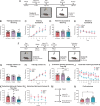
Fig. 1. Chemogenetic activation of the DP/DTT increases anxiety-like behavior in the open field and elevated plus maze tests.
A, B Experimental timeline for the OFT. Mice previously injected with hM3D, hM4D or control mCherry constructs in the DP/DTT were injected with the DREADD agonist C21 one hour prior to being placed in an open field. The time spent in the center zone (red) and the outer zone (white) was measured. C The time spent in the center zone of the open field was significantly reduced in hM3D mice compared to mCherry and hM4D mice (no difference between mCherry and hM4D, p = 0.965). D The amount of time spent in the outer zone of the open field arena was significantly greater in the hM3D mice compared to mCherry and hM4D mice (mCherry versus hM4D, p = 0.938). E The total distance traveled during the test did not differ between groups (one-way ANOVA, F2,38 = 2.292, p = 0.115). F Representative track and heat maps. G, H Experimental timeline for the EPM test. Mice previously injected with hM3D, hM4D or control mCherry constructs in the DP/DTT were injected with the DREADD agonist C21 one hour prior to being placed in an elevated plus maze. The time spent in the open arms vs closed arms was measured. I The time spent in the open arms of the elevated plus maze was significantly reduced in hM3D mice compared to mCherry and hM4D mice (no difference between mCherry and hM4D mice, p = 0.993). J The number of open arm entries was significantly reduced in hM3D mice compared to hM4D mice (no difference between hM3D and mCherry, p = 0.136; or hM4D and mCherry, p = 0.430). K The total distance traveled during the test did not differ between groups (one-way ANOVA, F2,38 = 0.022, p = 0.98). While we found that hM4D males traveled less than hM4D females (see results), separation of the data by sex did not reveal group differences (One-way ANOVA, females F2,16 = 0.3571, p = 0.7051; males F2,19 = 0.093, p = 0.9117). L Representative track and heat maps. M Schematic of IL viral targeting. Mice previously injected with hM3D or control mCherry constructs into the IL were injected with the DREADD agonist C21 one hour prior to undergoing the OFT (N, O) or EPM test (PR). N IL-hM3D mice spent the same amount of time in the center zone of the OFT as IL-mCherry mice (t14 = 0.2502, p = 0.8061). O OFT distance traveled did not differ between groups (t14 = 1.689, p = 0.1134). P IL-hM3D mice spent the same amount of time in the open arms of the EPM as IL-mCherry mice (t14 = 1.916, p = 0.076). Q IL-hM3D and IL-mCherry mice displayed the same number of open arm entries in the EPM (t14 = 0.8721, p = 0.3979). R EPM distance traveled did not differ between groups (t14 = 1.999, p = 0.0654). *p < 0.05, **p < 0.01. Male (square) and female (circle) individual datapoints are displayed for transparency (see text for details). Some figure diagrams were created with the assistance of BioRender.com.
Fig. 2
Fig. 2. Chemogenetic activation of the DP/DTT increases immobility in the tail suspension test, but has no effect on forced swim behaviors.
A, B TST experimental timeline. Mice previously injected with hM3D, hM4D or control mCherry constructs in the DP/DTT were injected with the DREADD agonist C21 one hour prior to undergoing the TST. C Representative raster plots for each mouse indicating periods of immobility (black) and mobility (white) over the tail suspension test duration. D The average percent of time spent immobile during the TST was significantly greater in hM3D mice compared to mCherry and hM4D mice (but not different between mCherry and hM4D, p = 0.838). E, F Effects of IL DREADD soma manipulation on the TST. Mice previously injected with hM3D or control mCherry constructs in the IL were injected with the DREADD agonist C21 one hour prior to undergoing the TST. E Representative raster plots for each mouse indicating periods of immobility (black) and mobility (white) over the TST duration. F IL-hM3D mice spent the same amount of time immobile in the TST as IL-mCherry mice (t14 = 1.102, p = 0.2892). G, H FST experimental timeline. Mice previously injected with hM3D, hM4D or control mCherry constructs in the DP/DTT were injected with the DREADD agonist C21 one hour prior to undergoing the forced swim test. I Representative raster plots for each mouse indicating periods of immobility (black) and mobility (white) over the FST duration. J There was no effect of treatment on percent immobility in the forced swim test (one-way ANOVA, F2,38 = 1.722, p = 0.192). While we found that hM3D females had a lower percent time spent immobile compared to males (see results), separation of the data by sex did not reveal group differences (One-way ANOVA, females F2,16 = 2.816, p = 0.0896; males F2,19 = 1.042, p = 0.3722). *p < 0.05. Male (square) and female (circle) individual datapoints are displayed for transparency (see text for details). Some figure diagrams were created with the assistance of BioRender.com.
Fig. 3
Fig. 3. Chemogenetic inhibition of the DP/DTT has a modest effect on auditory fear acquisition, but activation of the DP/DTT facilitates within-session extinction, extinction retrieval and increases serum corticosterone levels.
AE DP/DTT manipulation during auditory fear acquisition. A Experimental timeline. Mice previously injected with hM3D, hM4D or control mCherry constructs in the DP/DTT were injected with the DREADD agonist C21 one hour prior to undergoing auditory fear conditioning. During acquisition, mice were placed in a sound-attenuated fear conditioning chamber and received 6 tone-shock pairings (0.5 mA). Twenty-four hours later, auditory fear memory was evaluated in a different context in the absence of shocks. B During fear acquisition, DP/DTT manipulations did not affect average tone freezing (one-way ANOVA, F2,38 = 2.828, p = 0.072). C hM4D mice displayed significantly more freezing than hM3D mice at tone presentations 4 and 6 during acquisition. Two-way rmANOVA found no main effect of treatment F2,38 = 2.828, p = 0.072, but a significant main effect of tone (F5,190 = 100.6, p < 0.001, and no treatment by tone interaction: F10,190 = 1.633, p = 0.998). During fear retrieval, DP/DTT manipulations did not affect freezing during average tone (D; one-way ANOVA, F2,38 = 0.507, p = 0.607). Individual tone analysis (E) showed no main effect of treatment (Two-way rmANOVA, F2,38 = 0.507, p = 0.607), but a significant main effect of tone (F5,190 = 4.00, p = 0.002, no treatment by tone interaction: F10,190 = 1.213, p = 0.285), likely driven by within-session extinction. formula image indicates a significant difference between hM3D and hM4D groups (p < 0.05). F–L DP/DTT manipulation during fear extinction. F Experimental timeline. Mice previously injected with hM3D, hM4D or control mCherry constructs in the DP/DTT (no prior behavior) underwent fear acquisition (as previously; 6 tone-shock pairings). The following day, mice were injected with the DREADD agonist C21 one hour prior to undergoing extinction training. During extinction training, mice were placed in an alternate context (context B) and received 12 tone presentations. The following day, fear memory was evaluated during extinction retrieval, in the absence of C21. During extinction retrieval, mice were placed in context B and received 5 tone presentations. During auditory fear acquisition, mice from all groups displayed similar average tone freezing (G; one-way ANOVA F2,31 = 0.5886, p = 0.5612) but decreased freezing in tone 6 in the hM3D group (H; Two-way rmANOVA significant effect of tone F5,155 = 74.69, p < 0.0001 and interaction F10,155 = 1.984, p = 0.0385; but no main effect of treatment F2,31 = 0.8356, p = 0.4431; tone 6 mCherry vs hM3D p = 0.0023 and hM3D vs hM4D p = 0.0109). During extinction training, 1 h after C21 treatment, hM3D mice showed decreased average tone freezing (I; Two-way rmANOVA main effect of treatment and tone but no interaction F22,341 = 1.072, p = 0.3758) and tone-by-tone freezing (J) compared to hM4D and mCherry control mice. During extinction retrieval, hM3D mice froze less compared to hM4D and mCherry control mice in the average tone (K) and tone-by-tone (L; Two-way rmANOVA significant main effect of treatment and tone but no interaction F8,124 = 0.6037, p = 0.7733) analyses. Minimal sex differences were found in this dataset (see Supplementary Materials). M Experimental timeline for serum corticosterone assay. Mice previously injected with hM3D, hM4D or control mCherry constructs in the DP/DTT were injected with the DREADD agonist C21 and euthanized 90 minutes later. Blood was extracted at the time of perfusion for corticosterone measurements. N Serum corticosterone was significantly higher in hM3D mice compared to mCherry mice (but no change between mCherry and hM4D, p = 0.440, hM4D vs hM3D, p = 0.103). formula image indicates a significant group difference between mCherry and hM3D conditions; formula image indicates a significant group difference between hM3D and hM4D conditions; formula image indicates a significant difference between hM3D and all other groups. *p < 0.05. Male (square) and female (circle) individual datapoints are displayed for transparency. Some figure diagrams were created with the assistance of BioRender.com.
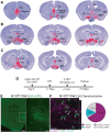
Fig. 4
Fig. 4. Mapping of DP/DTT downstream projections and characterization of activity-tagged DP/DTT neurons following the OFT.
A–C Representative projections of the DP/DTT in anterior, intermediate, and posterior sections. A In anterior sections, we observed notable viral expression in the indusium griseum (IG), septum, bed nucleus of the stria terminalis (BNST), dorsal endopiriform nucleus (DEn), nucleus reuniens (RE) and paraventricular thalamus (PVT). B In intermediate sections, we observed continued viral expression in the PVT and RE. Viral expression was also observed in the paraventricular nucleus (PVN) submedius thalamic nucleus (Sub), dorsomedial hypothalamus (DMH), lateral hypothalamus (LH), and posterior hypothalamus (PH). C In the most posterior sections we examined, viral expression was detected in the periaqueductal gray (PAG), retromammillary nuclei (RM), PVT, LH and PH. In posterior brainstem sections, we also observed DP/DTT projections to the raphe nuclei (data not shown). DF Characterization and downstream projection patterns of activity-tagged DP/DTT neurons following the OFT. D Experimental timeline. TRAP2 mice were infused with AAV expressing a DIO-YFP construct in the DP/DTT. Seven days later, animals underwent the OFT and were injected with tamoxifen immediately afterwards. Ten days later, animals were perfused and their brains processed for immunohistochemistry against AMPA-type glutamate receptor subunits 2/3 (GluR2/3) and glutamate decarboxylase 67 (GAD67). E Representative image of DP/DTT activity tagged neurons (green). Scale bars: 400 µm (left), 100 µm (right inset). F Left: Representative image showing DP/DTT OFT activity-tagged neurons (green) co-stained with GluR2/3 (magenta; arrows) or GAD67 (cyan; arrow heads). Scale bar: 50 µm. Right: Distribution of DP/DTT OFT activity-tagged neurons indicates that these cells predominantly co-label with GluR2/3 (67.9%), with 13.9% of neurons co-labeled with GAD67, 18.2% negative for either marker, and a minority of cells (2.5%) co-labeled for both GluR2/3 and GAD67 (n = 6).
Fig. 5
Fig. 5. Optogenetic activation of the DP/DTT-DMH pathway acutely suppresses freezing but has no effect on affective behavior.
A Experimental timeline. Mice were injected with ChR2 or control GFP AAV constructs in the DP/DTT and implanted with an optic fiber in the DMH. Ten days later, animals underwent behavior, starting with the OFT. Three days later, animals went through the EPM and, following another 3 days, one of two auditory fear conditioning protocols (see below). B–D Behavior in the OFT. B Light activation did not affect the time spent in the center of the open field between groups (Two-way rmANOVA significant main effect of stimulation F1,18 = 5.657, p = 0.0287; but no main effect of treatment F1, 18 = 0.1118, p = 0.7420 or interaction F1,18 = 0.02443, p = 0.8775; no significant posthoc tests, p > 0.17). C Light activation did not affect the time spent in the outer zone of the open field between groups (Two-way rmANOVA significant main effect of stimulation F1,18 = 6.549, p = 0.0197; but no main effect of treatment F1, 18 = 0.11240, p = 0.7288 or interaction F1,18 = 0.1744, p = 0.6811; no significant posthoc tests, p > 0.097). D The total distance traveled during the test was not affected by light activation (Two-way rmANOVA no main effect of treatment F1, 18 = 2.724, p = 0.1162; stimulation F1,18 = 3.054, p = 0.0976; or interaction F1,18 = 2.728, p = 0.1160). E–G Behavior in the EPM. Light activation did not affect the time spent in the open arms of the EPM (E; Two-way rmANOVA no main effect of treatment F1,14 = 0.9766, p = 0.3398; stimulation F1,14 = 2.631, p = 0.1271; or interaction F1,14 = 0.0100, p = 0.9217), the number of entries to the open arms of the EPM (F; we note a non-significant trend toward a decrease in open arm entries in the EPM for the ChR2 animals: Two-way rmANOVA significant effect of stimulation F1,14 = 10.53, p = 0.0059; but no main effect of treatment F1,14 = 3309, p = 0.5743 or interaction F1,14 = 0.1645, p = 0.6912; Šídák’s multiple comparisons test, ChR2 p = 0.057, GFP p = 0.097) or the total distance traveled (G; Two-way rmANOVA no main effect of treatment F1,14 = 1.207, p = 0.2904; stimulation F1,14 = 0.0676, p = 0.7986; or interaction F1,14 = 0.00511, p = 0.0940). H–L Effect of DP/DTT-DMH activation on fear extinction and extinction retrieval. H Experimental timeline for the fear extinction cohort. Three days after the EPM, mice were trained in auditory fear conditioning as described previously (6 tone-shock pairings). We note a significant effect of tone on fear training in this cohort (data not shown; Two-way rmANOVA significant effect of tone F3.537, 35.37 = 50.62, p < 0.0001; but no main effect of treatment F1,10 = 1.792, p = 0.2103 or interaction F5,50 = 0.5963, p = 0.7029). The following day, mice underwent extinction training in context B (12 tones, as previously) with light activation for the duration of each tone. On the next day, they underwent extinction retrieval in context B (5 tones, same protocol as before) in the absence of light. I During extinction training, DP/DTT-DMH optogenetic activation reduced average freezing (I) and freezing across tones (J). K, L During extinction retrieval in the absence of light, average tone freezing (K; t10 = 0.1392, p = 0.8921) and freezing across tones (L; Two-way rmANOVA no significant main effect of treatment F1,10 = 0.0193, p = 0.8921; tone F2.707,27.07 = 2.575, p = 0.0798 or interaction F4,40 = 1.592, p = 0.1953) were similar between groups. M–O Effect of DP/DTT-DMH activation on baseline fear after conditioning. M Experimental timeline for the fear baseline cohort. Three days after the EPM, a separate cohort of mice was trained in auditory fear conditioning as previously (6 tone-shock pairings). The following day, mice were placed in context B in the absence of tones, and after 2 min of baseline exploration the light was turned on for 2 min. N DP/DTT-DMH optogenetic activation decreased freezing in the absence of tones. Two-way rmANOVA significant main effect of stimulation and interaction, but not virus F1,11 = 0.006, p = 0.9396; Šídák’s multiple comparisons test, light OFF minus Light ON ChR2 p = 0.0011, GFP p = 0.7607. O DP/DTT-DMH optogenetic activation increased rearing instances. Two-way rmANOVA significant main effect of stimulation and interaction, but not virus F1,11 = 0.8904, p = 0.3656; Šídák’s multiple comparisons test, light OFF minus Light ON ChR2 p = 0.0008 GFP p = 0.8609. *p < 0.05. Male (square) and female (circle) individual datapoints are displayed for transparency. Some figure diagrams were created with the assistance of BioRender.com.

References

    1. Knight RT, Grabowecky MF, Scabini D. Role of human prefrontal cortex in attention control. Adv Neurol. 1995;66:21–26. - PubMed
    1. Rossi AF, Pessoa L, Desimone R, Ungerleider LG. The prefrontal cortex and the executive control of attention. Exp Brain Res. 2009;192:489–97. - PMC - PubMed
    1. Bechara A, Tranel D, Damasio H. Characterization of the decision-making deficit of patients with ventromedial prefrontal cortex lesions. Brain. 2000;123:2189–202. - PubMed
    1. Euston DR, Gruber AJ, McNaughton BL. The role of medial prefrontal cortex in memory and decision making. Neuron. 2012;76:1057–70. - PMC - PubMed
    1. Bahmani Z, Clark K, Merrikhi Y, Mueller A, Pettine W, Isabel Vanegas M, et al. Prefrontal Contributions to Attention and Working Memory. Curr Top Behav Neurosci. 2019;41:129–53. - PMC - PubMed

Publication types

MeSH terms

LinkOut - more resources