

Molecular image-guided surgery in gynaecological cancer: where do we stand?
- PMID: 38233609
- PMCID: PMC11300493
- DOI: 10.1007/s00259-024-06604-1
Molecular image-guided surgery in gynaecological cancer: where do we stand?
Abstract
Purpose: The aim of this review is to give an overview of the current status of molecular image-guided surgery in gynaecological malignancies, from both clinical and technological points of view.
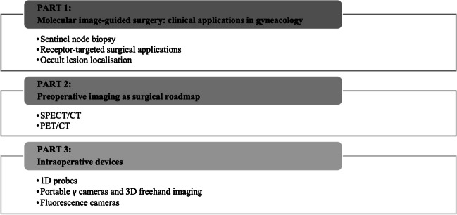
Methods: A narrative approach was taken to describe the relevant literature, focusing on clinical applications of molecular image-guided surgery in gynaecology, preoperative imaging as surgical roadmap, and intraoperative devices.
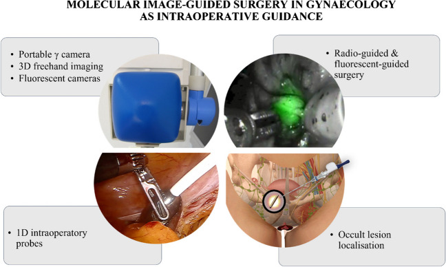
Results: The most common clinical application in gynaecology is sentinel node biopsy (SNB). Other promising approaches are receptor-target modalities and occult lesion localisation. Preoperative SPECT/CT and PET/CT permit a roadmap for adequate surgical planning. Intraoperative detection modalities span from 1D probes to 2D portable cameras and 3D freehand imaging.
Conclusion: After successful application of radio-guided SNB and SPECT, innovation is leaning towards hybrid modalities, such as hybrid tracer and fusion of imaging approaches including SPECT/CT and PET/CT. Robotic surgery, as well as augmented reality and virtual reality techniques, is leading to application of these innovative technologies to the clinical setting, guiding surgeons towards a precise, personalised, and minimally invasive approach.
Keywords: Gynaecological cancers; Hybrid tracer; Image-guided surgery; Robotic surgery; Sentinel node biopsy.
© 2024. The Author(s).
Conflict of interest statement
MF is consultant/speaker for Stryker and received research funding from AKesoBio. NAR is supported in part by the MSK National Cancer Institute/National Institutes of Health Cancer Center Support Grant (P30 CA008748); he reports research funding (paid to the institution) from GRAIL. MSK has equity in GRAIL. The other authors declare no conflict of interest.
Figures
Similar articles
-
The GOSTT concept and hybrid mixed/virtual/augmented reality environment radioguided surgery.Q J Nucl Med Mol Imaging. 2014 Jun;58(2):207-15. Q J Nucl Med Mol Imaging. 2014. PMID: 24835294 Review.
-
Clinical Usefulness of a Novel Freehand 3D Imaging Device for Radio-Guided Intraoperative Sentinel Lymph Node Detection in Malignant Melanoma.Clin Nucl Med. 2015 Sep;40(9):e436-40. doi: 10.1097/RLU.0000000000000882. Clin Nucl Med. 2015. PMID: 26164178
-
Image-guided surgery in gynecologic oncology.Future Oncol. 2017 Nov;13(26):2321-2328. doi: 10.2217/fon-2017-0253. Epub 2017 Nov 10. Future Oncol. 2017. PMID: 29121779 Review.
-
Radioguided surgery and the GOSTT concept: From pre-operative image and intraoperative navigation to image-assisted excision.Rev Esp Med Nucl Imagen Mol. 2017 May-Jun;36(3):175-184. doi: 10.1016/j.remn.2016.09.004. Epub 2016 Oct 25. Rev Esp Med Nucl Imagen Mol. 2017. PMID: 27793632 English, Spanish.
-
Continuous innovation in precision radio-guided surgery.Rev Esp Med Nucl Imagen Mol (Engl Ed). 2024 Jan-Feb;43(1):39-54. doi: 10.1016/j.remnie.2023.11.001. Epub 2023 Nov 12. Rev Esp Med Nucl Imagen Mol (Engl Ed). 2024. PMID: 37963516
Cited by
-
ASO Authors Reflections: Innovations for Intraoperative Lymph Node Imaging in the Era of Digital Surgery in Gynecological Cancers.Ann Surg Oncol. 2025 May;32(5):3469-3470. doi: 10.1245/s10434-025-17047-9. Epub 2025 Feb 22. Ann Surg Oncol. 2025. PMID: 39987386 No abstract available.
References
-
- van Leeuwen FWB, Schottelius M, Brouwer OR, Vidal-Sicart S, Achilefu S, Klode J, et al. Trending: radioactive and fluorescent bimodal/hybrid tracers as multiplexing solutions for surgical guidance. J Nucl Med. 2020;61:13–9. - PubMed
-
- Valdés Olmos RA, Rietbergen DDD, Rubello D, Pereira Arias-Bouda LM, Collarino A, Colletti PM, et al. Sentinel node imaging and radioguided surgery in the era of SPECT/CT and PET/CT: toward new interventional nuclear medicine strategies. Clin Nucl Med. 2020;45:771–7. - PubMed
-
- Giammarile F, Bozkurt MF, Cibula D, Pahisa J, Oyen WJ, Paredes P, et al. The EANM clinical and technical guidelines for lymphoscintigraphy and sentinel node localization in gynaecological cancers. Eur J Nucl Med Mol Imaging. 2014;41:1463–77. - PubMed
-
- Oonk MHM, van de Nieuwenhof HP, de Hullu JA, van der Zee AGJ. The role of sentinel node biopsy in gynecological cancer: a review. Curr Opin Oncol. 2009;21:425–32. - PubMed
Publication types
MeSH terms
Grants and funding
LinkOut - more resources
Full Text Sources
