

Regulatory function and mechanism research for m6A modification WTAP via SUCLG2-AS1- miR-17-5p-JAK1 axis in AML
- PMID: 38233760
- PMCID: PMC10795285
- DOI: 10.1186/s12885-023-11687-4
Regulatory function and mechanism research for m6A modification WTAP via SUCLG2-AS1- miR-17-5p-JAK1 axis in AML
Abstract
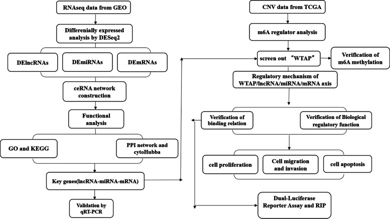
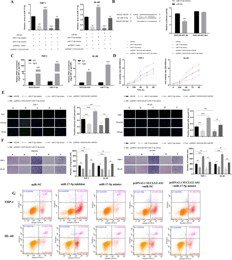
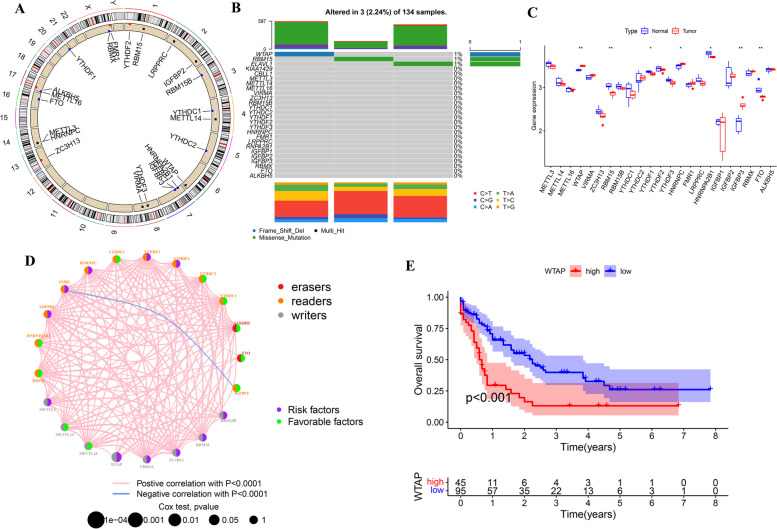
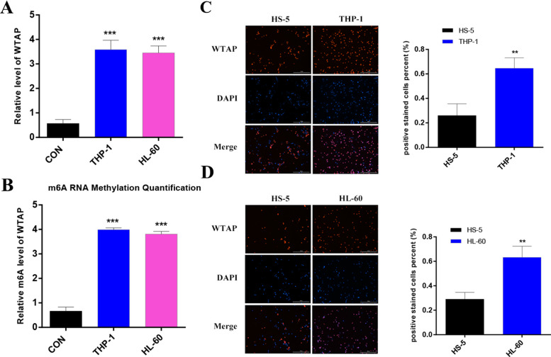
Acute myeloid leukemia (AML), characterized by the abnormal accumulation of immature marrow cells in the bone marrow, is a malignant tumor of the blood system. Currently, the pathogenesis of AML is not yet clear. Therefore, this study aims to explore the mechanisms underlying the development of AML. Firstly, we identified a competing endogenous RNA (ceRNA) SUCLG2-AS1-miR-17-5p-JAK1 axis through bioinformatics analysis. Overexpression of SUCLG2-AS1 inhibits proliferation, migration and invasion and promotes apoptosis of AML cells. Secondly, luciferase reporter assay and RIP assay validated that SUCLG2-AS1 functioned as ceRNA for sponging miR-17-5p, further leading to JAK1 underexpression. Additionally, the results of MeRIP-qPCR and m6A RNA methylation quantification indicted that SUCLG2-AS1(lncRNA) had higher levels of m6A RNA methylation compared with controls, and SUCLG2-AS1 is regulated by m6A modification of WTAP in AML cells. WTAP, one of the main regulatory components of m6A methyltransferase complexes, proved to be highly expressed in AML and elevated WTAP is associated with poor prognosis of AML patients. Taken together, the WTAP-SUCLG2-AS1-miR-17-5p-JAK1 axis played essential roles in the process of AML development, which provided a novel therapeutic target for AML.
Keywords: AML; JAK1; WTAP; lncRNA SUCLG2-AS1; miR-17-5p.
© 2024. The Author(s).
Conflict of interest statement
The authors declare no competing interests.
Figures


Similar articles
-
Chidamide inhibits cell glycolysis in acute myeloid leukemia by decreasing N6-methyladenosine-related GNAS-AS1.Daru. 2024 Jun;32(1):11-24. doi: 10.1007/s40199-023-00482-y. Epub 2023 Nov 6. Daru. 2024. PMID: 37926762 Free PMC article.
-
Long noncoding RNA SBF2-AS1 act as a ceRNA to modulate cell proliferation via binding with miR-188-5p in acute myeloid leukemia.Artif Cells Nanomed Biotechnol. 2019 Dec;47(1):1730-1737. doi: 10.1080/21691401.2019.1608221. Artif Cells Nanomed Biotechnol. 2019. PMID: 31062614
-
Long non-coding RNA AGAP2-AS1, functioning as a competitive endogenous RNA, upregulates ANXA11 expression by sponging miR-16-5p and promotes proliferation and metastasis in hepatocellular carcinoma.J Exp Clin Cancer Res. 2019 May 14;38(1):194. doi: 10.1186/s13046-019-1188-x. J Exp Clin Cancer Res. 2019. Retraction in: J Exp Clin Cancer Res. 2022 Nov 2;41(1):317. doi: 10.1186/s13046-022-02521-z. PMID: 31088485 Free PMC article. Retracted.
-
WTAP-Involved the m6A Modification of lncRNA FAM83H-AS1 Accelerates the Development of Gastric Cancer.Mol Biotechnol. 2024 Aug;66(8):1883-1893. doi: 10.1007/s12033-023-00810-2. Epub 2023 Jul 21. Mol Biotechnol. 2024. PMID: 37477820
-
AGAP2-AS1 promotes the assembly of m6A methyltransferases and activation of the IL6/STAT3 pathway by binding with WTAP in the carcinogenesis of gastric cancer.FASEB J. 2023 Dec;37(12):e23302. doi: 10.1096/fj.202301249R. FASEB J. 2023. PMID: 37983949
Cited by
-
Identification and Characterization of the RNA Modifying Factors PUS7 and WTAP as Key Components for the Control of Tumor Biological Processes in Renal Cell Carcinomas.Curr Issues Mol Biol. 2025 Apr 9;47(4):266. doi: 10.3390/cimb47040266. Curr Issues Mol Biol. 2025. PMID: 40699665 Free PMC article.
-
Role of MicroRNAs in Acute Myeloid Leukemia.Genes (Basel). 2025 Apr 11;16(4):446. doi: 10.3390/genes16040446. Genes (Basel). 2025. PMID: 40282406 Free PMC article. Review.
References
-
- Wang XN, Studzinski GP. Activation of extracellular signal-regulated kinases (ERKs) defines the first phase of 1,25-dihydroxyvitamin D-3-induced differentiation of HL60 cells. J Cell Biochem. 2001;80:471–82. https://doi.org/10.1002/1097-4644(20010315)80:4<471::Aid-jcb1001>3.... - PubMed
-
- Yan H, Wen L, Tan D, Xie P, Pang FM, Zhou HH, Zhang W, Liu ZQ, Tang J, Li X, Chen XP. Association of a cytarabine chemosensitivity related gene expression signature with survival in cytogenetically normal acute myeloid leukemia. Oncotarget. 2017;8:1529–1540. doi: 10.18632/oncotarget.13650. - DOI - PMC - PubMed
MeSH terms
Substances
Grants and funding
LinkOut - more resources
Full Text Sources
Medical
Research Materials
Miscellaneous
