. 2024 Jan 17;22(1):46.
doi: 10.1186/s12964-023-01366-z.

ZDHHC5-mediated S-palmitoylation of FAK promotes its membrane localization and epithelial-mesenchymal transition in glioma

Affiliations

ZDHHC5-mediated S-palmitoylation of FAK promotes its membrane localization and epithelial-mesenchymal transition in glioma

Yang Wang et al. Cell Commun Signal. .

Abstract

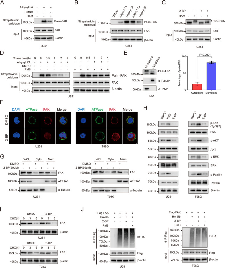
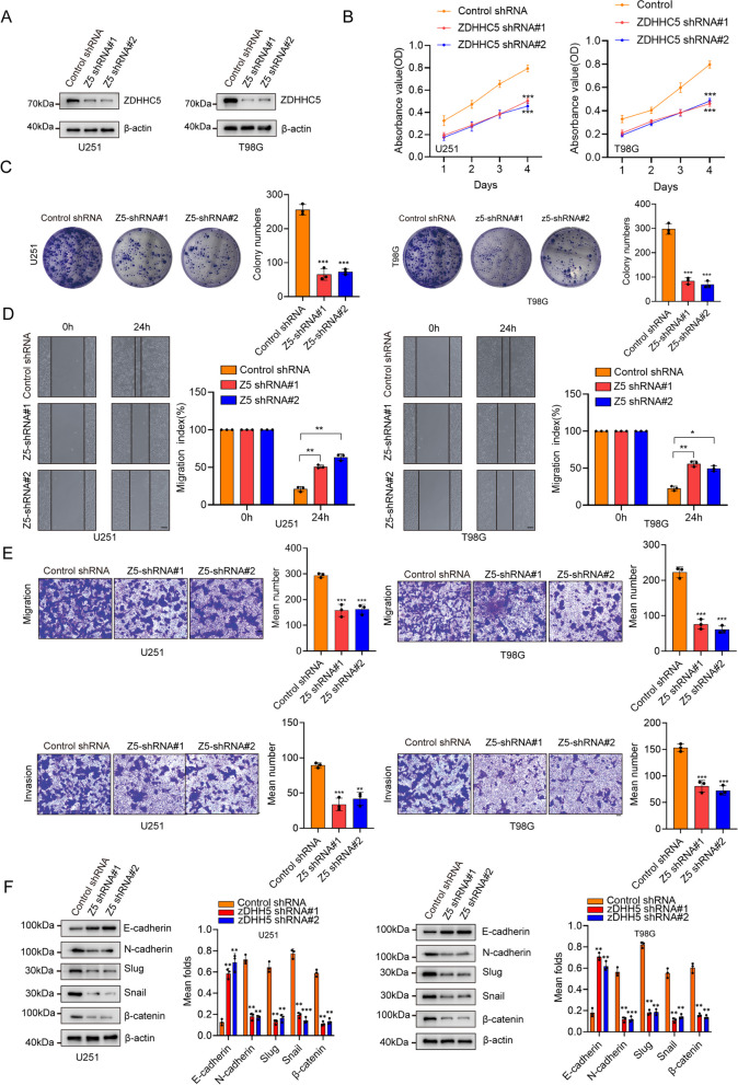
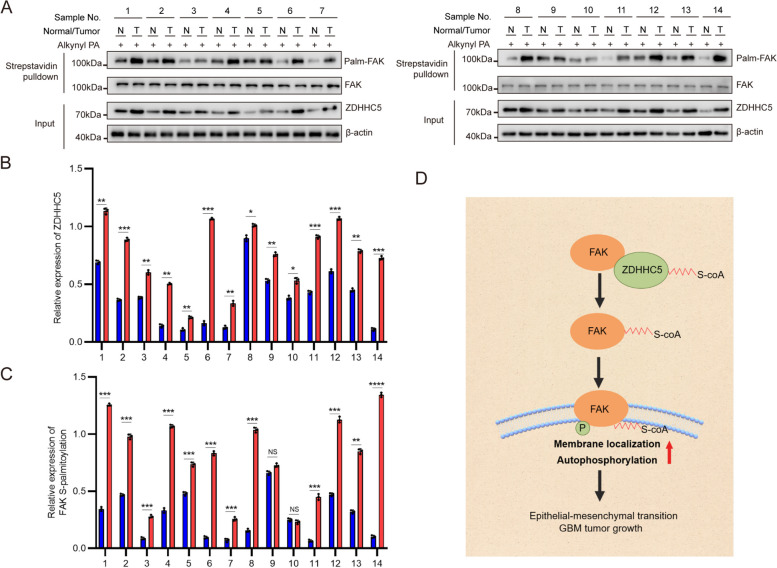
Background: Abnormal activation of FAK is associated with tumor development and metastasis. Through interactions with other intracellular signalling molecules, FAK influences cytoskeletal remodelling, modulation of adhesion signalling, and activation of transcription factors, promoting migration and invasion of tumor cells. However, the exact mechanism that regulates these processes remains unresolved. Herein, our findings indicate that the S-palmitoylation of FAK is crucial for both its membrane localization and activation.

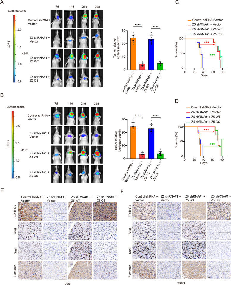
Methods: The palmitoylation of FAK in U251 and T98G cells was assessed by an acyl-PEG exchange (APE) assay and a metabolic incorporation assay. Cellular palmitoylation was inhibited using 2-bromopalmitate, and the palmitoylation status and cellular localization of FAK were determined. A metabolic incorporation assay was used to identify the potential palmitoyl acyltransferase and the palmitoylation site of FAK. Cell Counting Kit-8 (CCK8) assays, colony formation assays, and Transwell assays were conducted to assess the impact of ZDHHC5 in GBM. Additionally, intracranial GBM xenografts were utilized to investigate the effects of genetically silencing ZDHHC5 on tumor growth.

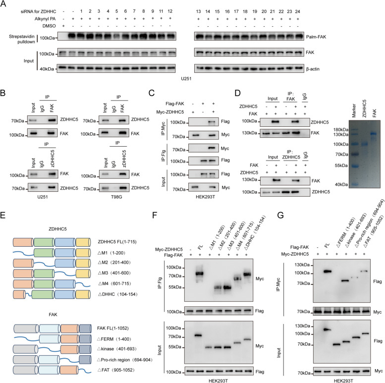
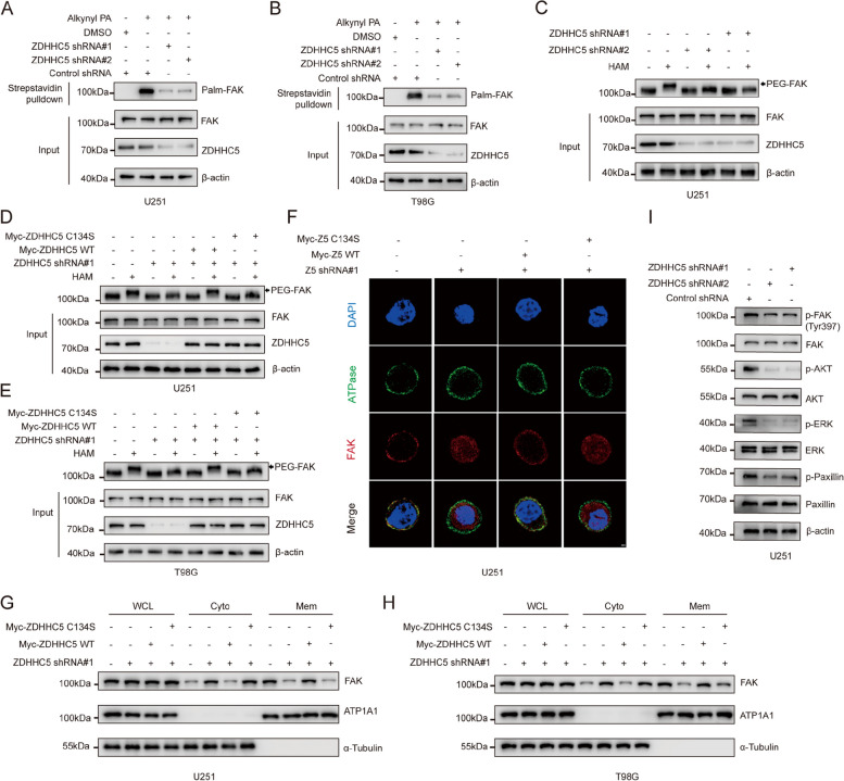
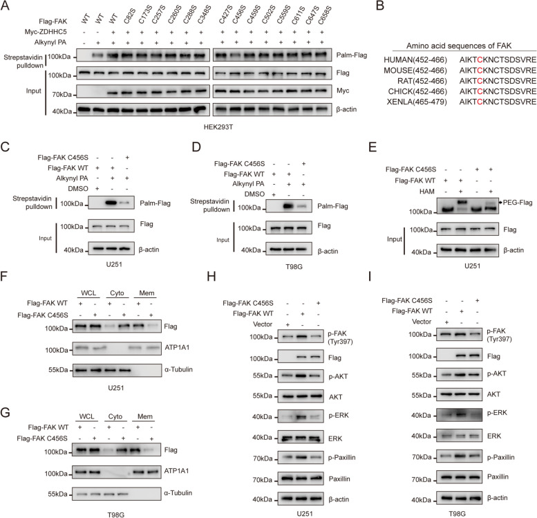
Results: Inhibiting FAK palmitoylation leads to its redistribution from the membrane to the cytoplasm and a decrease in its phosphorylation. Moreover, ZDHHC5, a protein-acyl-transferase (PAT), catalyzes this key modification of FAK at C456. Knockdown of ZDHHC5 abrogates the S-palmitoylation and membrane distribution of FAK and impairs cell proliferation, invasion, and epithelial-mesenchymal transition (EMT). Taken together, our research reveals the crucial role of ZDHHC5 as a PAT responsible for FAK S-palmitoylation, membrane localization, and activation.

Conclusions: These results imply that targeting the ZDHHC5/FAK axis has the potential to be a promising strategy for therapeutic interventions for glioblastoma (GBM). Video Abstract.

Keywords: FAK ZDHHC5 S; Mesenchymal transition; Palmitoylation glioma epithelial.


Conflict of interest statement

The authors declare no potential competing interests.

Figures

Fig. 1
Fig. 1
S-palmitoylation maintains the membrane localization of FAK (A) FAK palmitoylation was examined in lysates obtained from U251 cells that were metabolically treated with a palmitoylation probe (50 μM alkynyl palmitic acid [PA]) for a duration of 4 h. The analysis was carried out through click reaction and streptavidin bead pulldown, both in the absence and presence of hydroxylamine (HAM). The subsequent immunoblotting (IB) was performed using the indicated antibodies. B FAK fatty acylation levels were investigated using different chemical reporters of fatty acylation, ranging from Alk-C14 to Alk-C20. The analysis involved the use of streptavidin bead pulldown to isolate acylated FAK, followed by IB. C APE assays were conducted to examine the levels of FAK palmitoylation in U251 cells following treatment with 50 μM 2-BP, both in the absence and presence of HAM. D FAK palmitoylation studies were conducted in U251 cells that were metabolically labeled with 50 µM alkynyl PA for a duration of 4 h. The cells were treated either in the absence or presence of PalB (5 μM). After the specified time period, the cells were collected for further analysis of FAK palmitoylation. E FAK palmitoylation was analyzed using the APE assay after fractionation to distinguish between the cytoplasmic and membrane fractions. ATP1A1 and α-tubulin were used as controls for the membrane and cytoplasmic fractions, respectively. Quantification of FAK palmitoylation percentage in the cytoplasm and membrane in APE assays. All the data are presented as the mean ± SD, n = three independent experiments, and two-tailed Student’s t test. F U251 and T98G cells were treated with dimethyl sulfoxide (DMSO) or 50 μM 2-BP for 8 h, and the cellular localization of endogenous FAK was visualized using immunofluorescence staining. Scale bar, 1 μm. G U251 and T98G cells were subjected to treatment with either DMSO or 50 μM 2-BP for 8 h. The levels of FAK in the membrane and cytoplasmic fractions were evaluated using IB with the indicated antibodies. ATP1A1 and α-tubulin were used as membranal and cytoplasmic fraction controls, respectively. WCL refers to the whole-cell lysate, Mem refers to the membrane, and Cyto refers to the cytoplasm. H U251 and T98G cells were treated with DMSO, 50 μM 2-BP, or 2.5 μM PalB. Then, IB was performed for the indicated proteins. I U251 and T98G cells were pretreated with 2-BP or DMSO as a control and then incubated with cycloheximide (CHX). Immunoblotting was used to analyze the FAK and β-actin levels at the specified time points. J U251 and T98G cells were cotransfected with HA-Ub and treated with DMSO, 50 μM 2-BP, or 5 μM PalB for 8 h. U251 and T98G cells were subjected to IP-IB and IB for the indicated proteins
Fig. 2
Fig. 2
ZDHHC5 regulates FAK S-palmitoylation by directly binding to FAK (A) U251 cells infected with siRNAs targeting different zDHHC proteins. The cells were metabolically labeled with alkynyl PA (50 µM) for 4 h. FAK palmitoylation levels were assessed by click reaction and streptavidin bead pulldown, followed by IB. B Cell lysates from U251 and T98G cells were analyzed by IP using antibodies against FAK and ZDHHC5 and then subjected to IB analysis. IgG was used as the isotype control. C HEK293T cells transfected with Flag-FAK and/or Myc-ZDHHC5 were subjected to IP-IB and IB using the indicated antibodies. D In vitro protein binding assays with purified ZDHHC5 and FAK protein. The purities of ZDHHC5 and FAK were examined by SDS-PAGE and Coomassie Blue Staining. E Schematic representations of ZDHHC5, FAK, and their shortened mutants. F HEK293T cells were cotransfected with Flag-FAK and FL Myc-ZDHHC5 or its deletion mutants. The cell lysates were subjected to Flag bead pulldown, followed by IB using specific antibodies. G HEK293T cells were cotransfected with Myc-ZDHHC5 and FL Flag-FAK or its deletion mutants. Cell lysates were analyzed by Flag bead pulldown, followed by IB using the indicated antibodies
Fig. 3
Fig. 3
ZDHHC5 maintains FAK membrane localization and activation via S-palmitoylation (A) and (B) U251 and T98G cells were transduced with lentiviruses carrying either control shRNA or ZDHHC5 shRNAs. Subsequently, the cells were metabolically labeled with 50 μM alkynyl PA for a duration of 4 h. The levels of FAK palmitoylation were analyzed by performing a click reaction and streptavidin bead pulldown, followed by IB. C An APE assay was performed to analyze FAK palmitoylation in ZDHHC5-knockdown U251 cells. D and E ZDHHC5-knockdown U251 and T98G cells were rescued with Myc-ZDHHC5 WT or ZDHHC5 C134S. FAK palmitoylation levels were analyzed by APE assay. F ZDHHC5-knockdown U251 cells were rescued with Myc-ZDHHC5 WT or ZDHHC5 C134S, and endogenous FAK cellular localization was visualized by immunofluorescence staining using antibodies against FAK. Scale bar, 1 μm. Z5, ZDHHC5 (G), and (H) ZDHHC5-knockdown U251 cells were rescued with Myc-ZDHHC5 WT or ZDHHC5 C134S. The levels of FAK in the membrane and cytoplasmic fractions were evaluated using IB with the indicated antibodies. I U251 cells were infected with lentiviruses expressing control shRNA or ZDHHC5 shRNAs and then subjected to IB for the indicated proteins
Fig. 4
Fig. 4
FAK is palmitoylated at Cys456 (A) U251 cells were cotransfected with Flag-FAK WT or the indicated mutants and/or Myc-ZDHHC5. The cells were metabolically labeled with 50 µM alkynyl PA for 4 h. The levels of FAK palmitoylation were analyzed by performing a click reaction and streptavidin bead pulldown, followed by IB. B Amino acid sequences around the cysteine 456 residue of the FAK protein across different species. C and D U251 and T98G cells were transfected with Flag-FAK WT or Flag-FAK C456S and then metabolically labeled with 50 M alkynyl PA for 4 h. FAK palmitoylation levels were assessed by click reaction and streptavidin bead pulldown, followed by IB. E U251 and T98G cells were transfected with Flag-FAK WT or Flag-FAK C456S. The APE assay was conducted to analyze FAK palmitoylation in U251 cells with the specified modifications. F and G U251 and T98G cells were transfected with Flag-FAK WT or Flag-FAK C456S. The levels of FAK in the membrane and cytoplasmic fractions were evaluated using IB with the indicated antibodies. H and I U251 and T98G cells were transfected with Flag-FAK WT or Flag-FAK C456S. Then, IB was performed with the indicated antibodies
Fig. 5
Fig. 5
ZDHHC5-mediated FAK S-palmitoylation promotes cell proliferation, cell invasion and EMT in vitro. A IB for ZDHHC5 in U251 and T98G cells transfected with ZDHHC5 shRNAs or shRNA control. B CCK8 assays of U251 and T98G cells transfected with ZDHHC5 shRNAs or shRNA control. C Colony formation assay of U251 and T98G cells transfected with ZDHHC5 shRNAs or shRNA control. D Wound-healing assay of ZDHHC5 shRNAs or shRNA control. scale bars: 25 μm. E Invasion of U251 and T98G cells transfected with ZDHHC5 shRNAs or shRNA control; scale bars: 100 μm. (F) IB for the indicated proteins in U251 and T98G cells transfected with ZDHHC5 shRNAs or shRNA control. Data are represented as the mean ± SD (n = 3). Statistical analysis was performed using Student’s t test, *p < 0.05; **p < 0.01; ***p < 0.001; ****p < 0.0001
Fig. 6
Fig. 6
ZDHHC5 promotes GBM development in vivo. A and B Luciferase-expressing ZDHHC5-knockdown U251 and T98G cells were injected into athymic nude mice (n = 10). The mice were subjected to IVIS scanning on days 7,14,21,28 after the injection of tumor cells. Representative bioluminescent images of intracranial GBM xenografts are shown on the left. Quantification of bioluminescent images (day = 28) is shown on the right. C and D Kaplan‒Meier survival curves of mice intracranially injected with U251 and T98G cells with the indicated modifications (n = 10). P values were calculated using the two-tailed Student's t test for (A, B) and the log-rank (Mantel‒Cox) test for (C, D). **P < 0.01, ***P < 0.001, ****P < 0.0001. E and F IHC images of the indicated proteins are shown in consecutive brain sections. Scale bars, 20 µm
Fig. 7
Fig. 7
ZDHHC5 expression positively correlates with FAK expression in clinical glioma samples. A The S-palmitoylation of FAK in glioma samples (T) and their paired normal tissues (N) were analyzed by metabolic incorporation assay. B and C Relative expression levels of ZDHHC5 (B) and S-palmitoylation of FAK (C) from (A). Data are represented as the mean ± SD (n = 3). Statistical analysis was performed using Student’s t test, *p < 0.05; **p < 0.01; ***p < 0.001; ****p < 0.0001. D Schematic representation of the study model. ZDHHC5-catalyzed palmitoylation of FAK leads to its membrane localization and activation, subsequently promoting EMT and the development of GBM

Similar articles

Cited by

References

    1. Sampson JH, Gunn MD, Fecci PE, Ashley DM. Brain immunology and immunotherapy in brain tumours. Nat Rev Cancer. 2020;20:12–25. doi: 10.1038/s41568-019-0224-7. - DOI - PMC - PubMed
    1. Venkataramani V, Yang Y, Schubert MC, Reyhan E, Tetzlaff SK, Wissmann N, Botz M, Soyka SJ, Beretta CA, Pramatarov RL, et al. Glioblastoma hijacks neuronal mechanisms for brain invasion. Cell. 2022;185:2899–2917 e2831. doi: 10.1016/j.cell.2022.06.054. - DOI - PubMed
    1. Omuro A, DeAngelis LM. Glioblastoma and other malignant gliomas: a clinical review. JAMA. 2013;310:1842–1850. doi: 10.1001/jama.2013.280319. - DOI - PubMed
    1. Lim M, Xia Y, Bettegowda C, Weller M. Current state of immunotherapy for glioblastoma. Nat Rev Clin Oncol. 2018;15:422–442. doi: 10.1038/s41571-018-0003-5. - DOI - PubMed
    1. Aldape K, Brindle KM, Chesler L, Chopra R, Gajjar A, Gilbert MR, Gottardo N, Gutmann DH, Hargrave D, Holland EC, et al. Challenges to curing primary brain tumours. Nat Rev Clin Oncol. 2019;16:509–520. doi: 10.1038/s41571-019-0177-5. - DOI - PMC - PubMed

Publication types