. 2024 Jan 17;24(1):33.
doi: 10.1186/s12935-024-03224-5.

USP29 activation mediated by FUBP1 promotes AURKB stability and oncogenic functions in gastric cancer

Affiliations

USP29 activation mediated by FUBP1 promotes AURKB stability and oncogenic functions in gastric cancer

Rongfu Tu et al. Cancer Cell Int. .

Abstract

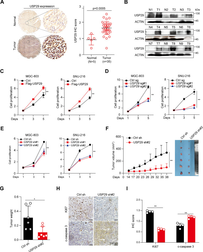
Background: Gastric cancer is a highly prevalent cancer type and the underlying molecular mechanisms are not fully understood. Ubiquitin-specific peptidase (USP) 29 has been suggested to regulate cell fate in several types of cancer, but its potential role in gastric carcinogenesis remains unclear.

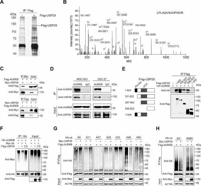
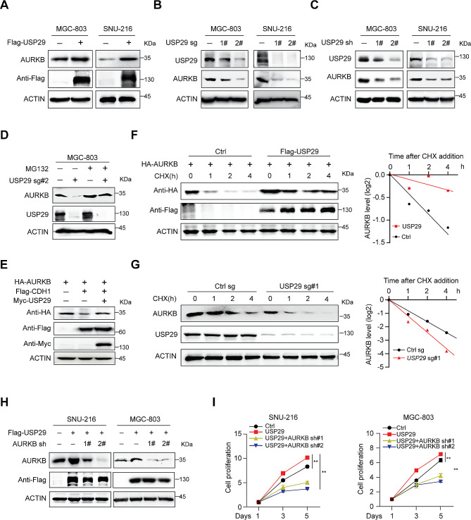
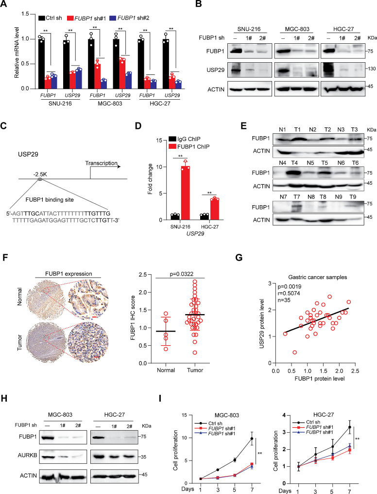
Methods: The expression of USP29 in normal and gastric cancer tissues was analyzed by bioinformatics analysis, immunohistochemistry and immunoblot. Gene overexpression, CRISPR-Cas9 technology, RNAi, and Usp29 knockout mice were used to investigate the roles of USP29 in cell culture, xenograft, and benzo[a]pyrene (BaP)-induced gastric carcinogenesis models. We then delineated the underlying mechanisms using mass spectrometry, co-immunoprecipitation (Co-IP), immunoblot, ubiquitination assay, chromatin immunoprecipitation (ChIP), quantitative real-time PCR (qRT-PCR), and luciferase assays.

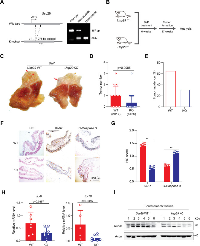
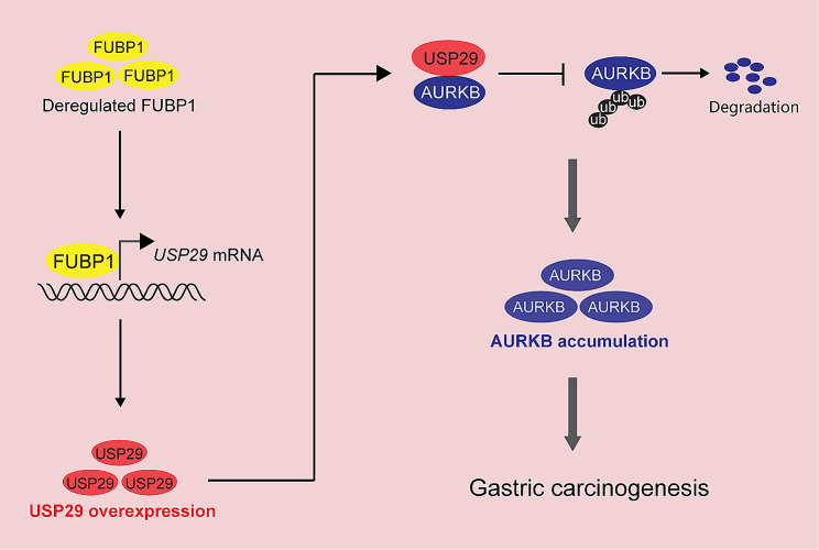
Results: In this study, we found that USP29 expression was significantly upregulated in gastric cancers and associated with poor patient survival. Ectopic expression of USP29 promoted, while depletion suppressed the tumor growth in vitro and in vivo mouse model. Mechanistically, transcription factor far upstream element binding protein 1 (FUBP1) directly activates USP29 gene transcription, which then interacts with and stabilizes aurora kinase B (AURKB) by suppressing K48-linked polyubiquitination, constituting a FUBP1-USP29-AURKB regulatory axis that medicates the oncogenic role of USP29. Importantly, systemic knockout of Usp29 in mice not only significantly decreased the BaP-induced carcinogenesis but also suppressed the Aurkb level in forestomach tissues.

Conclusions: These findings uncovered a novel FUBP1-USP29-AURKB regulatory axis that may play important roles in gastric carcinogenesis and tumor progression, and suggested that USP29 may become a promising drug target for cancer therapy.

Keywords: AURKB; FUBP1; Gastric Cancer; Targeted therapy; USP29.


Conflict of interest statement

The authors declare no competing interests.

Figures

Fig. 1
Fig. 1
Overexpression and an oncogenic role of USP29 in gastric cancers. (A) Left: Representative immunohistological images of USP29 in 5 normal gastric tissues and 35 tumors. Scale bar: 20 μm; right: Quantifications of the immunohistochemistry in the left are shown. (B) USP29 protein expression in 9 gastric tumors and paired non-cancerous tissues analyzed using Western blot. (C) Cell proliferation of MGC-803 and SNU-216 cells with or without USP29 overexpression. (D) Cell proliferation of MGC-803 and SNU-216 cells with or without USP29 depletion by specific sgRNAs. (E) Cell proliferation of MGC-803 and SNU-216 cells with or without USP29 knockdown by specific shRNAs. Data shown were obtained from mean ± SD of technical triplicates (C-E). (F) MGC-803 xenograft tumor growth curves and tumor image (n = 6). (G) Tumor weights of MGC-803 xenografts (n = 6). Data shown were obtained from mean ± SD of technical triplicates (F, G). (H) Representative immunohistological images of Ki-67 and cleaved-caspase-3 in MGC-803 xenograft tumors. Scale bar: 100 μm. (I) Quantifications of the immunohistochemistry in H. **p < 0.01
Fig. 2
Fig. 2
USP29 interacts with AURKB and removes its K48-linked polyubiquitination. (A) Immunoprecipitation of Flag-USP29 using anti-Flag antibody. Total cell proteins extracted from MGC-803 cells expressing Flag-tagged USP29 or vector alone were subjected to immunoprecipitation using anti-Flag beads, which were resolved by SDS-PAGE and visualized by silver staining. (B) Liquid chromatography-tandem mass spectrometry analysis of the Flag-USP29-associated peptides corresponding to AURKB. (C) 293T cells overexpressing Myc-USP29 and/ or Flag-AURKB were subjected to reciprocal Co-IP to detect protein interaction. (D) Lysates from MGC-803 and HGC-27 cells were subjected to immunoprecipitation using AURKB antibodies, and USP29 was detected by immunoblot. (E) Left: A schematic representation of various Flag-USP29 truncations; right: lysates from 293T cells overexpressing HA-AURKB and respective Flag-USP29 truncation were subjected to Co-IP and immunoblot. (F) 293T cells were co-transfected with HA-AURKB, Myc-Ub and Flag-USP29/USP29 C294S mutant (CS), and polyubiquitination analysis of AURKB was shown. G and H. 293T cells were co-transfected with indicated Flag-AURKB, Myc-USP29, HA-Ub (WT) and various HA-Ub mutant plasmids, polyubiquitination analysis of AURKB was analyzed. The experiments were independently repeated three times with similar results (A, C-D, right of E, F–H)
Fig. 3
Fig. 3
USP29 sustains AURKB protein stability. (A) Total cell lysates were extracted from MGC-803 and SNU-216 cells with or without USP29 overexpression, endogenous AURKB level was analyzed by immunoblotting. (B) Cells were infected with control or USP29 sgRNAs, USP29 and AURKB protein expression were detected using immunoblot. (C) Cells were infected with control or USP29 shRNAs, USP29 and AURKB protein expression were detected using immunoblot. (D) MGC-803 cells with or without USP29 depletion were treated with MG132 (10 µM) for 6 h prior to harvest. AURKB and USP29 proteins were analyzed by immunoblot. (E) 293T cells were co-transfected with epitope-tagged AURKB, CDH1 and USP29, immunoblot of respective proteins are shown. The experiments were independently repeated three times with similar results (AE). (F) 293T cells were transfected with indicated plasmids for 24 h, and treated with 100 µg/mL CHX; protein expression was analyzed with immunoblot and protein quantifications are shown. (G) MGC-803 cells with or without USP29 depletion were treated with 100 µg/mL CHX and harvested at the indicated time, endogenous AURKB protein were detected by immunoblot and protein quantifications are shown. Data shown were obtained from averages of three independent experiments (F, G). (H) Gastric cancer cells were infected with Flag-USP29 and/or AURKB shRNA viruses, the USP29 overexpression and AURKB knockdown were measured by immunoblots. The experiments were independently repeated three times with similar results. (I) Proliferation of stable cell lines in H. Data shown were obtained from mean ± SD of technical triplicates. **p < 0.01
Fig. 4
Fig. 4
FUBP1 overexpression transcriptionally activates USP29 in gastric cancers. (A and B). Gastric cancer cell lines were infected with control or FUBP1 specific shRNAs, the mRNA (A) and protein (B) expression of FUBP1 and USP29 were analyzed using qRT-PCR or immunoblot. Graph shows mean ± SD from triplicates; significance was determined by unpaired two-tailed Student’s t-test (A). The experiments were independently repeated three times with similar results (B). (C). A Schematic presentation of potential FUBP1 binding site on the USP29 promoter. (D). ChIP analysis of FUBP1 binding to the USP29 promoter in SNU-216 and HGC-27 cells. Averages of fold enrichment between the FUBP1 antibodies and IgG were shown. Graph shows mean ± SD from triplicates. (E). FUBP1 protein expression in 9 gastric tumors and paired non-cancerous tissues were analyzed using immunoblot. (F). left: Representative immunohistological images of FUBP1 in 5 normal gastric tissues and 35 tumors. Scale bar: 20 μm; right: quantifications of the immunohistochemistry are shown. (G). Correlation between protein levels of FUBP1 and USP29 in 35 gastric tumors. (H). MGC-803 and HGC-27 cells were infected with control or FUBP1 specific shRNAs, and the expression of FUBP1 and AURKB were analyzed using immunoblots. The experiments were independently repeated three times with similar results. (I). Proliferation of MGC-803 and HGC-27 cells with or without FUBP1 knockdown. Data shown were obtained from mean ± SD of technical triplicates. **p < 0.01
Fig. 5
Fig. 5
Usp29 knockout inhibits gastric tumor formation induced by BaP Treatment. (A) Left: sgRNA targeting strategy to knockout Usp29 allele in mice; right: representative genotyping of wildtype and Usp29 knockout mice. (B) Schematic representation of BaP-induced forestomach carcinogenesis. (C) Representative images showing gross morphology of stomachs harboring BaP-induced tumors (23 weeks) from wildtype and Usp29−/− mice. D and E. Tumor number (D) and tumor incidence (E) in stomachs of WT and Usp29−/− mice after administration of BaP (23 weeks). (F and G). Representative images (F) and quantification (G) of HE, Ki-67, and cleaved caspase-3 staining in WT and USP29−/− tumors. (H). IL-8 and IL-1β mRNA levels in WT and Usp29−/− stomachs harboring BaP treatment were analyzed by RT-qPCR. Graph shows mean ± SD from triplicates; significance was determined by unpaired two-tailed Student’s t-test. (I). AURKB protein expression in WT and Usp29−/− stomachs harboring BaP treatment were analyzed by immunoblots. **p < 0.01
Fig. 6
Fig. 6
A proposed model depicting regulation of malignant proliferation and tumor progression by the FUBP1-USP29-AURKB axis. Deregulated FUBP1 directly activates USP29 transcription, leading to the overexpression of USP29 in gastric cancer, which subsequently deubiquitinates and stabilizes AURKB, forming a FUBP1-USP29-AURKB regulatory axis. See text for more details

References

    1. Smyth EC, Nilsson M, Grabsch HI, van Grieken NC, Lordick F. Gastric cancer. Lancet. 2020;396(10251):635–48. doi: 10.1016/S0140-6736(20)31288-5. - DOI - PubMed
    1. Yeoh KG, Tan P. Mapping the genomic diaspora of gastric cancer. Nat Rev Cancer. 2022;22(2):71–84. doi: 10.1038/s41568-021-00412-7. - DOI - PubMed
    1. Tan P, Yeoh KG. Genetics and Molecular Pathogenesis of gastric adenocarcinoma. Gastroenterology. 2015;149(5):1153–62. doi: 10.1053/j.gastro.2015.05.059. - DOI - PubMed
    1. Borah NA, Reddy MM. Aurora kinase B inhibition: a potential therapeutic strategy for Cancer. Molecules 2021, 26(7). - PMC - PubMed
    1. Crosio C, Fimia GM, Loury R, Kimura M, Okano Y, Zhou H, Sen S, Allis CD, Sassone-Corsi P. Mitotic phosphorylation of histone H3: spatio-temporal regulation by mammalian Aurora kinases. Mol Cell Biol. 2002;22(3):874–85. doi: 10.1128/MCB.22.3.874-885.2002. - DOI - PMC - PubMed