. 2024 Jan 17;22(1):50.
doi: 10.1186/s12964-023-01379-8.

Neutrophil extracellular traps mediated by platelet microvesicles promote thrombosis and brain injury in acute ischemic stroke

Affiliations

Neutrophil extracellular traps mediated by platelet microvesicles promote thrombosis and brain injury in acute ischemic stroke

Xin Gao et al. Cell Commun Signal. .

Abstract

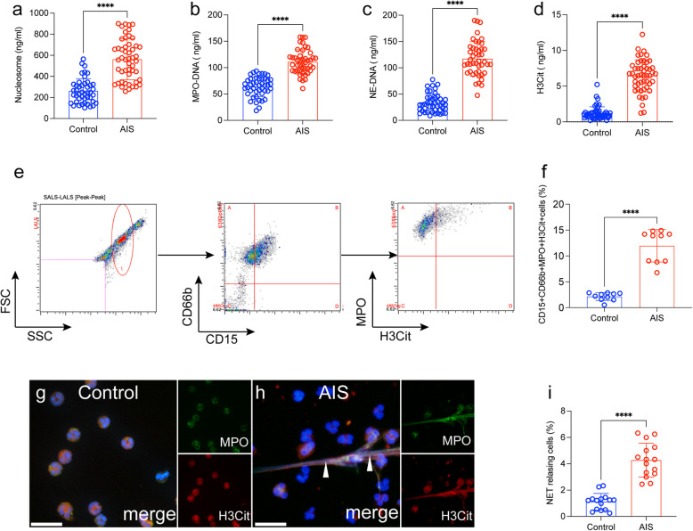
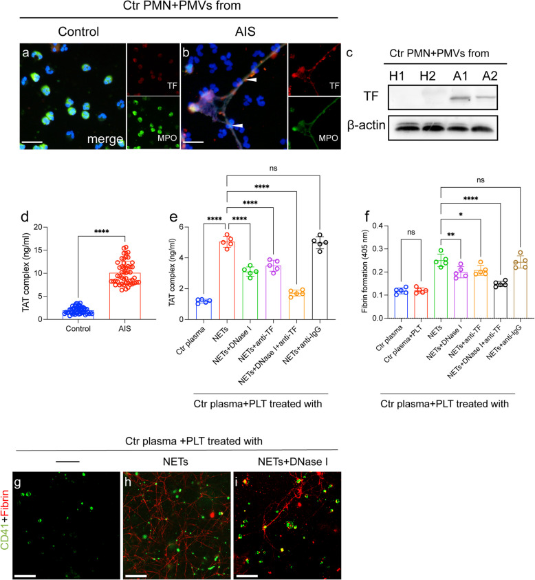
Aims: Neutrophil extracellular traps (NETs) have been implicated in thrombotic diseases. There is no definitive explanation for how NETs form during acute ischemic strokes (AIS). The purpose of our study was to investigate the potential mechanism and role of NETs formation in the AIS process.

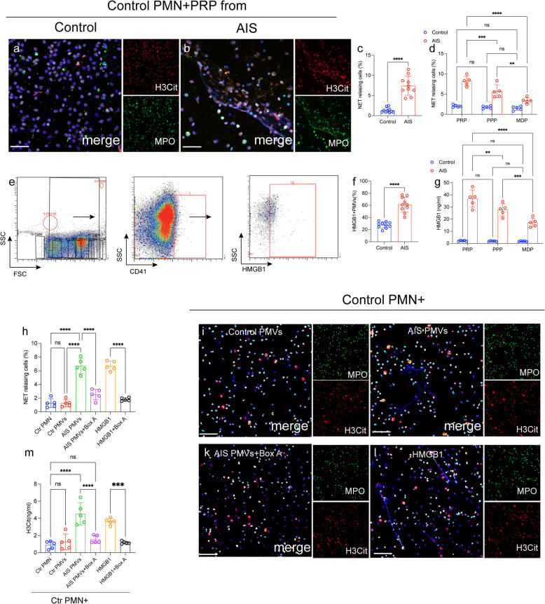
Methods: As well as 45 healthy subjects, 45 patients with AIS had ELISA tests performed to detect NET markers. Expression of high-mobility group box 1 (HMGB1) on platelet microvesicles (PMVs) was analyzed by flow cytometry in healthy subjects and AIS patients' blood samples. We established middle cerebral artery occlusion (MCAO) mice model to elucidate the interaction between PMPs and NETs.

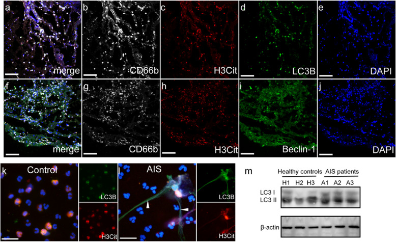
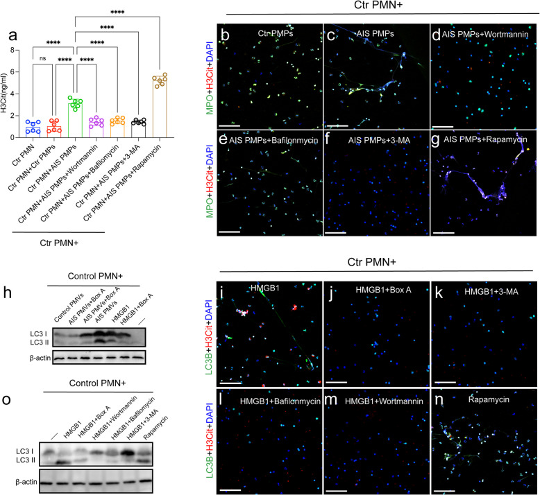
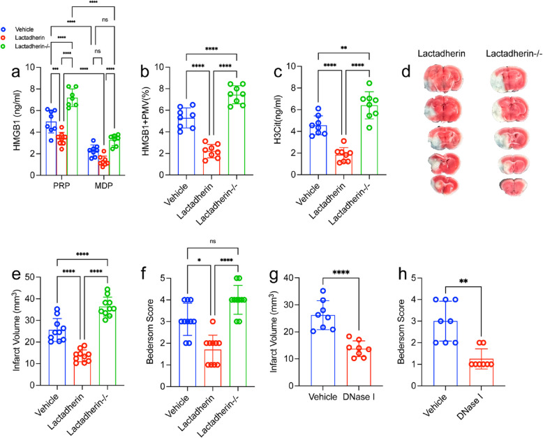
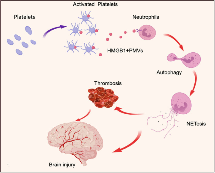
Results: A significant elevation in NET markers was found in patient plasma in AIS patients, and neutrophils generated more NETs from patients' neutrophils. HMGB1 expression was upregulated on PMVs from AIS patients and induced NET formation. NETs enhanced Procoagulant activity (PCA) through tissue factor and via platelet activation. Targeting lactadherin in genetical and in pharmacology could regulate the formation of NETs in MCAO model.

Conclusions: NETs mediated by PMVs derived HMGB1 exacerbate thrombosis and brain injury in AIS. Video Abstract.

Keywords: HMGB1; Microvesicles; Neutrophil extracellular traps; Platelet; Stroke; Thrombosis.


Conflict of interest statement

The authors declare no competing interests.

Figures

Fig. 1
Fig. 1
NET markers in stroke patients before and after thrombolysis. NET markers nucleosome (a), MPO-DNA (b), NE-DNA (c) and H3Cit-DNA (d) were measured in plasma samples from healthy subjects (n = 45) and ischemic stroke patients (n = 45) by ELISA. e NET releasing cells, defined as CD15 + CD66b + MPO + H3Cit + groups and detected in whole blood samples from healthy subjects (n = 10) and AIS patients (n = 45). The flow cytometry gate is showed. f The rate of NET releasing cells in whole blood samples from each group. Neutrophils from healthy subjects (g) and AIS patients (h) were stained for MPO (green) and H3Cit (red) and observed by confocal microscopy. i The rate of NETosis in neutrophils from each group. The inset scale bars ine, f and h, Iiare 40 μm. Statistics, Mann-Whitney test (a-d, f, i). Data are presented as the mean ± SD. ****P < 0.0001
Fig. 2
Fig. 2
HMGB1 from PMPs induces NET formation. a-c Neutrophils from healthy individuals were incubated with PRP from AIS patients (n = 10) and healthy controls (n = 10). Treated neutrophils were co-stained with MPO (green) and H3Cit (red) and observed by confocal microscopy. d NET releasing cells in neutrophils incubated with PPR, PFP and MDP. e The flow cytometry gated of HMGB1 + PMPs is showed. f HMGB1 + PMPs in samples from healthy subjects (n = 10) and AIS patients (n = 10) was detected by flow cytometry. Neutrophils were incubated with PMPs in the presence of Box A (the competitive HMGB1 antagonist). The levels of H3Cit in the supernatant from each group was measured by ELISA (g) and neutrophils were stained with MPO (green) and H3Cit (red) analyzed by confocal microscopy (h-l). The inset scale bars in a, b and h-k are 40 μm. Statistics, Mann-Whitney test (c, f) and ordinary ANOVA (d, g, l). Data are presented as the mean ± SD. ***P < 0.001, and ****P < 0.0001. PMN, polymorphonuclear
Fig. 3
Fig. 3
NET markers were colocalized with autophagy associated protein in stroke thrombi and neutrophils. a-j Stroke thrombi were stained with CD66b (white), H3Cit (red), LC3B (green), Beclin-1 (green) and DAPI (blue). Neutrophils from healthy controls (k) and AIS patients (l) were stained with H3Cit (red) and LC3B (green) and observed by confocal microscopy. m The expression of LC3B on neutrophils from healthy controls (n = 3) and AIS patients (n = 3). The inset scale bars are 20 μm in a-j
Fig. 4
Fig. 4
HMGB1 from PMPs promote NETosis through autophagy. Control neutrophils were incubated with PMPs in the presence of with or without wortmannin (150 nM), 3-MA (5 mM), bafilomycin A1 (1µM), rapamycin (100 nM), Box A. The levels of H3Cit in the supernatant from each group was measured by ELISA (a) and neutrophils were stained with MPO (green) and LC3B (red) analyzed by confocal microscopy (b-g). Control neutrophils were incubated with PMPs in the presence of with or without Box A. h The expression of LC3B on treated neutrophils were detected by western blotting and neutrophils were stained with MPO (green) and LC3B (red) analyzed by confocal microscopy (i-n). o Control neutrophils were incubated with recombinant HMGB1 in the presence of with or without wortmannin (150 nM), 3-MA (5 mM), bafilomycin A1 (1µM), rapamycin (100 nM) and the expression of LC3B on treated neutrophils were detected by western blotting. The inset scale bars in b-g and i-n are 40 μm. Statistics, ordinary one-way ANOVA. Data are presented as the mean ± SD. ****P < 0.0001. PMN, polymorphonuclear
Fig. 5
Fig. 5
NETs induce procoagulant activity in the AIS process. Control neutrophils were incubated with PMPs from healthy controls (a) and AIS patients (b) and stained with TF (red) and MPO (green) and observed by confocal microscopy. The expression of TF on neutrophils healthy controls (n = 2) and AIS patients (n = 2) detected by western blots. d Levels of TAT complexes in plasma from healthy subjects (n = 45) and AIS patients (n = 45) were detected by ELISA. e Control plasma was incubated with isolated NETs (0.5 µg DNA/ml) and treated with an anti-TF antibody and DNase I in inhibition assays. f Control plasma was incubated PLTs treated with NETs and in the presence of DNase I. g-i PLTs were stained with CD41 (green) and fibrin (red) in each group. Statistics, Mann-Whitney test (d) and ordinary ANOVA (e, f). Data are presented as the mean ± SD. *P < 0.05, **P < 0.01 and ****P < 0.0001
Fig. 6
Fig. 6
Targeting PMVs could regulate NETs formation and brain injury in MCAO model. a PMVs were isolated from lactadherin+/+ or lactadherin-/- mice and incubated for 2 h with neutrophils from WT mice. b Plasma HMGB1 levels were measured by ELISA. c NET releasing cells from each group. Lactadherin+/+ (WT; n = 10) or lactadherin-/-mice or (KO; n = 10) mice were subjected to 1 h of transient middle cerebral artery occlusion followed by 23 h of reperfusion. d NETs were quantified using an H3Cit ELISA from each group. Plasma was isolated and brains were analyzed for ischemic stroke brain damage by TTC staining, 24 h after stroke onset. e Upon TTC staining, live brain tissue will stain red, while dead brain tissue will remain white (outlined with black dotted line). f Infarct size was determined by TTC staining and planimetric analysis. g Neurological score was measured 24 h after stroke using the Bederson Test. Statistics, Brown-Forsythe and Welch’s ANOVA test (g) and ordinary ANOVA (b-df). Data are presented as the mean ± SD. *P < 0.05, **P < 0.01, ***P < 0.001 and ****P < 0.0001
Fig. 7
Fig. 7
Proposed mechanism of NETs and PMPs contribute to brain injury and thrombosis in AIS. Activated platelets release PMPs in acute phage of AIS. PMPs contain HMGB1 induce neutrophils autophagy associated NETosis and contribute to brain injury and thrombosis in AIS

References

    1. Benjamin EJ, Virani SS, Callaway CW, Chamberlain AM, Chang AR, Cheng S, Chiuve SE, Cushman M, Delling FN, Deo R, et al. Heart disease and stroke statistics-2018 update: a report from the American Heart Association. Circulation. 2018;137:e67–492. - PubMed
    1. Campbell BCV, Khatri P. Stroke. Lancet. 2020;396:129–42. - PubMed
    1. Powers WJ. Acute ischemic stroke. N Engl J Med. 2020;383:252–60. - PubMed
    1. Yamagami S, Tamura M, Hayashi M, Endo N, Tanabe H, Katsuura Y, Komoriya K. Differential production of MCP-1 and cytokine-induced neutrophil chemoattractant in the ischemic brain after transient focal ischemia in rats. J Leukoc Biol. 1999;65:744–9. - PubMed
    1. Price CJ, Menon DK, Peters AM, Ballinger JR, Barber RW, Balan KK, Lynch A, Xuereb JH, Fryer T, Guadagno JV, Warburton EA. Cerebral neutrophil recruitment, histology, and outcome in acute ischemic stroke: an imaging-based study. Stroke. 2004;35:1659–64. - PubMed

Publication types