

Diagnostic immunohistochemistry of primary and secondary central nervous system neoplasms of dogs and cats
- PMID: 38234003
- PMCID: PMC10929637
- DOI: 10.1177/10406387231221858
Diagnostic immunohistochemistry of primary and secondary central nervous system neoplasms of dogs and cats
Abstract
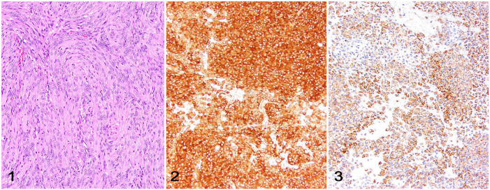
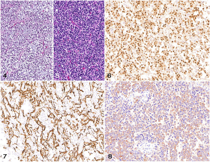
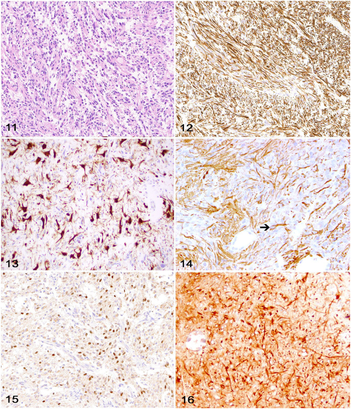
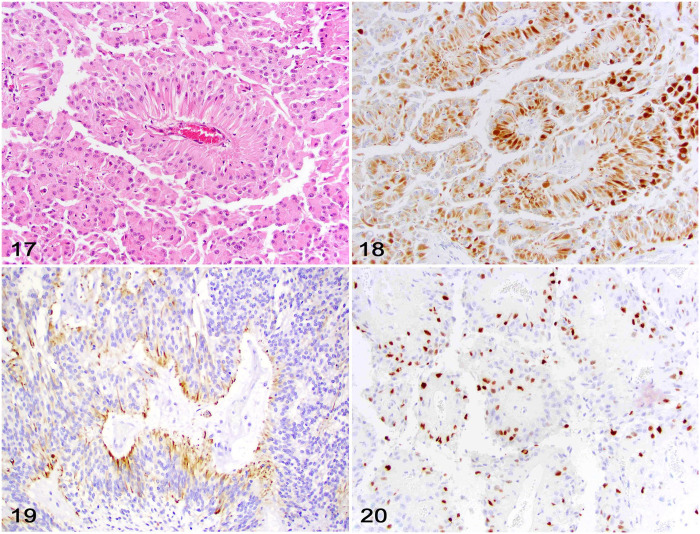
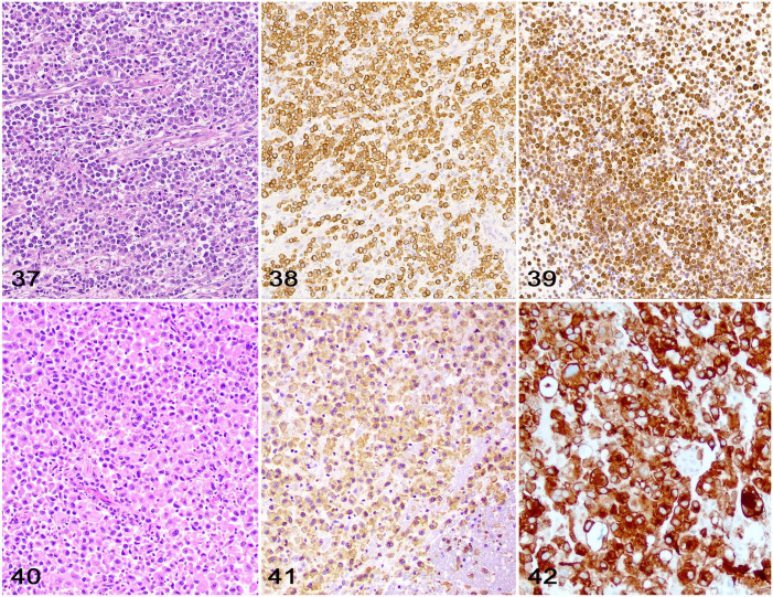
The diagnosis of primary and secondary CNS neoplasms of dogs and cats relies on histologic examination of autopsy or biopsy samples. In addition, many neoplasms must be further characterized by immunohistochemistry (IHC) for a more refined diagnosis in specific cases. Given the many investigations assessing the diagnostic and prognostic IHC profile of CNS neoplasms in the veterinary literature, it may be difficult for the diagnostic pathologist or pathology trainee to narrow the list of reliable diagnostic IHCs when facing a challenging case. Here we compile a comprehensive list of the most diagnostically relevant immunomarkers that should be utilized for the diagnostic support or confirmation of the most common primary and secondary CNS neoplasms of dogs and cats.
Keywords: CNS tumors; cats; dogs; glioma; immunohistochemistry, meningioma; neuropathology.
Conflict of interest statement
Declaration of conflicting interestsThe authors declared no potential conflicts of interest with respect to the research, authorship, and/or publication of this article.
Figures

Similar articles
-
Immunohistochemical detection of CD34, E-cadherin, claudin-1, glucose transporter 1, laminin, and protein gene product 9.5 in 28 canine and 8 feline meningiomas.Vet Pathol. 2010 Jul;47(4):725-37. doi: 10.1177/0300985810364528. Epub 2010 Apr 19. Vet Pathol. 2010. PMID: 20403881
-
Squash-prep cytology in the diagnosis of canine and feline nervous system lesions: a study of 42 cases.Vet Clin Pathol. 2006 Jun;35(2):208-14. doi: 10.1111/j.1939-165x.2006.tb00116.x. Vet Clin Pathol. 2006. PMID: 16783715
-
Overview of the Veterinary Cancer Guidelines and Protocols for CNS neoplasms of dogs and cats.J Vet Diagn Invest. 2023 Nov;35(6):593-594. doi: 10.1177/10406387231194323. Epub 2023 Aug 23. J Vet Diagn Invest. 2023. PMID: 37608762 Free PMC article. No abstract available.
-
A review of primary central nervous system neoplasms of cats.Vet Pathol. 2023 May;60(3):294-307. doi: 10.1177/03009858231155400. Epub 2023 Feb 20. Vet Pathol. 2023. PMID: 36803009 Review.
-
Neuroradiation oncology.Vet Clin North Am Small Anim Pract. 1997 Jan;27(1):95-100. doi: 10.1016/s0195-5616(97)50008-8. Vet Clin North Am Small Anim Pract. 1997. PMID: 9002169 Review.
Cited by
-
Canine cystic astrocytomas: 7 cases.J Vet Diagn Invest. 2025 Mar;37(2):398-403. doi: 10.1177/10406387241312898. Epub 2025 Jan 27. J Vet Diagn Invest. 2025. PMID: 39866072 Free PMC article.
-
Case Report: Primary intracranial lymphoma and meningioma manifesting as a composite tumor in a cat.Front Vet Sci. 2025 Aug 7;12:1619792. doi: 10.3389/fvets.2025.1619792. eCollection 2025. Front Vet Sci. 2025. PMID: 40852427 Free PMC article.
References
-
- Barnhart KF, et al.. Immunohistochemical staining patterns of canine meningiomas and correlation with published immunophenotypes. Vet Pathol 2002;39:311–321. - PubMed
-
- Belluco S, et al.. Standardisation of canine meningioma grading: inter-observer agreement and recommendations for reproducible histopathologic criteria. Vet Comp Oncol 2022;20:509–520. - PubMed
-
- Blümcke I, et al.. Microtubule-associated protein-2 immunoreactivity: a useful tool in the differential diagnosis of low-grade neuroepithelial tumors. Acta Neuropathol 2004;108:89–96. - PubMed
-
- Boozer LB, et al.. Characterization of immune cell infiltration into canine intracranial meningiomas. Vet Pathol 2012;49:784–795. - PubMed
-
- Brewer DM, et al.. Spinal cord nephroblastoma in dogs: 11 cases (1985–2007). J Am Vet Med Assoc 2011;238:618–624. - PubMed
Publication types
MeSH terms
LinkOut - more resources
Full Text Sources
Miscellaneous
