. 2024 Feb 20;123(4):478-488.
doi: 10.1016/j.bpj.2024.01.013. Epub 2024 Jan 17.

Targeting protein-protein interaction interfaces with antiviral N protein inhibitor in SARS-CoV-2

Affiliations

Targeting protein-protein interaction interfaces with antiviral N protein inhibitor in SARS-CoV-2

Jhen-Yi Hong et al. Biophys J. .

Abstract

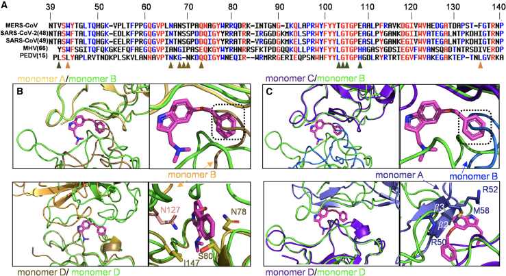
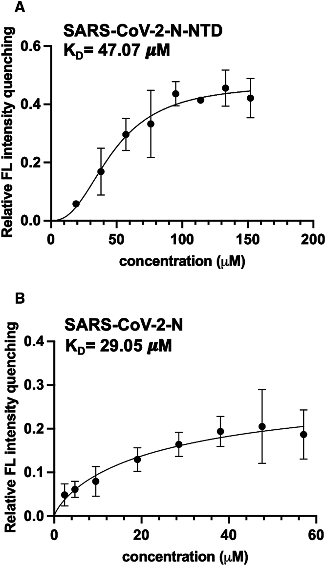
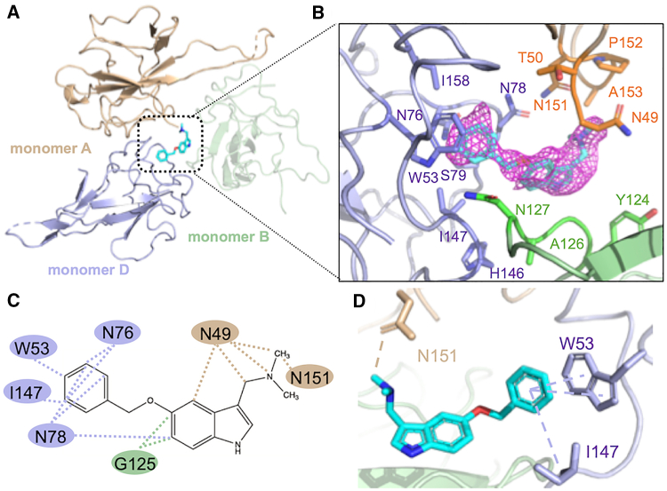
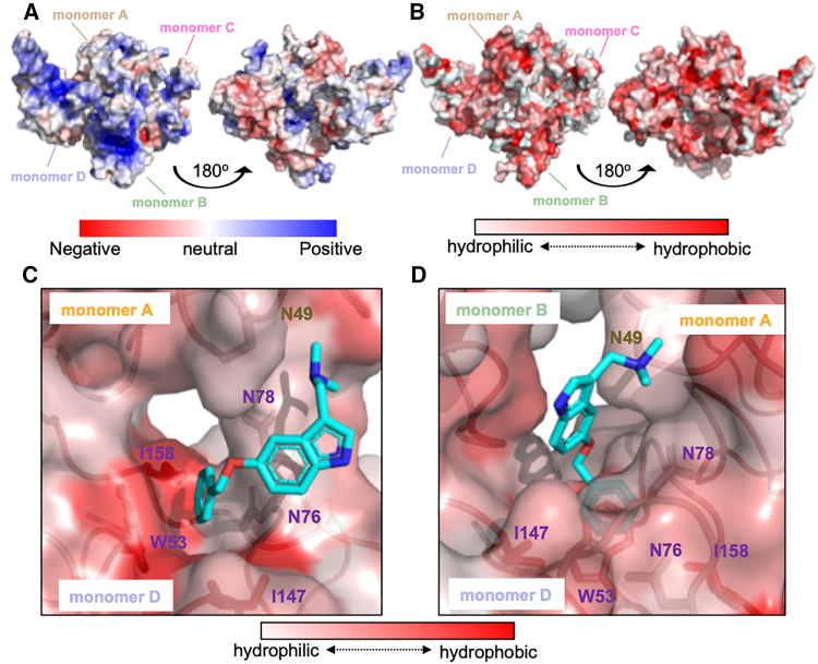
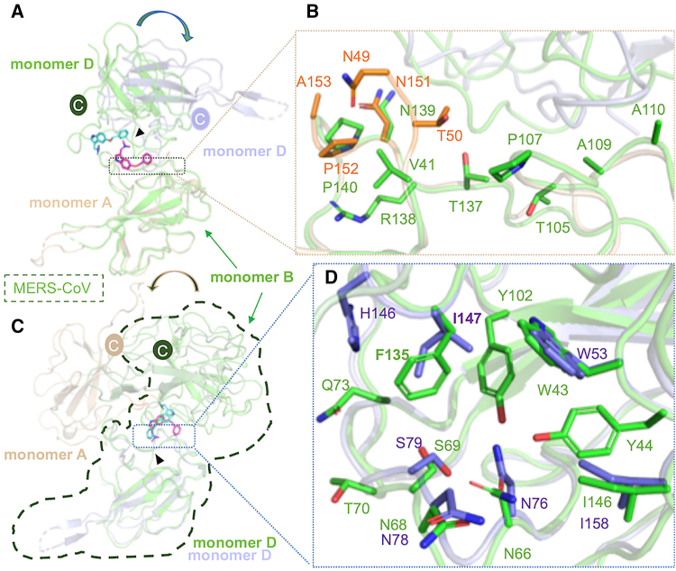
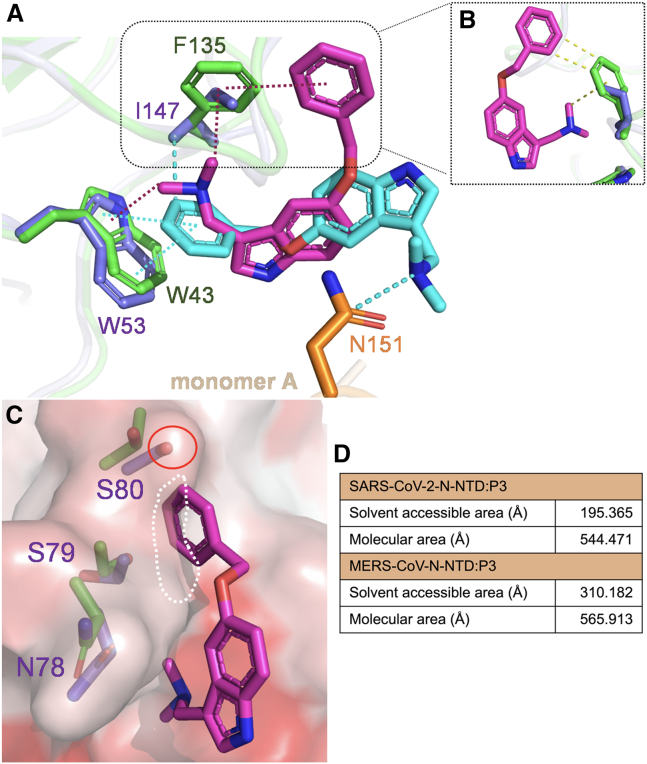
Coronaviruses not only pose significant global public health threats but also cause extensive damage to livestock-based industries. Previous studies have shown that 5-benzyloxygramine (P3) targets the Middle East respiratory syndrome coronavirus (MERS-CoV) nucleocapsid (N) protein N-terminal domain (N-NTD), inducing non-native protein-protein interactions (PPIs) that impair N protein function. Moreover, P3 exhibits broad-spectrum antiviral activity against CoVs. The sequence similarity of N proteins is relatively low among CoVs, further exhibiting notable variations in the hydrophobic residue responsible for non-native PPIs in the N-NTD. Therefore, to ascertain the mechanism by which P3 demonstrates broad-spectrum anti-CoV activity, we determined the crystal structure of the SARS-CoV-2 N-NTD:P3 complex. We found that P3 was positioned in the dimeric N-NTD via hydrophobic contacts. Compared with the interfaces in MERS-CoV N-NTD, P3 had a reversed orientation in SARS-CoV-2 N-NTD. The Phe residue in the MERS-CoV N-NTD:P3 complex stabilized both P3 moieties. However, in the SARS-CoV-2 N-NTD:P3 complex, the Ile residue formed only one interaction with the P3 benzene ring. Moreover, the pocket in the SARS-CoV-2 N-NTD:P3 complex was more hydrophobic, favoring the insertion of the P3 benzene ring into the complex. Nevertheless, hydrophobic interactions remained the primary stabilizing force in both complexes. These findings suggested that despite the differences in the sequence, P3 can accommodate a hydrophobic pocket in N-NTD to mediate a non-native PPI, enabling its effectiveness against various CoVs.


Conflict of interest statement

Declaration of interests The authors declare no competing interests.

Figures

Figure 1
Figure 1
Comparison of the structure and sequence of the N-terminal domain (N-NTD) between SARS-CoV-2 and MERS-CoV. (A) Sequence alignment of the N-terminal region of the N protein from various CoVs. Alignment was performed using Multalin (35). The results were color-coded based on amino acid identity, with distinct colors indicating variations in polarity or charge. Orange triangles highlight hydrophobic residues that participate in the mediating of non-native PPI; dark-green and brown triangles represent the interface residues between monomers B and D on MERS-CoV and P3, respectively. (B) Superimposition of monomers A and D of SARS-CoV-2-N-NTD (PDB: 6m3m) to monomers B and D of MERS-CoV-N-NTD (PDB: 6KL6), respectively. The root-mean-square deviation (RMSD) values for both comparisons were 0.623 (98 to 98 atoms) and 0.779 (100 to 100 atoms), respectively. (C) Superimposition of monomers C and D of SARS-CoV-2-N-NTD (PDB: 7cdz) to monomers B and D of MERS-CoV-N-NTD (PDB: 6KL6). The RMSD values for both comparisons were 0.639 (100 to 100 atoms) and 0.661 (98 to 98 atoms), respectively. The two SARS-CoV-2-N-NTD are shown in brown and purple-blue, whereas MERS-CoV-N-NTD is in green. To see this figure in color, go online.
Figure 2
Figure 2
The dissociation constant for the interaction of P3 with the SARS-CoV-2 N protein and N-NTD. KD values for the binding of P3 with SARS-CoV-2 (A) N-NTD and (B) N protein. The x axis represents P3 concentration (μM), while the y axis represents relative fluorescence intensity. The data were derived from three independent experiments. SARS-CoV-2 N-NTD was purified in 20 mM Tris-HCl (pH 7.5) and 50 mM NaCl, while the SARS-CoV-2 N protein was purified in 20 mM Na3PO4 (pH 7.8) and 300 mM NaCl. Data are presented as the mean ± standard deviation.
Figure 3
Figure 3
The crystal structure and interfaces of the SARS-CoV-2-N-NTD:P3 complex. (A) Overall crystal structure of SARS-CoV-2 N-NTD:P3; the monomers A, B, and D are colored in wheat, pale green, and light blue, respectively (B) The residues involved in creating the BSA between P3 and SARS-CoV-2 N-NTD. The macromolecular interactions were analyzed using the PISA database. The interacting residues of the SARS-CoV-2 N-NTD are indicated. (C) The hydrophobic interactions between P3 and N-NTD are indicated by the dotted lines. The data were validated by analyzing the interactions using LigPlot. (D) The benzene ring of P3 forms π-π and π-alkyl interactions with residues W53 and I147 of chain D of N-NTD, whereas a carbon-hydrogen bond forms between N151 from chain A of N-NTD and the P3 moiety. The data were analyzed using Discovery Studio. To see this figure in color, go online.
Figure 4
Figure 4
The benzene ring of P3 compound is embedded within a hydrophobic pocket. (A) The electrostatic surface of the native N-NTD protein structure. (B) The hydrophobicity level of the native protein structure; hydrophobicity was evaluated and colored using PyMOL. (C) The hydrophobic surface between monomers A and D. P3 is located within the center of monomeric A, B, and D. The gramine moiety is located beneath a hydrophobic surface created by chain B. (D) Rotation of 90° of (C) shows that the benzene ring of P3 is inserted into a hydrophobic pocket within monomer D. To see this figure in color, go online.
Figure 5
Figure 5
Structural comparison of N-NTD:P3 complexes in MERS-CoV and SARS-CoV-2. (A) The structures of MERS-CoV N-NTD:P3 (PDB: 6KL6) and SARS-CoV-2 N-NTD:P3 were aligned by superimposing chains B and A from MERS-CoV and SARS-CoV-2 N-NTD:P3 complexes, respectively. The MERS-CoV N-NTD:P3 complex is shown in green. RMSD = 0.699 Å (103 to 103 atoms). (B) A close-up view of the interfaces between monomers B and A from MERS-CoV and SARS-CoV-2 N-NTD:P3 complexes, respectively; the interacting residues are represented by sticks. (C) The superimposition of monomer D in both complexes. RMSD = 0.616 Å (100 to 100 atoms). The triangle indicates the hydrophobic pocket created by the residues surrounding W53. (D) The interfaces between monomer D in both complexes. The interactions were analyzed using the PISA database. To see this figure in color, go online.
Figure 6
Figure 6
Analysis of the mechanism through which P3 interacts with various CoVs. (A) Superimposition of the MERS-CoV and SARS-CoV-2 N-NTD:P3 complexes. The structures of SARS-CoV-2 N-NTD:P3 and MERS-CoV N-NTD:P3 were aligned by superimposing the chain D; RMSD = 0.616 Å (100 to 100 atoms). The interacting residues on the SARS-CoV-2 and MERS-CoV N-NTD:P3 complexes are shown in slate blue, orange, and green, respectively. The magenta dashed line represents the π-cation interactions between P3 and MERS-CoV N-NTD; the cyan dashed line represents the π-π and π-alkyl interactions between P3 and SARS-CoV-2 N-NTD. (B) The distances between the two P3 moieties and the Phe benzene ring in MERS-CoV N-NTD and the Ile side chain in SARS-CoV-2 N-NTD are represented by green and purple colors, respectively. (C) The side chain containing residues N78–S80 is highlighted, and the surface predicted to clash with the benzene ring of P3 in the MERS-CoV N-NTD:P3 complex is shown. (D) The surface area (Å) within the pocket was calculated using PyMOL. To see this figure in color, go online.

Similar articles

References

    1. Coronaviridae Study Group of the International Committee on Taxonomy of Viruses The species severe acute respiratory syndrome-related coronavirus: classifying 2019-nCoV and naming it SARS-CoV-2. Nat. Microbiol. 2020;5:536–544. - PMC - PubMed
    1. Harvey W.T., Carabelli A.M., et al. Robertson D.L. SARS-CoV-2 variants, spike mutations and immune escape. Nat. Rev. Microbiol. 2021;19:409–424. - PMC - PubMed
    1. Hu J., Peng P., et al. Huang A.L. Increased immune escape of the new SARS-CoV-2 variant of concern omicron. Cell. Mol. Immunol. 2022;19:293–295. - PMC - PubMed
    1. McBride R., van Zyl M., Fielding B.C. The coronavirus nucleocapsid is a multifunctional protein. Viruses. 2014;6:2991–3018. - PMC - PubMed
    1. Chang C.K., Hou M.H., et al. Huang T.H. The SARS coronavirus nucleocapsid protein--forms and functions. Antivir. Res. 2014;103:39–50. - PMC - PubMed

Publication types

LinkOut - more resources