. 2024 Dec;46(1):2303396.
doi: 10.1080/0886022X.2024.2303396. Epub 2024 Jan 17.

Placenta-derived mesenchymal stem cells protect against diabetic kidney disease by upregulating autophagy-mediated SIRT1/FOXO1 pathway

Affiliations

Placenta-derived mesenchymal stem cells protect against diabetic kidney disease by upregulating autophagy-mediated SIRT1/FOXO1 pathway

Honghong Liu et al. Ren Fail. 2024 Dec.

Abstract

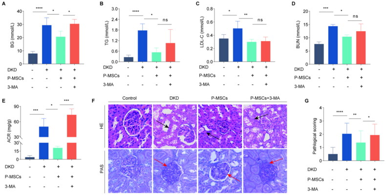
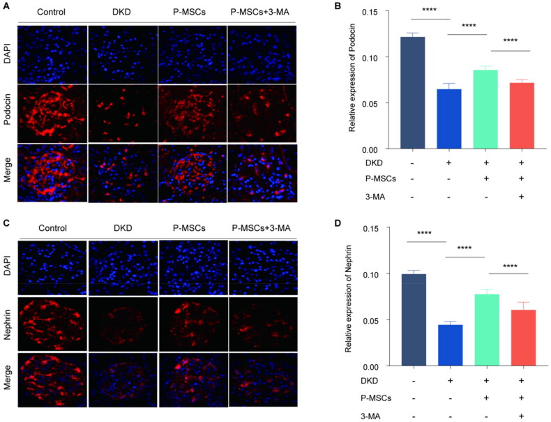
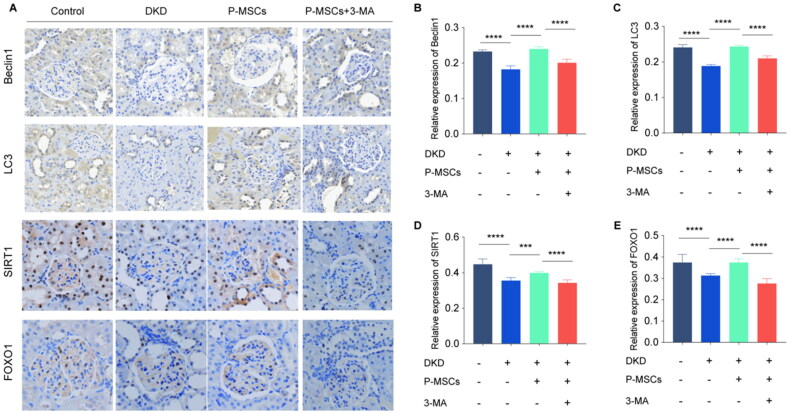
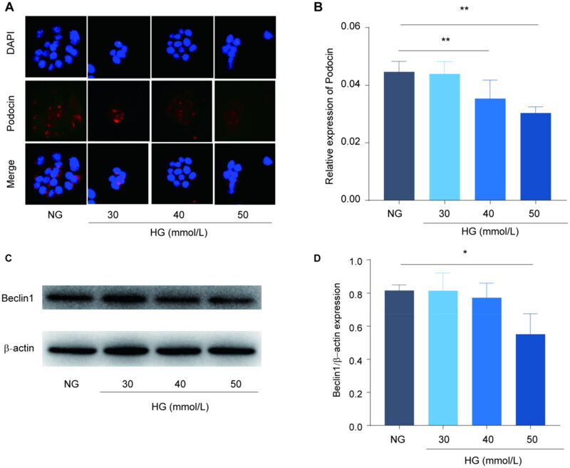
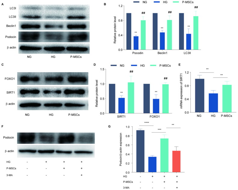
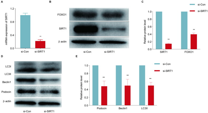
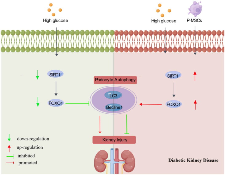
Diabetic kidney disease (DKD) is a common chronic microvascular complication of diabetes mellitus. Although studies have indicated the therapeutic potential of mesenchymal stem cells (MSCs) for DKD, the underlying molecular mechanisms remain unclear. Herein, we explored the renoprotective effect of placenta-derived MSCs (P-MSCs) and the potential mechanism of SIRT1/FOXO1 pathway-mediated autophagy in DKD. The urine microalbumin/creatinine ratio was determined using ELISA, and renal pathological changes were detected by special staining techniques. Immunofluorescence was used for detecting the renal tissue expression of podocin and nephrin; immunohistochemistry for the renal expression of autophagy-related proteins (LC3, Beclin-1, SIRT1, and FOXO1); and western blotting and PCR for the expression of podocyte autophagy- and pathway-related indicators. We found that P-MSCs ameliorated renal tubular injury and glomerular mesangial matrix deposition and alleviated podocyte damage in DKD rats. PMSCs enhanced autophagy levels and increased SIRT1 and FOXO1 expression in DKD rat renal tissue, whereas the autophagy inhibitor 3-methyladenine significantly attenuated the renoprotective effect of P-MSCs. P-MSCs improved HG-induced Mouse podocyte clone5(MPC5)injury, increased podocyte autophagy, and upregulated SIRT1 and FOXO1 expression. Moreover, downregulation of SIRT1 expression blocked the P-MSC-mediated enhancement of podocyte autophagy and improvement of podocyte injury. Thus, P-MSCs can significantly improve renal damage and reduce podocyte injury in DKD rats by modulating the SIRT1/FOXO1 pathway and enhancing podocyte autophagy.

Keywords: Placenta-derived mesenchymal stem cells; SIRT1/FOXO1 pathway; autophagy; diabetic kidney disease; podocyte injury.


Conflict of interest statement

No potential conflict of interest was reported by the author(s).

Figures

Figure 1.
Figure 1.
Effects of placenta-derived mesenchymal stem cells (P-MSCs) on blood glucose (BG), lipid, renal function, and renal pathological structure in diabetic rats. A: Effect of P-MSCs on BG in diabetic kidney disease (DKD) rats; B: Effect of P-MSCs on triglyceride (TG) in DKD rats; C: Effect of P-MSCs on low-density lipoprotein cholesterol (LDL-C) in DKD rats; D: Effect of P-MSCs on blood urea nitrogen (BUN) in DKD rats; E: Effect of P-MSCs on urinary albumin-to-creatinine ratio (UACR) in DKD rats; F: Effect of P-MSCs on renal tubule and glomerulus in DKD rats; G: Pathological score of renal tubular injury(there are 6 rats in each group, and 5 pathological pictures are randomly selected for each group, so there are 30 pictures in each group to analyze the pathological score.)(nsP >0.05, *p < 0.05, **p < 0.01, ***p < 0.001, ****p < 0.0001).
Figure 2.
Figure 2.
Effects of placenta-derived mesenchymal stem cells (P-MSCs) and 3-methyladenine (3-MA) on glomerular podocyte fissure membrane proteins podocin and nephrin in diabetic kidney disease (DKD) rats. A-B: Immunofluorescence was used to detect the expression of podocin in rat glomerulus. C-D: Effects of P-MSCs and 3-MA on nephrin in DKD rats. (****p < 0.0001).
Figure 3.
Figure 3.
Effects of placenta-derived mesenchymal stem cells (P-MSCs) and 3-methyladenine (3-MA) on autophagy-related proteins, SITR1 and FOXO1 in renal tissue of diabetic kidney disease (DKD) rats. A: Expression of autophagy-related proteins Beclin1, LC3, SITR1, and FOXO1 was evaluated by immunohistochemical staining in renal tissue of rats in each group; B: Effects of P-MSCs and 3-MA on Beclin1 in renal tissue of DKD rats; C: Effects of P-MSCs and 3-MA on LC3 in renal tissue of DKD rats D: Effects of P-MSCs and 3-MA on SITR1 in renal tissue of DKD rats; E: Effects of P-MSCs and 3-MA on FOXO1 in renal tissue of DKD rats. (***p < 0.001,****p < 0.0001).
Figure 4.
Figure 4.
Effects of high glucose on podocyte- and autophagy-related proteins Beclin1 of podocytes. A-B: Immunofluorescence was used to detect the expression of podocin expression in podocytes with different concentrations of high glucose; C-D: Western blot was used to detect the effects of different concentrations of high glucose on podocyte autophagy. (*p < 0.05, **p < 0.01).
Figure 5.
Figure 5.
Effects of placenta-derived mesenchymal stem cells (P-MSCs) and 3-methyladenine (3-MA) on autophagy-related proteins and SIRT1, FOXO1 in high glucose-induced MPC5 cells A-B: Effects of P-MSCs on podocin and autophagy-related proteins (Beclin1 and LC3) in high glucose-induced MPC5; C-D: Western blot was used to detect the expression of SIRT1 and FOXO1 in high glucose-induced MPC5 cells treated with P-MSCs; E: PCR was used to evaluate SIRT1 mRNA expression in high glucose-induced MPC5 cells by P-MSCs; F-G: Effects of P-MSCs and 3-MA on podocin in high glucose-induced MPC5 cells. (**p < 0.01, ***p < 0.001, ****p < 0.000, ## p < 0.001).
Figure 6.
Figure 6.
Effects of SIRT1 siRNA on SIRT1/FOXO1 and autophagy-related proteins in MPC5 cells treated with high glucose and placenta-derived mesenchymal stem cells (P-MSCs). A: SIRT1 siRNA inhibited P-MSCs from promoting SIRT1 mRNA expression in high glucose-induced MPC5 cells; B-C: Effects of SIRT1 siRNA on SIRT1 and FOXO1 expression in MPC5 cells treated with high glucose and P-MSCs; D-E: Effects of SIRT1 siRNA on podocin and autophagy-related proteins in MPC5 cells treated with high glucose and P-MSCs. (**p < 0.01).
Figure 7.
Figure 7.
Schematic diagram of the effects of P-MSCs on podocyte in DKD via enhancing SIRT1/FOXO1 pathway-mediated autophagy(visual abstract).

References

    1. Ogurtsova K, Da Rocha Fernandes JD, Huang Y, et al. IDF diabetes atlas: global estimates for the prevalence of diabetes for 2015 and 2040. Diabetes Res Clin Pract. 2017;128:1–12. doi: 10.1016/j.diabres.2017.03.024. - DOI - PubMed
    1. Koye DN, Magliano DJ, Nelson RG, et al. The global epidemiology of diabetes and kidney disease. Adv Chronic Kidney Dis. 2018;25(2):121–132. doi: 10.1053/j.ackd.2017.10.011. - DOI - PMC - PubMed
    1. Reidy K, Kang HM, Hostetter T, et al. Molecular mechanisms of diabetic kidney disease. J Clin Invest. 2014;124(6):2333–2340. doi: 10.1172/JCI72271. - DOI - PMC - PubMed
    1. Ye H, Bai X, Gao H, et al. Urinary podocalyxin positive-element occurs in the early stage of diabetic nephropathy and is correlated with a clinical diagnosis of diabetic nephropathy. J Diabetes Complications. 2014;28(1):96–100. doi: 10.1016/j.jdiacomp.2013.08.006. - DOI - PubMed
    1. Griffin TP, Martin WP, Islam N, et al. The promise of mesenchymal stem cell therapy for diabetic kidney disease. Curr Diab Rep. 2016;16(5):42. doi: 10.1007/s11892-016-0734-6. - DOI - PubMed