[Preprint]. 2023 Dec 27:2023.12.25.573323.
doi: 10.1101/2023.12.25.573323.

Towards a Brighter Constellation: Multi-Organ Neuroimaging of Neural and Vascular Dynamics in the Spinal Cord and Brain

Affiliations

Towards a Brighter Constellation: Multi-Organ Neuroimaging of Neural and Vascular Dynamics in the Spinal Cord and Brain

Dmitrijs Celinskis et al. bioRxiv. .

Update in

Abstract

Significance: Pain is comprised of a complex interaction between motor action and somatosensation that is dependent on dynamic interactions between the brain and spinal cord. This makes understanding pain particularly challenging as it involves rich interactions between many circuits (e.g., neural and vascular) and signaling cascades throughout the body. As such, experimentation on a single region may lead to an incomplete and potentially incorrect understanding of crucial underlying mechanisms.

Aim: Here, we aimed to develop and validate new tools to enable detailed and extended observation of neural and vascular activity in the brain and spinal cord. The first key set of innovations were targeted to developing novel imaging hardware that addresses the many challenges of multi-site imaging. The second key set of innovations were targeted to enabling bioluminescent imaging, as this approach can address limitations of fluorescent microscopy including photobleaching, phototoxicity and decreased resolution due to scattering of excitation signals.

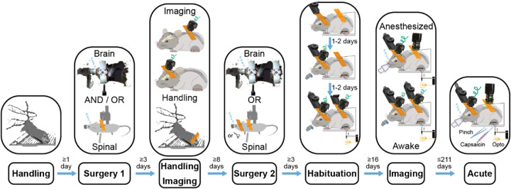
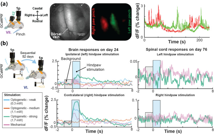
Approach: We designed 3D-printed brain and spinal cord implants to enable effective surgical implantations and optical access with wearable miniscopes or an open window (e.g., for one- or two-photon microscopy or optogenetic stimulation). We also tested the viability for bioluminescent imaging, and developed a novel modified miniscope optimized for these signals (BLmini).

Results: Here, we describe novel 'universal' implants for acute and chronic simultaneous brain-spinal cord imaging and optical stimulation. We further describe successful imaging of bioluminescent signals in both foci, and a new miniscope, the 'BLmini,' which has reduced weight, cost and form-factor relative to standard wearable miniscopes.

Conclusions: The combination of 3D printed implants, advanced imaging tools, and bioluminescence imaging techniques offers a new coalition of methods for understanding spinal cord-brain interactions. This work has the potential for use in future research into neuropathic pain and other sensory disorders and motor behavior.

Keywords: bioluminescence (BL); brain; fluorescence (FL); implantable window; miniscope; multi-organ imaging; sensory processing; spinal cord; two-photon.


Conflict of interest statement

Disclosures The authors declare no competing interests.

Figures

Fig. 1
Fig. 1. A Key Use Case for Multi-Organ Imaging: Studying Mechanisms of Nociception and Pain
Peripheral nociceptive input that drives pain engages a wide range of dynamic body systems. Integrated activity spans multiple organs, including skin, dorsal root ganglia (DRG), spinal cord and brain, and depends critically on a broad array of networks, including neural, vascular and immune signaling. State-of-the-art approaches for studying sensory information processing typically rely on in vivo imaging in one organ, and focuses on neurons. In the present work, we expand the in vivo methodological toolbox by developing procedures for multi-organ in vivo studies.
Fig. 2
Fig. 2. Imaging Bioluminescence (BL) Offers Advantages Over Fluorescence (FL), Including Decreased Scattering Artifacts and Simplified Optics
(a) Comparative advantages of BL and FL imaging. (b) BL offers up to 3x potential increase in imaging depth of discrete emitters (e.g., cells) compared to FL. Mouse somatosensory cortical layers and spinal dorsal horn laminae marked in red indicate the approximate deepest cortical layers and laminae, that can be imaged in an adult mouse via 1-photon FL and BL approaches. Imaging depths were determined from. The estimated increase in imaging depth is especially beneficial for spinal cord imaging because it is significantly constrained by photon scattering properties of myelin. The brain background image in (b) is from and is in the public domain, the spinal cord image in (b) is from, licensed under Public Domain Mark 1.0.
Fig. 3
Fig. 3. UPRIM Brain and Spinal Window Implants Consist of Off-the-Shelf Components and a 3D-Printed Implant Chamber
Identical UPRIM implant chambers can be used for brain and spinal imaging. The only material difference is the need for additional parts to attach 3D-printed chamber to the spinal cord. The specific design illustrated here is equipped with a mount for UCLA miniscope v4, but can also be readily adapted for any wearable microscope and is compatible with the benchtop microscopes with ≥ 5 mm objective working distance.
Fig. 4
Fig. 4. UPRIM Implants Can Be Used for Brain and Spinal Cord Imaging in Awake and Anesthetized Animals
(a) Images of the UPRIM 3D-printed implant attached to the brain and the spinal cord. (b) Images of use of the implants for imaging through multiple miniscopes (UCLA V4) and a benchtop microscope.
Fig. 5
Fig. 5. Timing of Steps that Enable Awake Simultaneous Brain-Spinal Cord Imaging
Awake imaging simultaneously in the brain and spinal cord to study sensory information processing requires careful design of a multi-step animal training protocol to maximize animal comfort and data quality.
Fig. 6
Fig. 6. Spinal Cord Photomodulation Approach for Integratied 2-Photon Imaging
(a) We performed optical stimulation in the spinal cord under isoflurane anesthesia through a chronically implanted spinal cord window using an optical fiber positioned adjacent to the imaging objective. (b) We imaged spinal cord vascular responses using 2-photon galvo-galvo scan mode. The use of 2-photon galvo-galvo allowed us to minimize the optical stimulation artifacts by flashing the stimulation LED only during the scanner flyback between the scan lines, see Voigts et al. (2020) for further details. LDT = line dwell time, SLI = scan line interval, PD = pulse duration, IPI = inter-pulse interval, ITI = inter-trial interval.
Fig. 7
Fig. 7. Bioluminescence (BL) Imaging Was Conducted in Anesthetized Animals With Direct Cortical Luciferin Application and In Awake Behaving Animals With Systemic Injection
(a) We performed acute BL imaging under isoflurane anesthesia with direct cortical administration of luciferin (Coelenterazine). The same preparation was used for imaging either using an emCCD camera or the BLmini. During the experiments testing novel BL calcium indicators (e.g., CaBLAM (APD3A6)) we stimulated vibrissae using a piezoelectric element. (b) For awake BL and FL (e.g., jRGeCO) imaging we administered luciferin intraperitonially (IP) before headpost-restraining the animal on a running wheel. We imaged BL and FL through the same cranial window sequentially to compare relative activation profiles.
Fig. 8
Fig. 8. Tolerance of the Multiple Implant Variants Tested
Across implantation approaches, animals were healthy by observation, and showed either maintenance of pre-surgical body weight or recovery after ~1–2 weeks.
Fig. 9
Fig. 9. UPRIM Spinal Cord Implants are Viable for Single Cell GCaMP Imaging
(a) Individual cell GCaMP6f calcium responses to hind paw pinch were imaged under anesthesia in an animal implanted with spinal cord UPRIM via surgical procedure # IV and using Miniscope v4. Calcium indicator was expressed via AAV2/9-CaMKIIa-GCaMP6f-P2A-nls-dTomato. (b) A pinch response map as the average of multiple trials, showing activation in multiple distinct neurons (pink, green and blue circles). (c) Time series show changes in signal for each color-matched cell. Grey shadings highlight the pinching time intervals, arrows emphasize responses that differed between 3 cells.
Fig. 10
Fig. 10. Calcium Activity in the Brain and Spinal Cord in the Same Mouse Using UPRIM
(a) Lateralized localization of responses to hindpaw pinch in the spinal cord using the UPRIM implant and 4x magnification benchtop imaging. The GCaMP6f indicator was expressed in Somatostatin-positive interneurons (SOM). Images in this panel from left to right show: The experimental setup with spinal cord reference axes for orientation, spinal cord brightfield image, GCaMP6f average response maps, and time courses showing the independence of relative light intensity signals for left (green) and right (red) cord regions. In this experiment, the mouse was implanted with a brain and spinal cord UPRIM (surgical procedure # VII). (b) UPRIM calcium responses in cortex to optogenetic and mechanical stimulation 24 days after implantation, and in the same mouse in the spinal cord 52 days after implanting and imaging brain UPRIM (surgical procedure # VI). Both data sets were acquired under anesthesia. This was a Trpv1ChR2eYFP+/− animal injected with a mixture of two non-flex AAVs to co-express GCaMP6f and jRGeCO both in the brain and spinal cord.
Fig. 11
Fig. 11. Vascular Dilations in the Spinal Cord in Wild Type Mice Driven by 2 sec Long Trains of Pulsed Light
(a) Map of spinal cord vasculature obtained with 2-photon imaging after intravenous 10 kD FITC-green dextran injection. Zoomed-in vessel inset in top right shows a single frame of a segmented vessel during stimulation. (b) Example of a responsive vessel segment.
Fig. 12
Fig. 12. Bioluminescent Imaging Requires Fewer System Components, Decreasing Miniscope Complexity and Weight
Conversion of UCLA miniscopes v3.2 or v4 from FL-centric to BL-centric design offers 22–27% reduction in mass, 32–45% reduction in the number of components and assembly labor, 19–58% reduction in cost/scope, 3–66 times higher sensitivity for optical signals that do not require filtering, 13–50% reduction in electrical power consumption.
Fig. 13
Fig. 13. Imaging Bioluminescent and Fluorescent Indicators Using the BLmini and FL
(a) Time courses of BL photon production detected by an emCCD (ANDOR) and two generations of BLmini miniscopes in three different mice. Luciferin (CTZ) was directly applied to the cortical surface, previously transduced with either LMO3 (emCCD data) or NCS2 (BLmini data). (b) BLmini v2.0 allows detection of photon production by the calcium indicator CaBLAM (APD3A6). (c) Time series for BL and FL calcium indicator output from a single imaging session, from CaBLAM (APD3A6-ISN) and jRGeCO (with full field excitation illumination) virally transduced in SI Vibrissal Barrel Neocortex (synapsin (Syn) promoter). In these data, vibrissal stimulation was applied during either BL (blue) or FL (green) imaging (25 trials in each condition). (d) The Region-of-Interest used to extract jRGeCO and CaBLAM (APD3A6-ISN) signals overlaid on the mean jRGeCO vibrissa response map (left), the mean red FL intensity map across all frames (center) and the mean green FL map, reflecting expression of mNeon-green (mNG) in the CaBLAM (APD3A6-ISN) construct.
Fig. 14
Fig. 14. Beyond Brain-Only Imaging Offers Many Opportunities for Experimental Designs and Hardware Combinations
(a) In this report we demonstrate feasibility of gaining chronic optical access to the brain and spinal cord within the same animal. Universal format implants described as part of this report allow sequential (b) and simultaneous (c) imaging of the brain and spinal cord within the same animal using benchtop and wearable microscopes via FL or BL. Wearable microscopes for BL imaging, and hence the implants, can be designed smaller, allowing animals to engage in more natural behaviors.

References

    1. Ayaz A. et al. , “Layer-specific integration of locomotion and sensory information in mouse barrel cortex,” Nat Commun 10(1), 2585 (2019) [doi: 10.1038/s41467-019-10564-8]. - DOI - PMC - PubMed
    1. Nelson N. A. et al. , “Imaging spinal cord activity in behaving animals,” Exp Neurol 320, 112974 (2019) [doi: 10.1016/j.expneurol.2019.112974]. - DOI - PMC - PubMed
    1. Fukui A. et al. , “Layer-specific sensory processing impairment in the primary somatosensory cortex after motor cortex infarction,” Sci Rep 10(1), 3771 (2020) [doi: 10.1038/s41598-020-60662-7]. - DOI - PMC - PubMed
    1. Borowska J. et al. , “Functional Subpopulations of V3 Interneurons in the Mature Mouse Spinal Cord,” J Neurosci 33(47), 18553–18565 (2013) [doi: 10.1523/JNEUROSCI.2005-13.2013]. - DOI - PMC - PubMed
    1. Raghuram A. et al. , “Determining the Depth Limit of Bioluminescent Sources in Scattering Media,” p. 2020.04.21.044982, bioRxiv (2020) [doi: 10.1101/2020.04.21.044982]. - DOI

Publication types