

Tissue adhesive, ROS scavenging and injectable PRP-based 'plasticine' for promoting cartilage repair
- PMID: 38235061
- PMCID: PMC10793072
- DOI: 10.1093/rb/rbad104
Tissue adhesive, ROS scavenging and injectable PRP-based 'plasticine' for promoting cartilage repair
Abstract
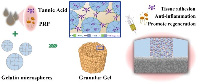
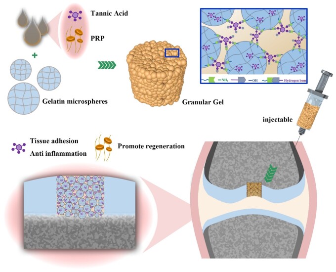
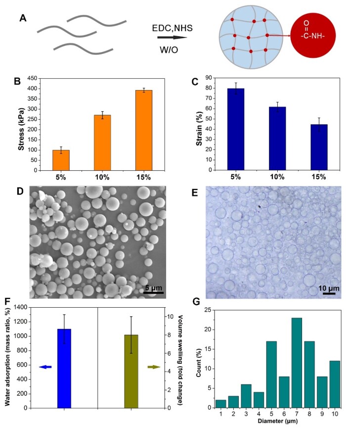
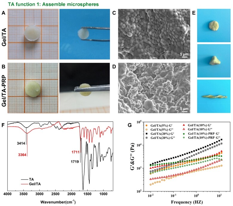
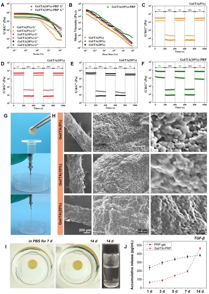
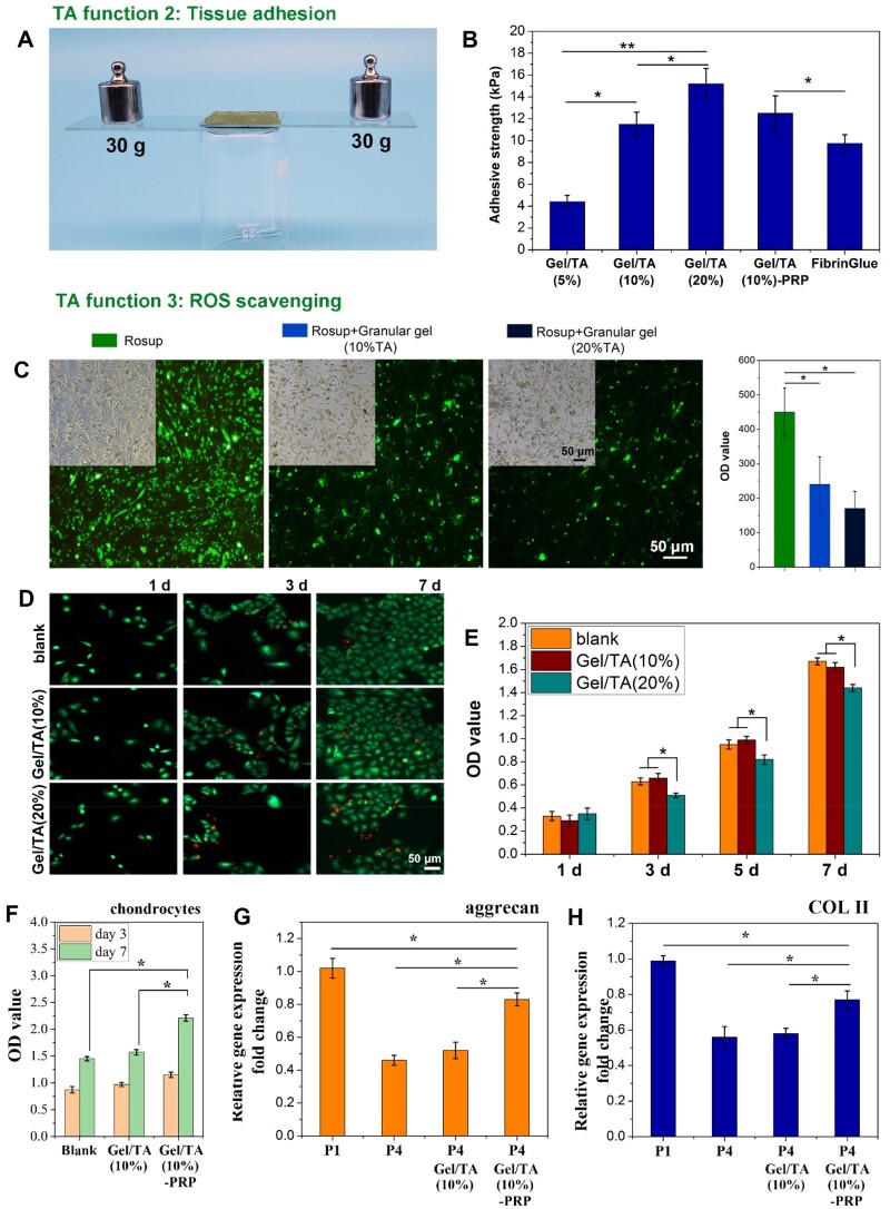
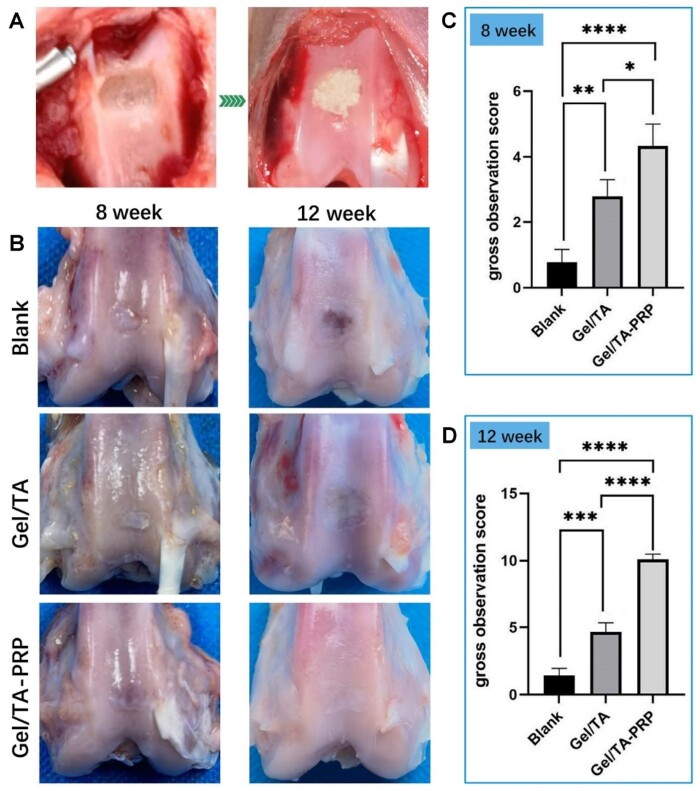
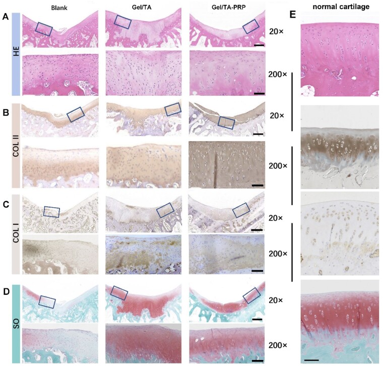
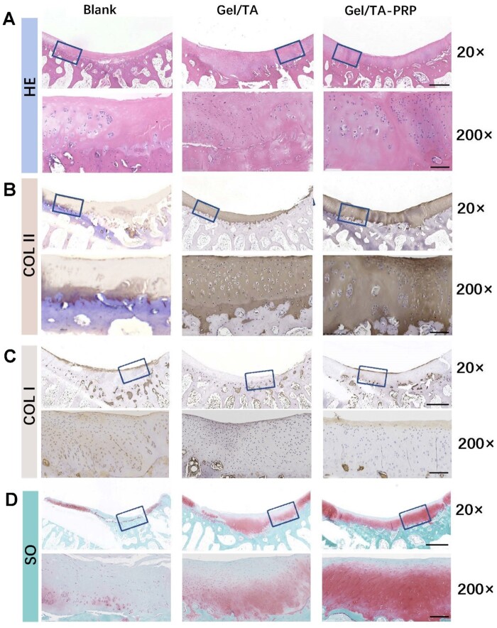
Platelet-rich plasma (PRP) that has various growth factors has been used clinically in cartilage repair. However, the short residence time and release time at the injury site limit its therapeutic effect. The present study fabricated a granular hydrogel that was assembled from gelatin microspheres and tannic acid through their abundant hydrogen bonding. Gelatin microspheres with the gelatin concentration of 10 wt% and the diameter distribution of 1-10 μm were used to assemble by tannic acid to form the granular hydrogel, which exhibited elasticity under low shear strain, but flowability under higher shear strain. The viscosity decreased with the increase in shear rate. Meanwhile, the granular hydrogel exhibited self-healing feature during rheology test. Thus, granular hydrogel carrying PRP not only exhibited well-performed injectability but also performed like a 'plasticine' that possessed good plasticity. The granular hydrogel showed tissue adhesion ability and reactive oxygen species scavenging ability. Granular hydrogel carrying PRP transplanted to full-thickness articular cartilage defects could integrate well with native cartilage, resulting in newly formed cartilage articular fully filled in defects and well-integrated with the native cartilage and subchondral bone. The unique features of the present granular hydrogel, including injectability, plasticity, porous structure, tissue adhesion and reactive oxygen species scavenging provided an ideal PRP carrier toward cartilage tissue engineering.
Keywords: PRP; cartilage regeneration; gelatin; granular hydrogel; tannic acid.
© The Author(s) 2023. Published by Oxford University Press.
Figures
Similar articles
-
An in situ photocrosslinkable platelet rich plasma - Complexed hydrogel glue with growth factor controlled release ability to promote cartilage defect repair.Acta Biomater. 2017 Oct 15;62:179-187. doi: 10.1016/j.actbio.2017.05.023. Epub 2017 May 10. Acta Biomater. 2017. PMID: 28501713
-
Dynamic hyaluronic acid hydrogel with covalent linked gelatin as an anti-oxidative bioink for cartilage tissue engineering.Biofabrication. 2021 Dec 31;14(1). doi: 10.1088/1758-5090/ac42de. Biofabrication. 2021. PMID: 34905737
-
Platelet-rich plasma combined with injectable hyaluronic acid hydrogel for porcine cartilage regeneration: a 6-month follow-up.Regen Biomater. 2020 Feb;7(1):77-90. doi: 10.1093/rb/rbz039. Epub 2019 Nov 21. Regen Biomater. 2020. PMID: 32153994 Free PMC article.
-
Development of Injectable Fucoidan and Biological Macromolecules Hybrid Hydrogels for Intra-Articular Delivery of Platelet-Rich Plasma.Mar Drugs. 2019 Apr 19;17(4):236. doi: 10.3390/md17040236. Mar Drugs. 2019. PMID: 31010247 Free PMC article.
-
Platelet Rich Plasma in the Repair of Articular Cartilage Injury: A Narrative Review.Cartilage. 2022 Jul-Sep;13(3):19476035221118419. doi: 10.1177/19476035221118419. Cartilage. 2022. PMID: 36086807 Free PMC article. Review.
Cited by
-
A facile nanopattern modification of silk fibroin electrospun scaffold and the corresponding impact on cell proliferation and osteogenesis.Regen Biomater. 2024 Oct 1;11:rbae117. doi: 10.1093/rb/rbae117. eCollection 2024. Regen Biomater. 2024. PMID: 39575301 Free PMC article.
-
Preparation of hydrogel microsphere and its application in articular cartilage injury.Mater Today Bio. 2025 Mar 8;31:101641. doi: 10.1016/j.mtbio.2025.101641. eCollection 2025 Apr. Mater Today Bio. 2025. PMID: 40130039 Free PMC article. Review.
-
Exosomes loaded a smart bilayer-hydrogel scaffold with ROS-scavenging and macrophage-reprogramming properties for repairing cartilage defect.Bioact Mater. 2024 Apr 27;38:137-153. doi: 10.1016/j.bioactmat.2024.04.017. eCollection 2024 Aug. Bioact Mater. 2024. PMID: 38699244 Free PMC article.
-
Porous PLLA microspheres dispersed in HA/collagen hydrogel as injectable facial fillers to enhance aesthetic effects.Regen Biomater. 2025 May 23;12:rbaf049. doi: 10.1093/rb/rbaf049. eCollection 2025. Regen Biomater. 2025. PMID: 40556787 Free PMC article.
References
-
- Goldring SR, Goldring MB.. Changes in the osteochondral unit during osteoarthritis: structure, function and cartilage-bone crosstalk. Nat Rev Rheumatol 2016;12:632–44. - PubMed
-
- Peng Z, Sun H, Bunpetch V, Koh YW, Wen Y, Wu DM, Ouyang HW.. The regulation of cartilage extracellular matrix homeostasis in joint cartilage degeneration and regeneration. Biomaterials 2021;268:120555. - PubMed
LinkOut - more resources
Full Text Sources
Research Materials
