

Bilateral peripheral ulcerative keratitis in a patient with known Sézary syndrome
- PMID: 38235438
- PMCID: PMC10792165
- DOI: 10.1016/j.ajoc.2023.101990
Bilateral peripheral ulcerative keratitis in a patient with known Sézary syndrome
Abstract
Purpose: To report a case of bilateral peripheral ulcerative keratitis (PUK) in a patient with underlying Sézary syndrome.
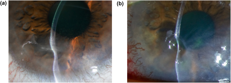
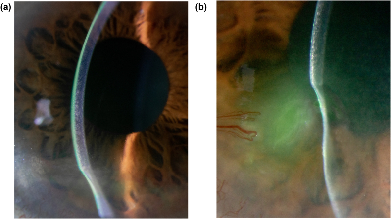
Observations: A 58-year-old male presented with bilateral corneal ulceration with stromal thinning and was diagnosed with PUK. He was actively being treated for Sézary syndrome, a cutaneous T-cell lymphoma. He had no lagophthalmos or other adnexal abnormalities that would lead to ocular surface breakdown. A systemic autoimmune and infectious workup for PUK was unremarkable. His keratitis resolved after treatment with oral prednisone.
Conclusions and importance: We describe a previously undocumented association of PUK with Sézary syndrome in a patient without adnexal disease.
Keywords: Mycosis fungoides; Peripheral ulcerative keratitis; Sézary syndrome.
© 2023 The Authors.
Conflict of interest statement
The authors declare that they have no known competing financial interests or personal relationships that could have appeared to influence the work reported in this paper.
Figures
References
-
- Stenson S., Ramsay D.L. Ocular findings in mycosis fungoides. JAMA Ophthalmol. 1981;99(February):272–277. - PubMed
Publication types
LinkOut - more resources
Full Text Sources
