. 2024 Jul;44(7):1145-1162.
doi: 10.1177/0271678X231224508. Epub 2024 Jan 18.

The role of serum/glucocorticoid-regulated kinase 1 in brain function following cerebral ischemia

Affiliations

The role of serum/glucocorticoid-regulated kinase 1 in brain function following cerebral ischemia

Celeste Yin-Chieh Wu et al. J Cereb Blood Flow Metab. 2024 Jul.

Abstract

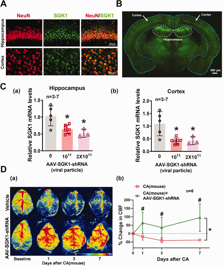
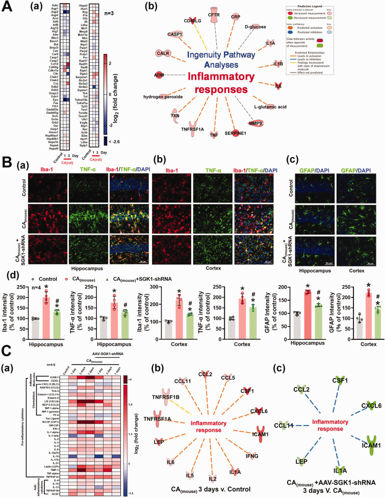
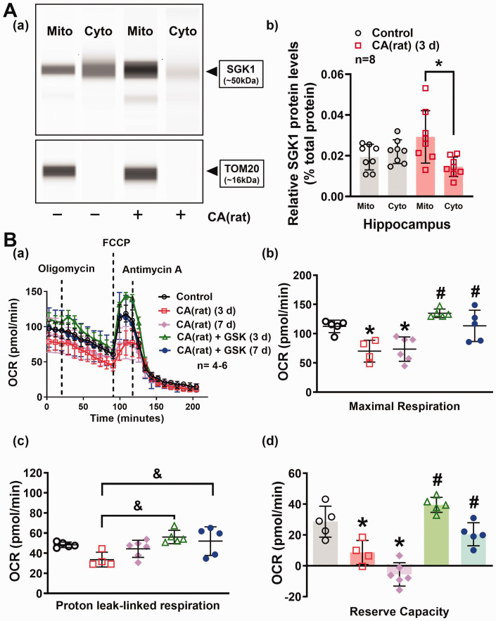
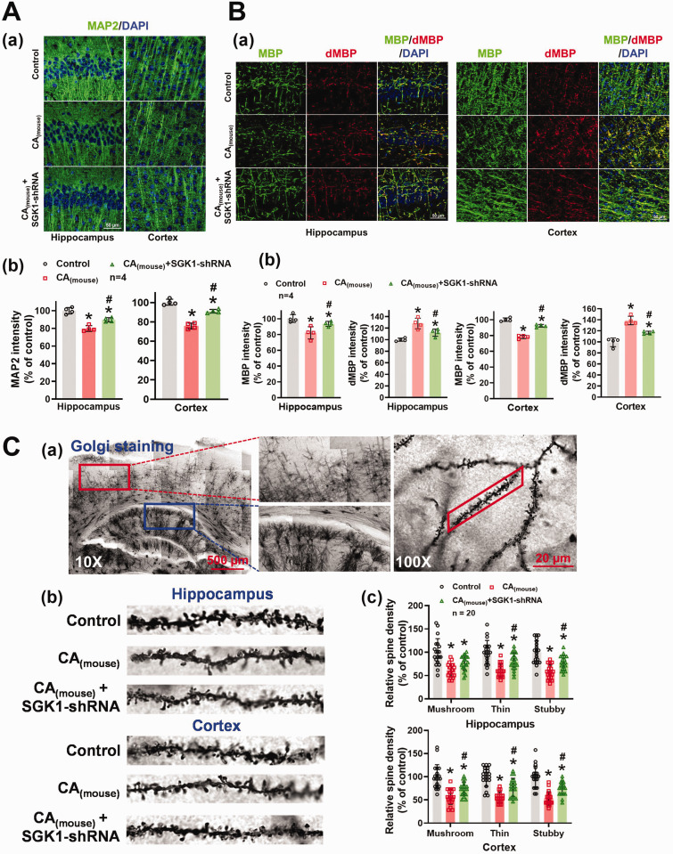
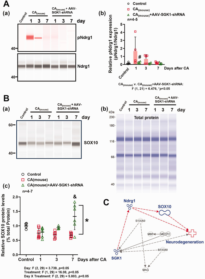
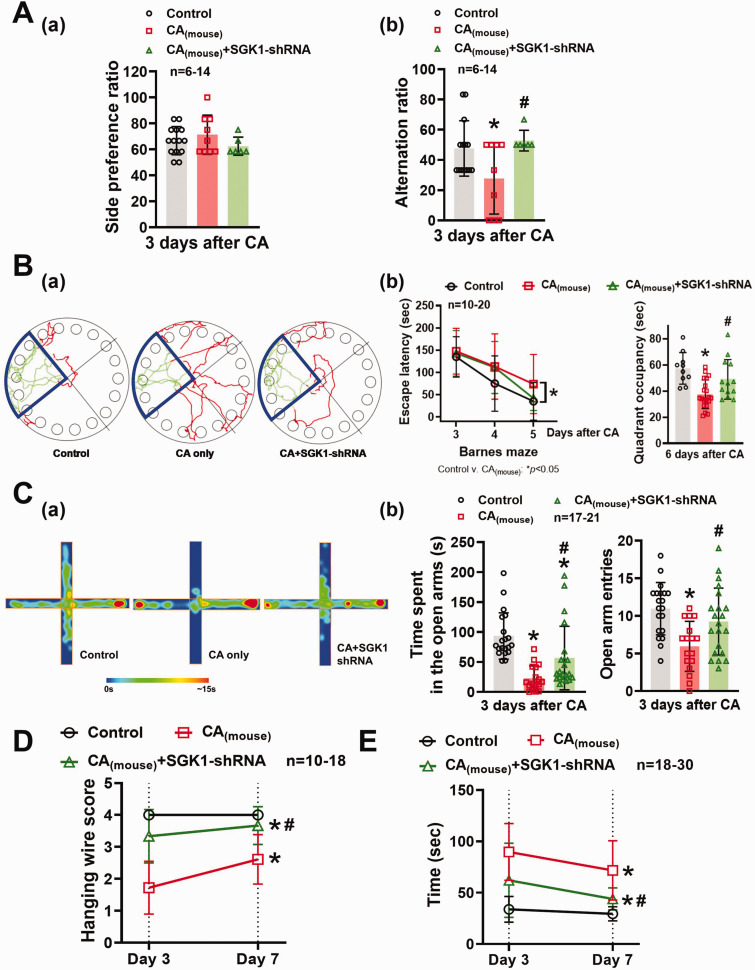
Cardiopulmonary arrest (CA) is a major cause of death/disability in the U.S. with poor prognosis and survival rates. Current therapeutic challenges are physiologically complex because they involve hypoperfusion (decreased cerebral blood flow), neuroinflammation, and mitochondrial dysfunction. We previously discovered novel serum/glucocorticoid-regulated kinase 1 (SGK1) is highly expressed in brain of neurons that are susceptible to ischemia (hippocampus and cortex). We inhibited SGK1 and utilized pharmacological (specific inhibitor, GSK650394) and neuron-specific genetic approaches (shRNA) in rodent models of CA to determine if SGK1 is responsible for hypoperfusion, neuroinflammation, mitochondrial dysfunctional, and neurological deficits after CA. Inhibition of SGK1 alleviated cortical hypoperfusion and neuroinflammation (via Iba1, GFAP, and cytokine array). Treatment with GSK650394 enhanced mitochondrial function (via Seahorse respirometry) in the hippocampus 3 and 7 days after CA. Neuronal injury (via MAP2, dMBP, and Golgi staining) in the hippocampus and cortex was observed 7 days after CA but ameliorated with SGK1-shRNA. Moreover, SGK1 mediated neuronal injury by regulating the Ndrg1-SOX10 axis. Finally, animals subjected to CA exhibited learning/memory, motor, and anxiety deficits after CA, whereas SGK1 inhibition via SGK1-shRNA improved neurocognitive function. The present study suggests the fundamental roles of SGK1 in brain circulation and neuronal survival/death in cerebral ischemia-related diseases.

Keywords: Serum/glucocorticoid-regulated kinase; cerebral ischemia; mitochondrial dysfunction; neuroinflammation; neurological deficits.


Conflict of interest statement

Declaration of conflicting interestsThe author(s) declared no potential conflicts of interest with respect to the research, authorship, and/or publication of this article.

Figures

Figure 1.
Figure 1.
Inhibition of SGK1 (via AAV-SGK1-shRNA) alleviated CA(mouse)-induced hypoperfusion. (A) Representative immunofluorescence images from control mice indicate that co-localization of NeuN (red) and SGK1 (green) in both cortex and hippocampus. Scale bar = 50 µm. (B) Representative images of coronal sections 30 days following virus administration. Scale bar = 500 µm. Green fluorescence indicates GFP-tagged AAV particles. (C) Relative SGK1 mRNA levels in the (a) hippocampus and (b) cortex. Results were expressed as mean ± SD. *p < 0.05 indicates significantly different from control mice. (D) (a) Representative flux images of cortical vasculature before (baseline) and 1 day, 3 days, and 7 days after CA. Results were summarized in panel (b). Changes in CBF were presented as percent change from the baseline (CBF 30 min before CA). Results were expressed as mean ± SD. *p < 0.05 indicates overall significantly different versus CA-only mice (via two-way ANOVA), #p < 0.05 versus respective days after CA.
Figure 2.
Figure 2.
Inhibition of SGK1 (via AAV-SGK1-shRNA) alleviated CA-induced neuroinflammation. (A) Total RNA was extracted from the rat hippocampus 1 and 3 days after CA(rat). mRNA levels of 84 stress and toxicity-related genes in the hippocampus were measured via RT2 Profiler™ PCR Array. (a) Heatmap of mRNA expression of the selected genes involved in cellular responses to stress and toxic compounds. (b) Results from PCR array were further analyzed by QIAGEN Ingenuity Pathway Analysis. (B) (a, b, c) Representative fluorescence images of Iba-1 (red), TNF-α (green), and GFAP (green) in the mouse hippocampus and cortex 7 days after CA(mouse). Relative immunofluorescence intensity of Iba-1, TNF-α, and GFAP was summarized in panel (d) (n = 4). (C) Heatmap of protein levels of the inflammatory cytokines involved in the activation of microglia and astrocytes. (a) Total protein was extracted from mice 1, 3, and 7 days after CA(mouse). Results from inflammatory array were analyzed by QIAGEN Ingenuity Pathway Analysis as shown in panels (b) and (c). Results were expressed as mean ± SD. *p < 0.05 versus control, #p < 0.05 versus respective days after CA.
Figure 3.
Figure 3.
Inhibition of SGK1 (via GSK650394) alleviated CA(rat)-induced mitochondrial dysfunction in the hippocampus. (A) Representative images of the computer-generated pseudo bands from the capillary-based immunoassay. (a) SGK1 and TOM20 band at 50 and 16 kDa, respectively. (b) Hippocampal SGK1 protein levels in the mitochondria (mito) and cytosolic fraction (cyto) in control and rats subjected to CA were normalized to the total protein. Results were expressed as mean ± SD, *p < 0.05 as compared to mitochondrial SGK1 protein levels 3 days after CA. (B) (a) Mitochondrial oxygen consumption rate (OCR) was measured in hippocampal slices 3 and 7 days after CA(rat) via a Seahorse XF24 analyzer. Results were summarized in panels (b, c, d). Treatment with GSK650394 (GSK) improved mitochondrial maximal respiration, proton leak-linked respiration, and reverse capacity 3 and 7 days after CA(rat). *p < 0.05 versus control, #p < 0.05 versus respective days after CA. &p < 0.05 versus 3 days after CA(rat). All data were expressed as mean ± SD.
Figure 4.
Figure 4.
Knockdown of SGK1 alleviated dendrisomatic neuronal injury, while preserving dendritic spine density after CA. (A) (a) Representative immunofluorescence images of MAP2 (green) in the CA1 region of the hippocampus and cortex 7 days after CA(mouse). Quantification of the results were summarized in panel (b). (B) Representative images of MBP (green) and degraded MBP (dMBP, red) staining in the CA1 region of the hippocampal and cortex were shown in (a). Quantification of the relative MBP and dMBP fluorescence intensity was shown in panel (b). (C) (a) Representative images of dendritic spines in the hippocampus (blue rectangle) and cortex (red rectangle) via Golgi staining. (b) Selected dendritic segments in the CA1 region of the hippocampus and cortex from control and mice subjected to CA surgery. The total number of mushroom, thin, and stubby spines was manually counted and summarized in panel (c). All data were expressed as mean ± SD. *p < 0.05 versus control, #p < 0.05 versus respective days after CA.
Figure 5.
Figure 5.
Hippocampal SGK1 levels and Ndrg1 phosphorylation were enhanced concurrently with decreased SOX10 levels after CA, while inhibition of SGK1 via AAV enhanced SOX10 levels. (A, B) Relative protein levels of total Ndrg1, phosphorylated Ndrg1 (pNdrg1), and SOX10 in the hippocampus were measured by capillary-based immunoassay. pNdrg1, total Ndrg1, and SOX10 band at 50, 48, and 55 kDa, respectively. Results were normalized with total Ndrg1 [A (a)] or total protein [B (b)] (for SOX10) and summarized in panels A (b) and B (c). *p < 0.05 versus CA only group evaluated by two-way ANOVA with Tukey's post-hoc. #p < 0.05 versus 1 day after CA. &p < 0.05 versus CA only group (1, 3, 7 days) and CA+AAV-SGK1-shRNA group (1 day). (C) Ingenuity Pathway Analyses indicates SGK1 can phosphorylate Ndrg1 to modulate SOX10.
Figure 6.
Figure 6.
SGK1 inhibition reversed CA-induced cognitive and motor deficits. (A) 3 days after CA, the mice were subjected to T-maze spontaneous alternation test for assessment of functional learning/memory. Results from T-maze test were summarized in panels (a, side preference ratio) and (b, alternation ratio). (B) Barnes maze was utilized to evaluate mouse’s long-term memory 3–6 days after CA. (a) Representative tracking plots of control, CA(mouse), and CA(mouse)+SGK1-shRNA groups during the last training day (day 6 after CA). (b) The escape latency to find the target box on days 3, 4, and 5 after CA, and the quadrant occupancy on day 6 after CA. (C) Anxiety-like behavior in mice 3 days after CA was evaluated by elevated plus-maze. The degree of anxiety was determined by the time spent and number of entries in the open arms. (a) Representative heat maps of the elevated plus-maze test from the control, CA only, and CA+SGK1-shRNA groups. Time spent in the open arms and the number of open arm entries were summarized in panel (b). Mouse’s motor function 3 and 7 days after CA was studied via hanging wire test (D) and adhesive removal test (E). Data were expressed as mean ± SD. *p < 0.05 versus control, #p < 0.05 versus CA only.

Similar articles

Cited by

References

    1. Virani SS, Alonso A, Aparicio HJ, et al.. Heart disease and stroke statistics-2021 update: a report from the American heart association. Circulation 2021; 143: e254–e743. - PubMed
    1. Neumar RW, Nolan JP, Adrie C, et al.. Post-cardiac arrest syndrome: epidemiology, pathophysiology, treatment, and prognostication. A consensus statement from the international liaison committee on resuscitation (American Heart Association, Australian and New Zealand Council on Resuscitation, European Resuscitation Council, Heart and Stroke Foundation of Canada, InterAmerican Heart Foundation, Resuscitation Council of Asia, and the Resuscitation Council of Southern Africa); the American Heart Association Emergency Cardiovascular Care Committee; the Council on Cardiovascular Surgery and Anesthesia; the Council on Cardiopulmonary, Perioperative, and Critical Care; the Council on Clinical Cardiology; and the Stroke Council. Circulation 2008; 118: 2452–2483. - PubMed
    1. Harukuni I, Bhardwaj A. Mechanisms of brain injury after global cerebral ischemia. Neurol Clin 2006; 24: 1–21. - PubMed
    1. Sabri M, Lass E, Macdonald RL. Early brain injury: a common mechanism in subarachnoid hemorrhage and global cerebral ischemia. Stroke Res Treat 2013; 2013: 394036. - PMC - PubMed
    1. Lang F, Bohmer C, Palmada M, et al.. (Patho)physiological significance of the serum- and glucocorticoid-inducible kinase isoforms. Physiol Rev 2006; 86: 1151–1178. - PubMed

MeSH terms

Substances

LinkOut - more resources