. 2024 Feb 20;98(2):e0121623.
doi: 10.1128/jvi.01216-23. Epub 2024 Jan 18.

An RNA replicon system to investigate promising inhibitors of feline coronavirus

Affiliations

An RNA replicon system to investigate promising inhibitors of feline coronavirus

Kimberly Schmied et al. J Virol. .

Abstract

Feline infectious peritonitis (FIP) is a fatal feline disease, caused by a feline coronavirus (FCoV), namely feline infectious peritonitis virus (FIPV). We produced a baby hamster kidney 21 (BHK) cell line expressing a serotype I FCoV replicon RNA with a green fluorescent protein (GFP) reporter gene (BHK-F-Rep) and used it as an in vitro screening system to test different antiviral compounds. Two inhibitors of the FCoV main protease (Mpro), namely GC376 and Nirmatrelvir, as well as the nucleoside analog Remdesivir proved to be effective in inhibiting the replicon system. Different combinations of these compounds also proved to be potent inhibitors, having an additive effect when combined. Remdesivir, GC376, and Nirmatrelvir all have a 50% cytotoxic concentration (CC50) more than 200 times higher than their half-maximal inhibitory concentrations (IC50), making them important candidates for future in vivo studies as well as clinically implemented drug candidates. In addition, results were acquired with a virus infection system, where Felis catus whole fetus 4 (Fcwf-4) cells were infected with a previously described recombinant GFP-expressing FIPV (based on the laboratory-adapted serotype I FIPV strain Black) and treated with the most promising compounds. Results acquired with the replicon system were comparable to the results acquired with the virus infection system, demonstrating that we successfully implemented the FCoV replicon system for antiviral screening. We expect that this system will greatly facilitate future screens for anti-FIPV compounds and provide a non-infectious system to study and evaluate drug-resistant mutations that may emerge in the FIPV genome.IMPORTANCEFIPV is of great significance in the cat population around the world, causing 0.3%-1.4% of feline deaths in veterinary practices (2). As there are neither effective preventive measures nor approved treatment options available, there is an urgent need to identify antiviral drugs against FIPV. Our FCoV replicon system provides a valuable tool for drug discovery in vitro. Due to the lack of cell culture systems for serotype I FCoVs (the serotype most prevalent in the feline population) (2), a different system is needed to study these viruses. A viral replicon system is a valuable tool for studying FCoVs. Overall, our results demonstrate the utility of the serotype I feline coronavirus replicon system for antiviral screening as well as to study this virus in general. We propose several compounds representing promising candidates for future clinical trials and ultimately with the potential to save cats suffering from FIP.

Keywords: antiviral agents; feline coronavirus; feline infectious peritonitis; replicon.


Conflict of interest statement

The authors declare no conflict of interest.

Figures

Fig 1
Fig 1
Illustration of the vrecFECVrep genome and function inside the cell including visualization and detection via flow cytometry. (A) vrecFECVrep genome compared to the FECV genome was illustrated from the 5′ UTR to the 3’UTR. The modifications introduced to the sequence upstream and downstream of ORF1ab are presented in solid and dashed rectangles, respectively. Kb = kilobases, ORF = open reading frame, S = spike, E = envelope, M = membrane, N = nucleocapsid, EGFP = enhanced green fluorescent protein, nsp = non-structural protein, T2A = TaV 2A-like element from Thosea asigna virus, NEO = neomycin selection gene, IRES = internal ribosome entry site, ATG = start codon, Upstr. N = part of the M protein upstream of N, TRS-N = transcription-regulating sequence of N. (B) Illustration of the BHK-F-Rep cell. The negative-sense vrecFECVrep RNA genome is replicated and transcribed into negative-sense full-length and subgenomic RNAs in the cytoplasm. The two subgenomic mRNAs encoding EGFP and N are then translated into proteins. (C) BHK-F-Rep cell colony expressing GFP was obtained after electroporation and selection. (D) Representation of the BHK-F-Rep cells in flow cytometry. (E) Expression of the RNA-dependent-RNA-Polymerase (RdRp), EGFP, subgenomic GFP (sgEGFP), and subgenomic nucleocapsid (sgN) gene as RT-qPCR results in BHK-F-Rep and BHK-21 cells. nd = not detected. Created with BioRender.com.
Fig 2
Fig 2
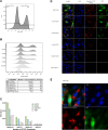
Representative flow cytometry analysis of BHK-F-Rep in response to inhibitors. The BHK-F-Rep were treated with the different compounds directly after seeding and kept at 37°C and 5% CO2 for 96 hours and subsequently analyzed with flow cytometry or immunofluorescence staining. (A) Untreated BHK-F-Rep with gating of GFP+ and GFP− cells. (B) An example of dose-response with different drug concentrations was analyzed with flow cytometry. The median GFP signal decreases with increasing GC376 concentration. (C) Establishment of the BHK-F-Repcured cell line with two treatments of the BHK-F-Rep cells with 20 µM of GC376. RdRp, EGFP, subgenomic EGFP (sgEGFP), and subgenomic N (sgN) expression before, during, and after the BHK-F-Rep cells were cured were analyzed with RT-qPCR. The BHK-F-Repcured cells were then used as a negative control for all experiments. nd = not detected. (D) An example of dose-response with different drug concentrations was analyzed with immunofluorescence to show the correlation between the GFP signal and the expression of the N-Protein. Blue = DAPI stain of the nucleus, green = GFP signal, red = Alexa Fluor 647 signal of the FCoV N-Protein staining, scale bar = 50 µm. (E) Close-up immunofluorescence image illustrating the different distribution of the expression of N-Protein versus GFP expression in the BHK-F-Rep cells. Blue = DAPI stain of the nucleus, green = GFP signal, red = Alexa Fluor 647 signal of the FCoV N-Protein staining, scale bar = 20 µm
Fig 3
Fig 3
Individual tested antiviral compounds. BHK-F-Rep cells treated with (A) Remdesivir, (B) GC376, (C) Nirmatrelvir, and (D) Amodiaquine for 96 hours and analyzed by flow cytometry for GFP expression as shown in Fig. 2. Relative inhibition was calculated by the median intensity of GFP expression in the cells treated with antiviral drugs or DMSO in comparison to mock control. In addition, IC50 values and CC50 values were calculated as explained in the methods and represented on the individual graphs. (A–C) Remdesivir, GC376, and Nirmatrelvir showed effective antiviral dose-response with IC50 values < 1 µM, at concentrations lower than the cytotoxic concentrations (D) Amodiaquine alone did not show any inhibitory effect at the concentrations that already affected the cell growth The black line in the graphs represents the mean value of the data collected from three independent experiments, and the gray area represents standard error of the mean.
Fig 4
Fig 4
Combined antiviral compounds. BHK-F-Rep cells treated with different mixtures of Remdesivir, GC376, Nirmatrelvir, and Amodiaquine for 96 hours and analyzed by flow cytometry for GFP expression as shown in Fig. 2. (A and B) Combining Remdesivir in a 1:1 mixture with GC376 or Nirmatrelvir had a positive effect on IC50 values, making it more efficient in inhibiting replication of the FECV replicon. (C) Combining either GC376 or Nirmatrelvir at their approximate IC50 values to Remdesivir to illustrate the additive effect of both Mpro inhibitors to Remdesivir. (D) Combining GC376 at its approximate IC50 value with different concentrations of Nirmatrelvir illustrates a highly increased additive effect when combining these two Mpro inhibitors. (E and F) Adding Amodiaquine to either GC376 or Nirmatrelvir did not have a positive effect on the dose-response of either primary inhibitor. The black and red lines in the graphs represent the mean value of the data collected from three independent experiments of individual drugs or their combination, respectively. The gray and red shaded areas represent the standard error of the mean.
Fig 5
Fig 5
Comparison to virus infection. Testing of three different compounds or combinations of compounds: GC376 (A), Remdesivir (B), and Remdesivir + GC376 in a 1:1 ratio (C) in recFCoV-GFP-infected Fcwf-4 cells with dose-response curve of recFCoV-GFP infected Fcwf-4 cells treated for 48 hours. Read-out as with the BHK-F-Rep cells done via flow cytometry and detection of GFP-positive cells (relative Inhibition) including IC50 values are shown in black with the left y-axis. As with the BHK-F-Rep cells, data shown on cell viability testing in the recFCoV-GFP infected Fcwf-4 cells are shown in black with the right y-axis including CC50 values. Additional data on viral titers of the recFCoV-GFP infected Fcwf-4 cells treated with the inhibitors and corresponding IC50 values are shown in red. The black and red lines in the graphs represent the mean value of the data collected from three independent experiments of individual drugs or their combination, respectively. The gray and red shaded areas represent the standard error of the mean.

References

    1. Thayer V, Gogolski S, Felten S, Hartmann K, Kennedy M, Olah GA. 2022. 2022 AAFP/EveryCat feline infectious peritonitis diagnosis guidelines. J Feline Med Surg 24:905–933. doi:10.1177/1098612X221118761 - DOI - PMC - PubMed
    1. Pedersen NC. 2014. An update on feline infectious peritonitis: virology and immunopathogenesis. Vet J 201:123–132. doi:10.1016/j.tvjl.2014.04.017 - DOI - PMC - PubMed
    1. Kennedy MA. 2020. Feline infectious peritonitis: update on pathogenesis, diagnostics, and treatment. Vet Clin North Am Small Anim Pract 50:1001–1011. doi:10.1016/j.cvsm.2020.05.002 - DOI - PubMed
    1. Pedersen NC. 2009. A review of feline infectious peritonitis virus infection: 1963-2008. J Feline Med Surg 11:225–258. doi:10.1016/j.jfms.2008.09.008 - DOI - PMC - PubMed
    1. de Groot RJ, Baker SC, Baric R, Enjuanes L, Gorbalenya AE, Holmes KV, Perlman S, Poon L, Rottier PJM, Talbot PJ. 2011. Family Coronaviridae

Publication types