

Bacteriophage P22 SieA-mediated superinfection exclusion
- PMID: 38236051
- PMCID: PMC10883804
- DOI: 10.1128/mbio.02169-23
Bacteriophage P22 SieA-mediated superinfection exclusion
Abstract
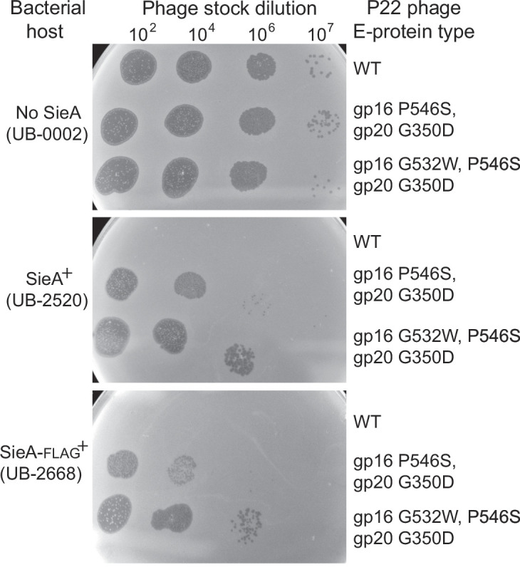
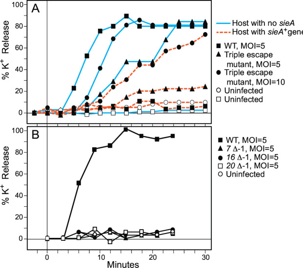
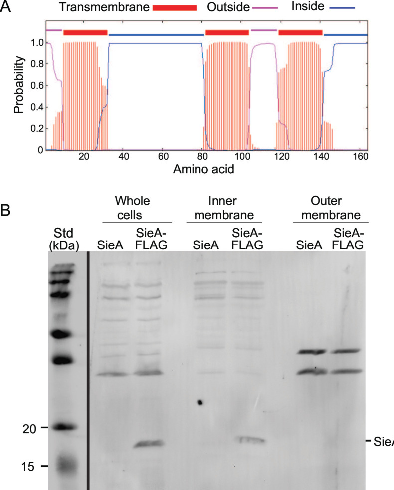
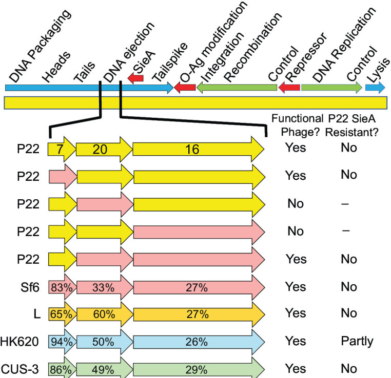
Many temperate phages encode prophage-expressed functions that interfere with superinfection of the host bacterium by external phages. Salmonella phage P22 has four such systems that are expressed from the prophage in a lysogen that are encoded by the c2 (repressor), gtrABC, sieA, and sieB genes. Here we report that the P22-encoded SieA protein is necessary and sufficient for exclusion by the SieA system and that it is an inner membrane protein that blocks DNA injection by P22 and its relatives, but has no effect on infection by other tailed phage types. The P22 virion injects its DNA through the host cell membranes and periplasm via a conduit assembled from three "ejection proteins" after their release from the virion. Phage P22 mutants that overcome the SieA block were isolated, and they have amino acid changes in the C-terminal regions of the gene 16 and 20 encoded ejection proteins. Three different single-amino acid changes in these proteins are required to obtain nearly full resistance to SieA. Hybrid P22 phages that have phage HK620 ejection protein genes are also partially resistant to SieA. There are three sequence types of extant phage-encoded SieA proteins that are less than 30% identical to one another, yet comparison of two of these types found no differences in phage target specificity. Our data strongly suggest a model in which the inner membrane protein SieA interferes with the assembly or function of the periplasmic gp20 and membrane-bound gp16 DNA delivery conduit.IMPORTANCEThe ongoing evolutionary battle between bacteria and the viruses that infect them is a critical feature of bacterial ecology on Earth. Viruses can kill bacteria by infecting them. However, when their chromosomes are integrated into a bacterial genome as a prophage, viruses can also protect the host bacterium by expressing genes whose products defend against infection by other viruses. This defense property is called "superinfection exclusion." A significant fraction of bacteria harbor prophages that encode such protective systems, and there are many different molecular strategies by which superinfection exclusion is mediated. This report is the first to describe the mechanism by which bacteriophage P22 SieA superinfection exclusion protein protects its host bacterium from infection by other P22-like phages. The P22 prophage-encoded inner membrane SieA protein prevents infection by blocking transport of superinfecting phage DNA across the inner membrane during injection.
Keywords: P22; SieA; bacteriophage; ejection proteins; superinfection exclusion.
Conflict of interest statement
The authors declare no conflict of interest.
Figures
Update of
-
Bacteriophage P22 SieA mediated superinfection exclusion.bioRxiv [Preprint]. 2023 Aug 16:2023.08.15.553423. doi: 10.1101/2023.08.15.553423. bioRxiv. 2023. Update in: mBio. 2024 Feb 14;15(2):e0216923. doi: 10.1128/mbio.02169-23. PMID: 37645741 Free PMC article. Updated. Preprint.
Similar articles
-
Bacteriophage P22 SieA mediated superinfection exclusion.bioRxiv [Preprint]. 2023 Aug 16:2023.08.15.553423. doi: 10.1101/2023.08.15.553423. bioRxiv. 2023. Update in: mBio. 2024 Feb 14;15(2):e0216923. doi: 10.1128/mbio.02169-23. PMID: 37645741 Free PMC article. Updated. Preprint.
-
The superinfection exclusion gene (sieA) of bacteriophage P22: identification and overexpression of the gene and localization of the gene product.J Bacteriol. 1995 Jun;177(11):3080-6. doi: 10.1128/jb.177.11.3080-3086.1995. J Bacteriol. 1995. PMID: 7768804 Free PMC article.
-
Superinfection exclusion and changes in cellular transport processes in phage infected Salmonella typhimurium.Mol Gen Genet. 1978 Feb 27;159(3):293-6. doi: 10.1007/BF00268265. Mol Gen Genet. 1978. PMID: 345099
-
Phage against the Machine: The SIE-ence of Superinfection Exclusion.Viruses. 2024 Aug 23;16(9):1348. doi: 10.3390/v16091348. Viruses. 2024. PMID: 39339825 Free PMC article. Review.
-
Phages and the evolution of bacterial pathogens: from genomic rearrangements to lysogenic conversion.Microbiol Mol Biol Rev. 2004 Sep;68(3):560-602, table of contents. doi: 10.1128/MMBR.68.3.560-602.2004. Microbiol Mol Biol Rev. 2004. PMID: 15353570 Free PMC article. Review.
Cited by
-
Insight into the natural regulatory mechanisms and clinical applications of the CRISPR-Cas system.Heliyon. 2024 Oct 18;10(20):e39538. doi: 10.1016/j.heliyon.2024.e39538. eCollection 2024 Oct 30. Heliyon. 2024. PMID: 39502233 Free PMC article. Review.
-
Lytic bacteriophages as alternative to overcoming antibiotic-resistant biofilms formed by clinically significant bacteria.Ther Adv Infect Dis. 2025 Jul 18;12:20499361251356057. doi: 10.1177/20499361251356057. eCollection 2025 Jan-Dec. Ther Adv Infect Dis. 2025. PMID: 40686849 Free PMC article. Review.
-
Pseudomonas aeruginosa as a model bacterium in antiphage defense research.FEMS Microbiol Rev. 2025 Jan 14;49:fuaf014. doi: 10.1093/femsre/fuaf014. FEMS Microbiol Rev. 2025. PMID: 40240293 Free PMC article. Review.
-
Exploring Viral Interactions in Clavibacter Species: In Silico Analysis of Prophage Prevalence and Antiviral Defenses.Life (Basel). 2025 Jan 27;15(2):187. doi: 10.3390/life15020187. Life (Basel). 2025. PMID: 40003596 Free PMC article.
-
Armed Phages: A New Weapon in the Battle Against Antimicrobial Resistance.Viruses. 2025 Jun 27;17(7):911. doi: 10.3390/v17070911. Viruses. 2025. PMID: 40733529 Free PMC article. Review.
References
MeSH terms
Substances
Grants and funding
LinkOut - more resources
Full Text Sources
Miscellaneous
