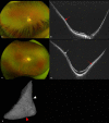
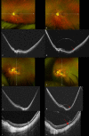
Characteristics and Prevalence of Staphyloma Edges at Different Ages in Highly Myopic Eyes
- PMID: 38236188
- PMCID: PMC10807494
- DOI: 10.1167/iovs.65.1.32
Characteristics and Prevalence of Staphyloma Edges at Different Ages in Highly Myopic Eyes
Abstract
Purpose: The purpose of this study was to determine the characteristics of staphyloma edges in highly myopic eyes and how they progress.
Methods: We conducted a cross-sectional analysis using baseline data and a longitudinal study with follow-up data from 256 patients (447 eyes) with high myopia, with a mean (SD) follow-up of 3.79 (0.78) years. Participants were divided into four age groups: children (<13), youth (13-24), mature (25-59), and elderly (>60). Ultrawide-field swept-source optical coherence tomography was used to analyze staphyloma edges, which were divided into four areas: nasal to the optic disc (OD), superior to the macula, inferior to the macula, and temporal to the macula.
Results: Staphylomas were significantly more prevalent in the mature (42.49%) and the elderly (51.35%) groups than in the children (13%) and youth (9%) groups. Staphyloma edges were predominantly superior to the macula in the mature and elderly groups. In contrast, staphylomas were rare in children and youth, with their edges mainly located nasal to the OD. The edges of staphylomas located superior and temporal to the macula were more likely to be associated with myopic traction maculopathy. During the follow-up period, 11 new staphyloma edges developed primarily in the mature group (64%). Additionally, 12 edges had an increased degree of protrusion over time, with most cases occurring in the mature (75%) group.
Conclusions: The prevalence and location of staphyloma edges show significant variations depending on age. As time progresses, staphyloma edges manifest at distinct sites and increase their protrusion, potentially playing a role in the emergence of fundus complications.
Conflict of interest statement
Disclosure:
Figures


Similar articles
-
[Occurrence of myopic maculopathy in eyes with staphylomas of various localization].Vestn Oftalmol. 2022;138(6):55-64. doi: 10.17116/oftalma202213806155. Vestn Oftalmol. 2022. PMID: 36573948 Russian.
-
Characteristics of Peripapillary Staphylomas Associated With High Myopia Determined by Swept-Source Optical Coherence Tomography.Am J Ophthalmol. 2016 Sep;169:138-144. doi: 10.1016/j.ajo.2016.06.033. Epub 2016 Jun 27. Am J Ophthalmol. 2016. PMID: 27365146
-
Posterior staphylomas and scleral curvature in highly myopic children and adolescents investigated by ultra-widefield optical coherence tomography.PLoS One. 2019 Jun 10;14(6):e0218107. doi: 10.1371/journal.pone.0218107. eCollection 2019. PLoS One. 2019. PMID: 31181108 Free PMC article.
-
Posterior staphyloma in pathologic myopia.Prog Retin Eye Res. 2019 May;70:99-109. doi: 10.1016/j.preteyeres.2018.12.001. Epub 2018 Dec 8. Prog Retin Eye Res. 2019. PMID: 30537538 Review.
-
IMI Pathologic Myopia.Invest Ophthalmol Vis Sci. 2021 Apr 28;62(5):5. doi: 10.1167/iovs.62.5.5. Invest Ophthalmol Vis Sci. 2021. PMID: 33909033 Free PMC article. Review.
Cited by
-
Development of posterior staphyloma in a child with retinitis pigmentosa and high myopia detected by Ultra-widefield Optical Coherence Tomography: A 4-year follow-up.Am J Ophthalmol Case Rep. 2025 Mar 5;38:102301. doi: 10.1016/j.ajoc.2025.102301. eCollection 2025 Jun. Am J Ophthalmol Case Rep. 2025. PMID: 40129886 Free PMC article.
-
Development of Deep Learning Models to Screen Posterior Staphylomas in Highly Myopic Eyes Using UWF-OCT Images.Transl Vis Sci Technol. 2025 Jun 2;14(6):25. doi: 10.1167/tvst.14.6.25. Transl Vis Sci Technol. 2025. PMID: 40504567 Free PMC article.
-
Polarization-Sensitive Optical Coherence Tomography Imaging of Posterior Staphyloma Edges in Eyes With High Myopia.Invest Ophthalmol Vis Sci. 2025 Aug 1;66(11):6. doi: 10.1167/iovs.66.11.6. Invest Ophthalmol Vis Sci. 2025. PMID: 40762541 Free PMC article.
-
Retinal curvature in Chinese children with myopia measured by ultra-widefield swept-source optical coherence tomography.Br J Ophthalmol. 2025 Mar 20;109(4):470-475. doi: 10.1136/bjo-2024-325704. Br J Ophthalmol. 2025. PMID: 39244223 Free PMC article.
References
-
- Ohno-Matsui K, Lai TY, Lai CC, Cheung CM.. Updates of pathologic myopia. Prog Retin Eye Res. 2016; 52: 156–187. - PubMed
-
- Spaide RF. Staphyloma: Part 1. In: Spaide RF, Ohno-Matsui K, Yannuzzi LA, eds. Pathologic Myopia . New York, NY: Springer New York; 2014: 167–176.
-
- Fang Y, Yokoi T, Nagaoka N, et al. .. Progression of myopic maculopathy during 18-year follow-up. Ophthalmology. 2018; 125: 863–877. - PubMed
-
- Jonas JB, Ohno-Matsui K, Holbach L, Panda-Jonas S. Association between axial length and horizontal and vertical globe diameters. Graefes Arch Clin Exp Ophthalmol. 2017; 255: 237–242. - PubMed
-
- Jonas JB, Xu L, Wei WB, et al. .. Retinal thickness and axial length. Invest Ophthalmol Vis Sci. 2016; 57: 1791–1797. - PubMed
MeSH terms
LinkOut - more resources
Full Text Sources
Research Materials

