. 2024 Jan 18;81(1):44.
doi: 10.1007/s00018-023-05058-2.

Restraining of glycoprotein VI- and integrin α2β1-dependent thrombus formation by platelet PECAM1

Affiliations

Restraining of glycoprotein VI- and integrin α2β1-dependent thrombus formation by platelet PECAM1

Natalie J Jooss et al. Cell Mol Life Sci. .

Abstract

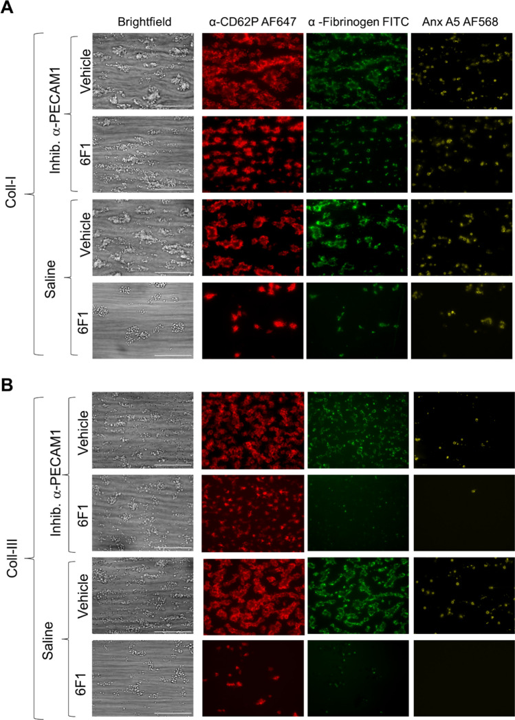
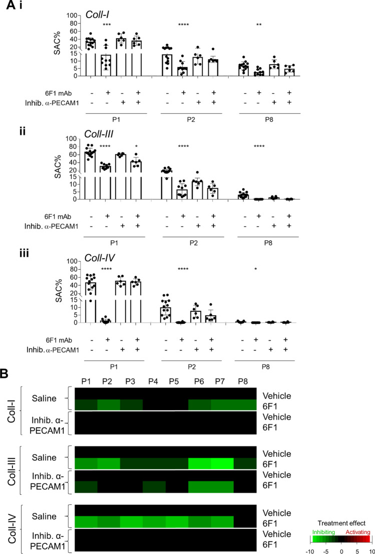
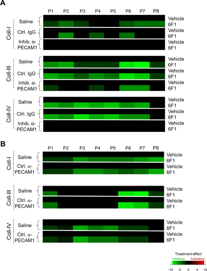
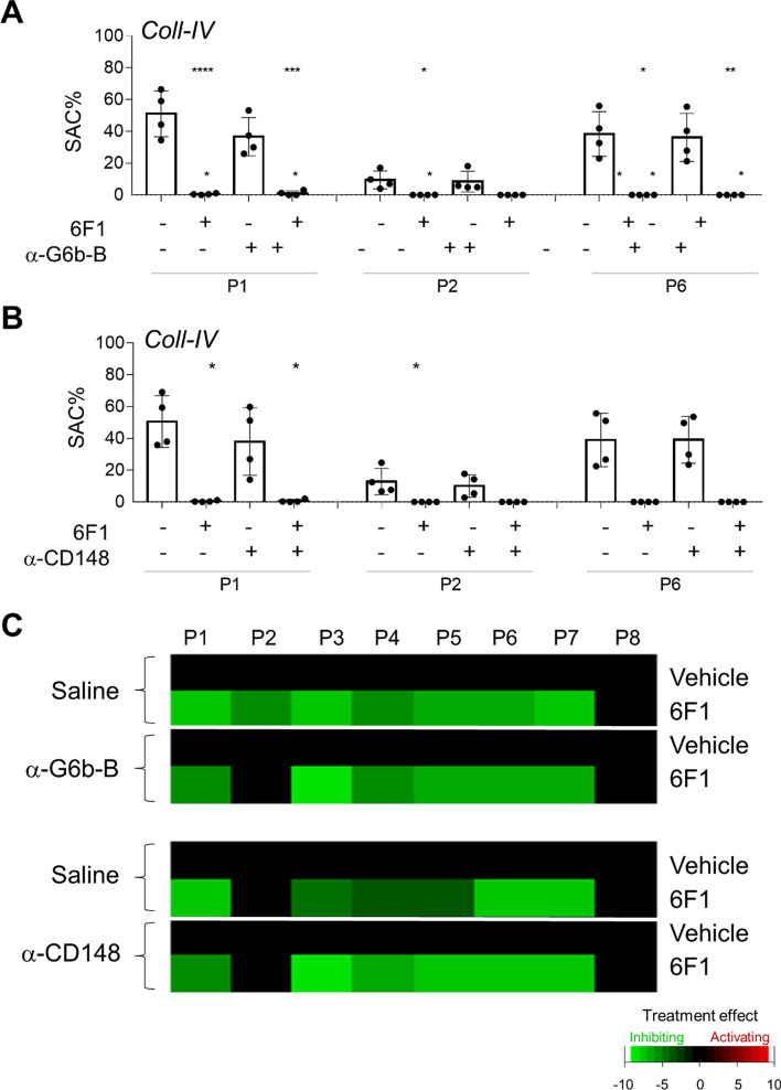
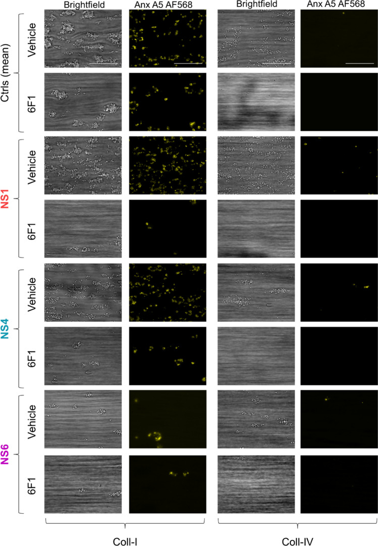
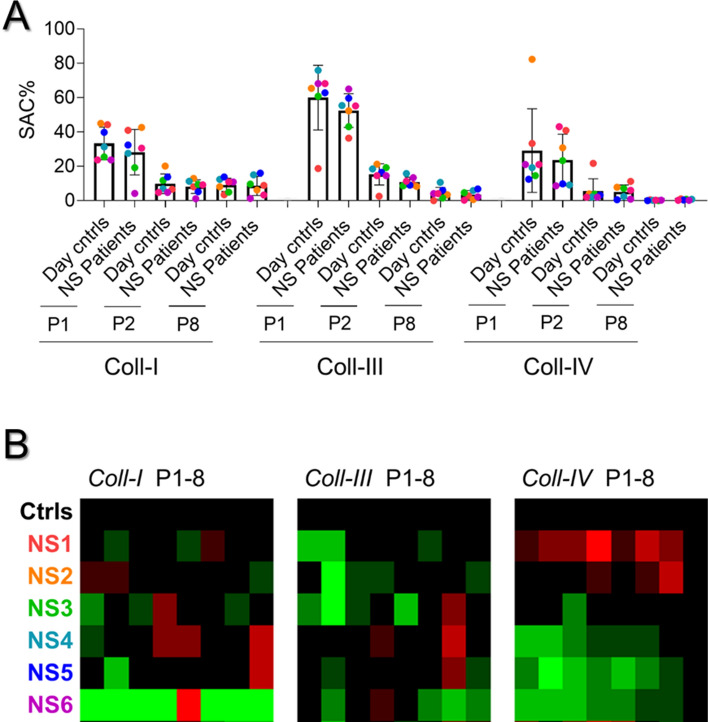
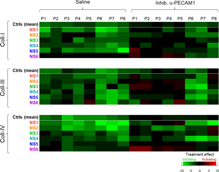
The platelet receptors, glycoprotein VI (GPVI) and integrin α2β1 jointly control collagen-dependent thrombus formation via protein tyrosine kinases. It is unresolved to which extent the ITIM (immunoreceptor tyrosine-based inhibitory motif) receptor PECAM1 and its downstream acting protein tyrosine phosphatase PTPN11 interfere in this process. Here, we hypothesized that integrin α2β1 has a co-regulatory role in the PECAM1- and PTPN11-dependent restraint of thrombus formation. We investigated platelet activation under flow on collagens with a different GPVI dependency and using integrin α2β1 blockage. Blood was obtained from healthy subjects and from patients with Noonan syndrome with a gain-of-function mutation of PTPN11 and variable bleeding phenotype. On collagens with decreasing GPVI activity (types I, III, IV), the surface-dependent inhibition of PECAM1 did not alter thrombus parameters using control blood. Blockage of α2β1 generally reduced thrombus parameters, most effectively on collagen IV. Strikingly, simultaneous inhibition of PECAM1 and α2β1 led to a restoration of thrombus formation, indicating that the suppressing signaling effect of PECAM1 is masked by the platelet-adhesive receptor α2β1. Blood from 4 out of 6 Noonan patients showed subnormal thrombus formation on collagen IV. In these patients, effects of α2β1 blockage were counterbalanced by PECAM1 inhibition to a normal phenotype. In summary, we conclude that the suppression of GPVI-dependent thrombus formation by either PECAM1 or a gain-of-function of PTPN11 can be overruled by α2β1 engagement.

Keywords: Collagens; Microfluidics; Microspots; Noonan syndrome; PTPN11; SHP2.


Conflict of interest statement

J.W.M.H. is a consultant at Synapse Research Institute. The other authors declare no relevant conflict of interest.

Figures

Fig. 1
Fig. 1
Enhanced collagen-induced thrombus formation by PECAM1 blockage in the absence of integrin α2β1. Blood samples were preincubated with anti-α2β1 6F1 mAb (20 µg/mL) or vehicle, and then perfused during 3.5 min at 1000 s−1 over microspots of collagen-I (A) or collagen-III (B); microspots were post-coated with saline or inhibitory anti-PECAM1 mAb (clone WM59, mouse IgG1). Brightfield and multicolor microscopic images were taken at end stage with an EVOS-FL microscope and 60 × oil objective. Platelet labeling was withAF647 anti-CD62P mAb, FITC anti-fibrinogen mAb and AF658 annexin A5. Shown are representative images for per collagen surface with or without inhibitory anti-PECAM1 mAb (n = 6–7). EVOS microscope; scale bars, 50 µm
Fig. 2
Fig. 2
Enhanced collagen-induced thrombus formation by inhibitory anti-PECAM1 antibody upon blockade of integrin α2β1. A Blood samples were preincubated with 6F1 mAb (20 µg/mL) or vehicle, and then perfused during 3.5 min at 1000/s over microspots of collagen I (i), collagen III (ii) or collagen IV (iii); microspots were post-coated with saline or inhibitory anti-PECAM1 mAb (WM59 at 1 µg/mL for 1 h). Multicolor microscopic images, as for Fig. 1, were analyzed for platelet adhesion, thrombus phenotype and platelet activation parameters (Suppl. Table 1). Shown are quantified data of platelet deposition (parameter P1), thrombus multilayer size (parameter P2), and phosphatidylserine exposure (parameter P8) in the presence or absence of 6F1 mAb and/or inhibitory anti-PECAM1 mAb. Representative images are shown in Suppl. Fig. 1. B Subtraction heatmap presentation of the 8 univariately scaled (0–10) parameters across surfaces, showing values of treatment effect per microspot type relative to vehicle containing blood samples. For the color coding, a relevance filter was applied of p < 0.05. Means ± SD (n = 6–7); one-way ANOVA *p < 0.05, **p < 0.005, ***p < 0.001, ****p < 0.0001. Raw data of P1-8 are in Suppl. Datafile 1
Fig. 3
Fig. 3
Selective effect of inhibitory anti-PECAM1 mAb in rescue of collagen-induced thrombus formation upon α2β1 inhibition. Blood samples were preincubated with 6F1 mAb (20 µg/mL) or vehicle, and then perfused during 3.5 min at 1000/s over microspots of collagen I, collagen III or collagen IV. Microspots were post-coated with saline, inhibitory anti-PECAM1 mAb (IgG1), mouse isotype control IgG1, or a control anti-PECAM mAb (clone MBC 78.2), as indicated. Multicolor microscopic images (EVOS-FL microscope, 60 × oil objective) were analyzed for eight parameters (Suppl. Table 1), which were univariate scaled. A, B Subtraction heatmaps for comparing per collagen type the effects of integrin α2β1 blockage (by 6F1 mAb) on parameters: P1, platelet deposition; P2, thrombus multilayer size; P3, thrombus morphological score; P4, thrombus multilayer score; P5, thrombus contraction score; P6, P-selectin expression; P7, integrin αIIbβ3 activation; P8, phosphatidylserine exposure. (A) Heatmapped effects of the 6F1 mAb on microspots of a collagen + inhibitory anti-PECAM1 mAb or the collagen + control IgG. B Heatmapped effects of 6F1 mAb on microspots of a collagen + control anti-PECAM1 mAb. Means ± SD (n = 3–7), tested for significance with one-way ANOVA. For color coding, a filter was applied of p < 0.05. Raw data are given in Suppl. Datafile 1
Fig. 4
Fig. 4
Inability of other antibodies against ITIM containing receptors to rescue collagen-induced thrombus formation upon α2β1 inhibition. Blood samples were preincubated with 6F1 mAb (20 µg/mL) or vehicle, and perfused during 3.5 min at 1000/s over microspots of collagen IV, as for Fig. 1. Microspots were post-coated with saline, anti-G6bB Ab (rabbit polyclonal) or anti-CD148 mAb (clone Ab1). Multicolor microscopic images, as for Fig. 1, were analyzed for eight parameters (Suppl. Table 1), which were univariate scaled. A, B Shown are quantified outcome data of collagen induced platelet deposition (parameter P1), thrombus multilayer size (parameter P2), and phosphatidylserine exposure (parameter P8) in the presence or absence of 6F1 mAb (in blood) and/or anti-G6bB Ab (A) or anti-CD148 mAb (B). In addition, subtraction heatmaps comparing the effects of integrin α2β1 blockage by 6F1 mAb on all 8 parameters (C). For color coding, a relevance filter was applied of p < 0.05. Means ± SD (n = 4), tested for significance with one-way ANOVA, *p < 0.05, **p < 0.005, ***p < 0.001 and ****p < 0.0001. Raw data are given in Suppl. Datafile 1
Fig. 5
Fig. 5
Alterations in collagen-induced thrombus formation for distinct Noonan syndrome patients. Whole blood from (day) control subjects (n = 13) and indicated patients (n = 6) was perfused at 1000 s−1 for 3.5 min over collagen surfaces (100 µg/mL), as described for Fig. 1. Collagen microspots were co-coated with saline or inhibitory anti-PECAM1 mAb (1 µg/mL). Blood samples contained vehicle solution or blocking α2β1 6F1 mAb (20 µg/mL). Shown are end stage brightfield (P1-5) and phosphatidylserine exposure (parameter P8) microscopic images obtained with blood from a representative day control subject and from patients NS1, NS4 and NS6 perfused over collagen-I or collagen-IV. As indicated, vehicle medium was present, or integrin α2β1 was blocked with 6F1 mAb. Scale bars, 50 µm
Fig. 6
Fig. 6
Alterations in collagen-induced thrombus formation with Noonan syndrome patients. Blood were analyzed from 13 control subjects, including 6 day controls, and 6 patients with Noonan syndrome (NS1-NS6), genotyped for a gain of function mutation in PTPN11. Blood samples were perfused over collagen I, collagen III or collagen IV, after which images were analyzed for 8 parameters (duplicate runs). A Quantification of platelet deposition (P1), thrombus multilayer size (P2), and phosphatidylserine exposure (P8) for individual day controls and patients per surface. B Heatmapped of scaled parameters per collagen type versus means of controls
Fig. 7
Fig. 7
Increased responsiveness to α2β1 blockage with Noonan syndrome patients Parameters of thrombus formation for 13 controls subjects and 6 patients were recorded as for Fig. 6. Collagen microspots contained vehicle or inhibitory anti-PECAM1 mAb; and blood flow was in the presence or absence of 6F1 mAb. Shown is a subtraction heatmap of 6F1 mAb effects (scaled parameters P1-8) per patient for collagen I, collagen III and collagen IV containing or not inhibitory anti-PECAM1 mAb. Means ± SD (n = 13 controls). For raw data, see Suppl. Datafile 1

References

    1. Nieswandt B, Watson SP. Platelet-collagen interaction: is GPVI the central receptor? Blood. 2003;102:449–461. doi: 10.1182/blood-2002-12-3882. - DOI - PubMed
    1. Lecut C, Schoolmeester A, Kuijpers MJ, Broers JL, van Zandvoort MA, Vanhoorelbeke K, Deckmyn H, Jandrot-Perrus M, Heemskerk JW. Principal role of glycoprotein VI in a2b1 and aIIbb3 activation during collagen-induced thrombus formation. Arterioscler Thromb Vasc Biol. 2004;24:1727–1733. doi: 10.1161/01.ATV.0000137974.85068.93. - DOI - PubMed
    1. Dumont B, Lasne D, Rothschild C, Bouabdelli M, Ollivier V, Oudin C, Ajzenberg N, Grandchamp B, Jandrot-Perrus M. Absence of collagen-induced platelet activation caused by compound heterozygous GPVI mutations. Blood. 2009;114:1900–1903. doi: 10.1182/blood-2009-03-213504. - DOI - PubMed
    1. Nagy M, van Geffen JP, Stegner D, Adams DJ, Braun A, de Witt SM, Elvers M, Geer MJ, Kuijpers MJ, Kunzelmann K, Mori J, Oury C, Pircher J, Pleines I, Poole AW, Senis YA, Verdoold R, Weber C, Nieswandt B, Heemskerk JW, Baaten CC. Comparative analysis of microfluidics thrombus formation in multiple genetically modified mice: link to thrombosis and hemostasis. Front Cardiovasc Med. 2019;6:99. doi: 10.3389/fcvm.2019.00099. - DOI - PMC - PubMed
    1. Clemetson KJ, Clemetson JM. Platelet GPIb-V-IX complex. Structure, function, physiology, and pathology. Semin Thromb Hemost. 1995;21:130–136. doi: 10.1055/s-2007-1000387. - DOI - PubMed