. 2024 Feb 27;8(4):991-1001.
doi: 10.1182/bloodadvances.2023011490.

Novel GNE missense variants impair de novo sialylation and cause defective angiogenesis in the developing brain in mice

Affiliations

Novel GNE missense variants impair de novo sialylation and cause defective angiogenesis in the developing brain in mice

Lulu Huang et al. Blood Adv. .

Abstract

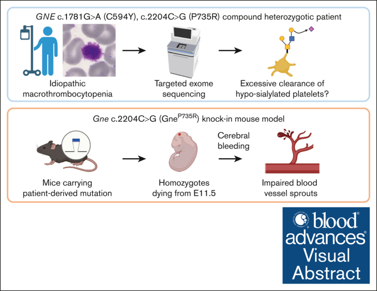
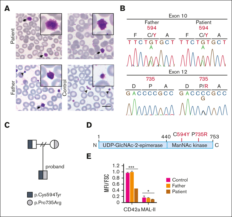
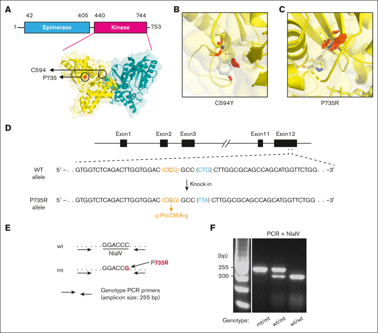
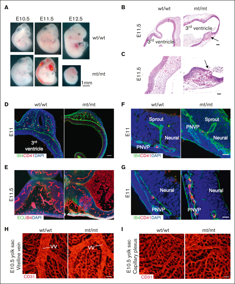
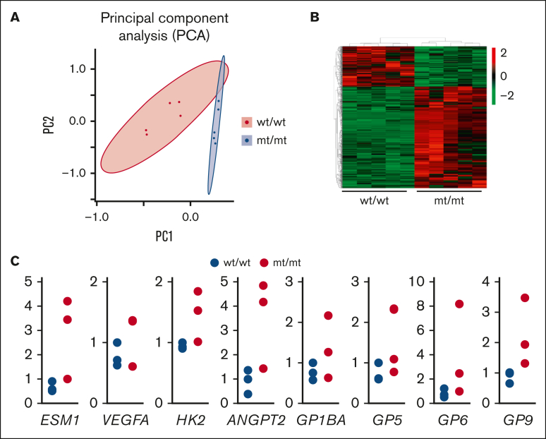
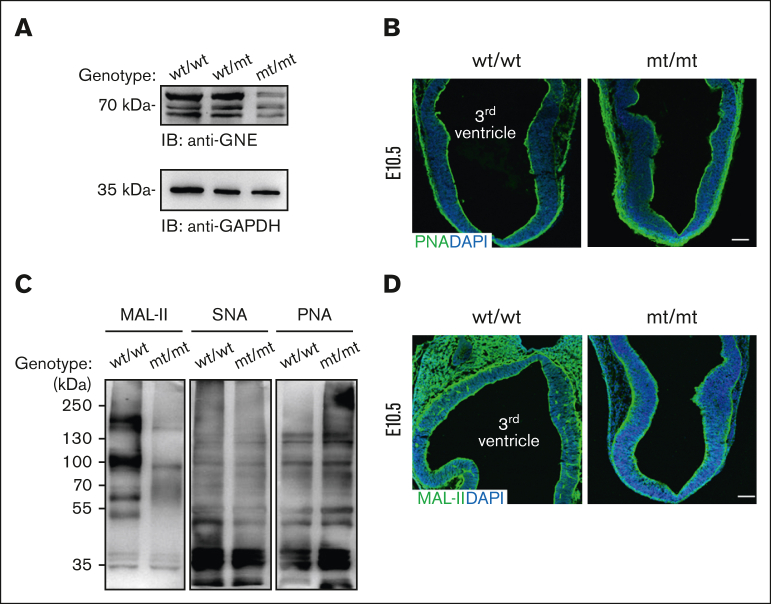
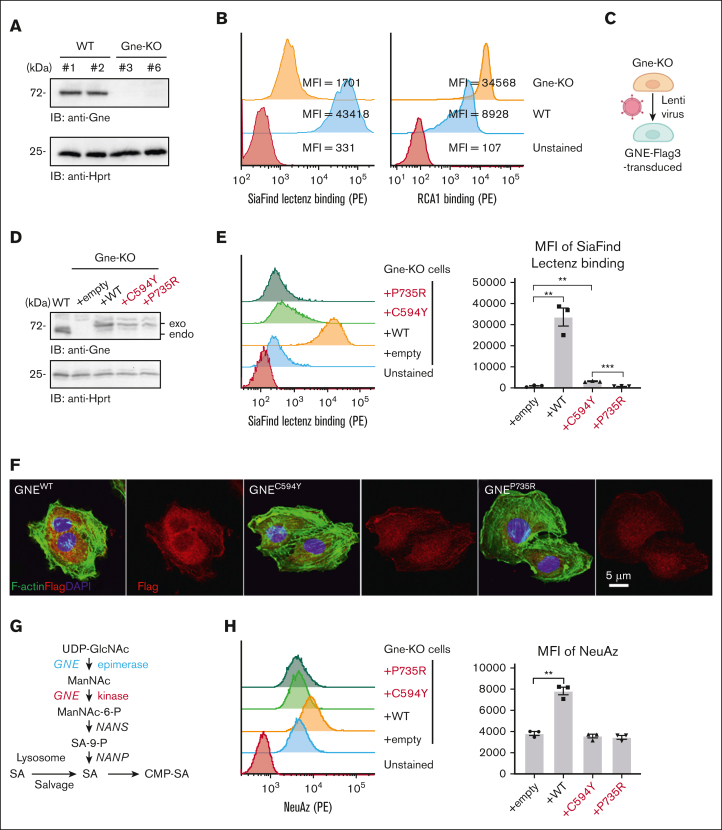
Glucosamine (UDP-N-acetyl)-2-epimerase and N-acetylmannosamine (ManNAc) kinase (GNE) is a cytosolic enzyme in de novo sialic acid biosynthesis. Congenital deficiency of GNE causes an autosomal recessive genetic disorder associated with hereditary inclusion body myopathy and macrothrombocytopenia. Here, we report a pediatric patient with severe macrothrombocytopenia carrying 2 novel GNE missense variants, c.1781G>A (p.Cys594Tyr, hereafter, C594Y) and c.2204C>G (p.Pro735Arg, hereafter, P735R). To investigate the biological significance of these variants in vivo, we generated a mouse model carrying the P735R mutation. Mice with homozygous P735R mutations exhibited cerebral hemorrhages as early as embryonic day 11 (E11), which subsequently progressed to large hemorrhages in the brain and spinal cord, and died between E11.5 and E12.5. Defective angiogenesis such as distended vascular sprouts were found in neural tissues and embryonic megakaryocytes were abnormally accumulated in the perineural vascular plexus in mutant mouse embryos. Furthermore, our in vitro experiments indicated that both C594Y and P735R are loss-of-function mutations with respect to de novo sialic acid biosynthesis. Overall, this study reveals a novel role for GNE-mediated de novo sialic acid biosynthesis in mouse embryonic angiogenesis.


Conflict of interest statement

Conflict-of-interest disclosure: The authors declare no competing financial interests.

Figures

None
Graphical abstract
Figure 1.
Figure 1.
Identification of GNE compound heterozygote variants in a patient with macrothrombocytopenia. (A) Representative images of Giemsa staining of peripheral blood smears from the patient (age 16), the patient’s father (age 40), and a healthy individual (age 28) as normal control who has no family relationship with the patient or his other family members. The size of patient’s platelets was nearly 3 times larger than that of the normal control. Arrows indicate enlarged platelets. Arrowheads indicate normal platelets. Black boxes indicate platelets with higher magnification. Scale bar, 10 μm. (B) Genomic DNA sequences of exon 10 and exon 12 of GNE in father- and patient-derived blood cells. Letters and numbers in red indicate mutated residues and sites in the GNE protein sequence, respectively. (C) Pedigree of the family. The filled square indicates the patient, who is the proband, and the half-filled square and circle indicate heterozygous father and mother, respectively. (D) Schematic domain structure of GNE carrying combined missense mutations in the patient. (E) MFI of MAL-II and platelet surface marker CD42a on peripheral platelets from the patient, patient’s father and a healthy individual as normal control (the same individual as in Figure 1A), respectively. MFI, mean fluorescence intensity; FSC, forward scatter; MAL-II, Maackia amurensis lectin II, binds to α-2,3 linked sialic acid. ∗P < .05; ∗∗∗P < .001.
Figure 2.
Figure 2.
Generation of P735R-knockin mice. (A) Schematic domain representation of UDP-GlcNAc-2-epimerase and ManNAc kinase in human GNE, and the structure of dimeric kinase domains (PDB ID: 2YHW). (B) Structural prediction of kinase domain carrying C594Y mutation, showing that 1 rotamer causes severe clashes in all 3 rotamers. (C) Structural prediction of kinase domain carrying P735R mutation, showing severe clashes for all the 22 rotamers. Mutagenesis and rotamer selection were performed using PyMOL, in which the gray sticks indicate the mutated residues and the red octagon disks indicate significant van der Waals overlap, meaning atoms are close to other atoms causing clashes. (D) Diagram of the targeted genomic sequence in the GneP735R/P735R (hereafter, mt/mt) mouse using the CRISPR-Cas9 system. The synonymous mutation p.Leu737 (CTG to TTA) was introduced as a blocking mutation to prevent re-cutting by Cas9 after homology-directed repair. (E) Illustration of the RFLP-based genotyping assay using PCR and restriction endonuclease NlaIV. (F) Genotyping results visualized using 2% agarose/TBE gel.
Figure 3.
Figure 3.
Homozygous P735R-knockin embryos develop distended vascular sprouts and spontaneous hemorrhages in the brain. (A) Comparison of wt/wt and mt/mt embryos at different developmental stages. Blood is visible in the hearts of the older embryos. Asterisks indicate hemorrhages in the brain ventricles, spinal cord, and spinal canal of E11.5 mt/mt embryos. The E11.5 and E12.5 mt/mt embryos are dead and appear pale as blood circulation has ceased. (B) Hematoxylin-eosin staining of coronal section of the wt/wt and mt/mt embryos’ heads at E11.5. Arrows indicate hemorrhagic lesions, which are shown at higher magnification in (C). Hemorrhages are visible in both the brain parenchyma as well as in the ventricles. Erythrocytes are nucleated at this stage of development. (D) Confocal images of angiogenic sprouting into the ventricle from PNVP of the diencephalon (coronal section) of E11 and (E) E11.5 embryos. (F) Confocal images of diencephalon distended sprouts and (G) accumulated eMKs in mt/mt embryos at E11. Neural, brain tissue. PNVP, perineural vascular plexus. ECL intensity reflects the degree of asialylation. In E11.5 mt/mt embryos, the hemorrhage penetrated the brain tissue and spread into the ventricle. IB4 marks the vasculature. CD41 marks eMKs and platelets (mostly eMKs at this stage). (H) Confocal microscopy images of wholemount stained E10.5 yolk sac with CD31 near the vitelline vein and (I) capillary plexus. Scale bar, 100 μm (B, D, F, H, I); 25 μm (C, E, G).
Figure 4.
Figure 4.
Bulk RNA sequencing of E11.5 embryos reveals significant differences in genome expression. (A) Principal component analysis displays the difference in global gene expression profiles between wt/wt and mt/mt embryos. N = 5 for each genotype. (B) Clustering analysis of differentially expressed genes of A shown as a heat map. (C) RT-qPCR of angiogenesis- and platelet-related differentially expressed genes scored by bulk RNA-seq. RNA was extracted from E11.5 embryonic heads. N = 3 for each genotype. All data were analyzed using t-tests.
Figure 5.
Figure 5.
Homozygotic P735R mutation of Gne causes hyposialylation in mice. (A) Western blotting of GNE and GAPDH as loading control using mouse tissue extracts at E10.5. (B) Confocal images of PNA staining in wt/wt and mt/mt E10.5 embryos. (C) Lectin blotting of E10.5 tissue extracts probed with MAL-II, which detects α2,3-sialylated glycans, SNA, which detects α2,6-sialylated glycans, and PNA, which detects non-sialylated core 1 O-glycans (also known as T-antigen). The blots are representative of 4 independent experiments. (D) Confocal images of MAL-II staining in wt/wt and mt/mt E10.5 embryos. MAL-II, Maackia amurensis lectin II; PNA, peanut agglutinin. N = 3 for each genotype in (B) and (D). Scale bar, 100 μm.
Figure 6.
Figure 6.
Both patient-derived GNE mutations severely affect de novo sialic acid biosynthesis. (A) Establishment of Gne-knockout mouse endothelial cell line MS1 validated by western blotting. Hprt, housekeeping protein. (B) Altered glycans on the cell surface of Gne-KO MS1 cells examined by flow cytometry using SiaFind Lectenz (pan-sialic acid probe) and RCA1 lectin (asialylated glycan lectin). The unstained control was used as a negative control. (C) Gne-KO MS1 cells were lentivirally infected with empty control or each GNE-Flag3 variant. (D) Expression analysis of each GNE protein by western blotting. Exogenously transduced GNE (exo) and endogenous Gne (endo) are distinguished by size based on Flag3 tag on transduced GNE. WT, wild type. (E) Infected cells were analyzed for SiaFind Lectenz binding by flow cytometry. (F) Expression of cytoplasmic GNE was reduced in MS1 cells transduced with mutant GNE imaged by confocal microscopy. Flag3, Flag3-tagged GNE; DAPI, nuclear marker. (G) Two pathways of sialic acid biosynthesis. (H) ManNAz-derived NeuAz on cell surface glycans of cells that were metabolically labeled with ManNAz for 3 days. ∗∗P < .01; ∗∗∗P < .001.

Similar articles

Cited by

References

    1. Macauley MS, Crocker PR, Paulson JC. Siglec-mediated regulation of immune cell function in disease. Nat Rev Immunol. 2014;14(10):653–666. - PMC - PubMed
    1. Smith BAH, Bertozzi CR. The clinical impact of glycobiology: targeting selectins, Siglecs and mammalian glycans. Nat Rev Drug Discov. 2021;20(3):217–243. - PMC - PubMed
    1. Li Y, Chen X. Sialic acid metabolism and sialyltransferases: natural functions and applications. Appl Microbiol Biotechnol. 2012;94(4):887–905. - PMC - PubMed
    1. Hinderlich S, Stäsche R, Zeitler R, Reutter W. A bifunctional enzyme catalyzes the first two steps in N- acetylneuraminic acid biosynthesis of rat liver. Purification and characterization of UDP-N-acetylglucosamine 2-epimerase/N-acetylmannosamine kinase. J Biol Chem. 1997;272(39):24313–24318. - PubMed
    1. Tanner ME. The enzymes of sialic acid biosynthesis. Bioorg Chem. 2005;33(3):216–228. - PubMed

Publication types

Substances