

Analysis of the Healthy Platelet Proteome Identifies a New Form of Domain-Specific O-Fucosylation
- PMID: 38237698
- PMCID: PMC10879016
- DOI: 10.1016/j.mcpro.2024.100717
Analysis of the Healthy Platelet Proteome Identifies a New Form of Domain-Specific O-Fucosylation
Abstract
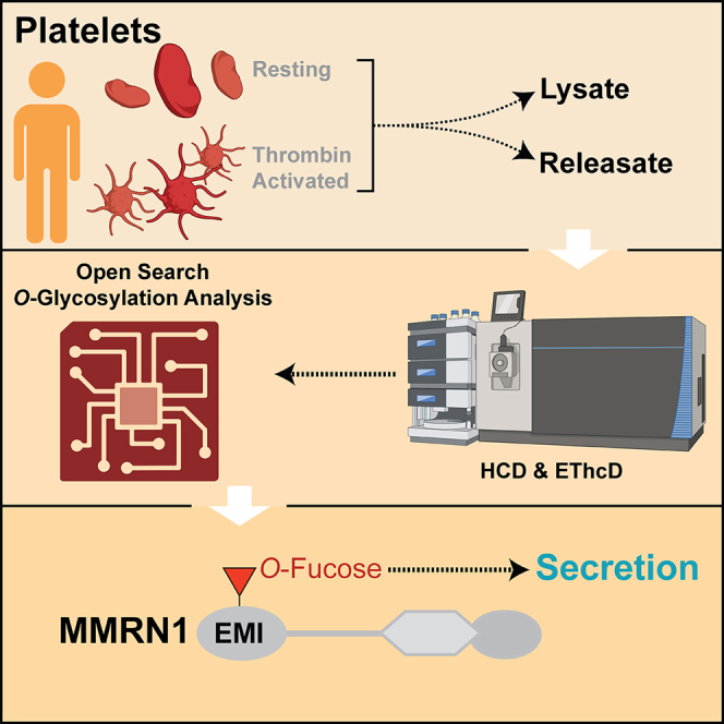
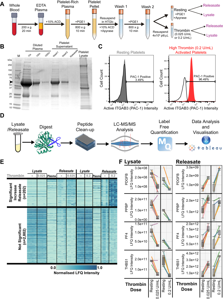
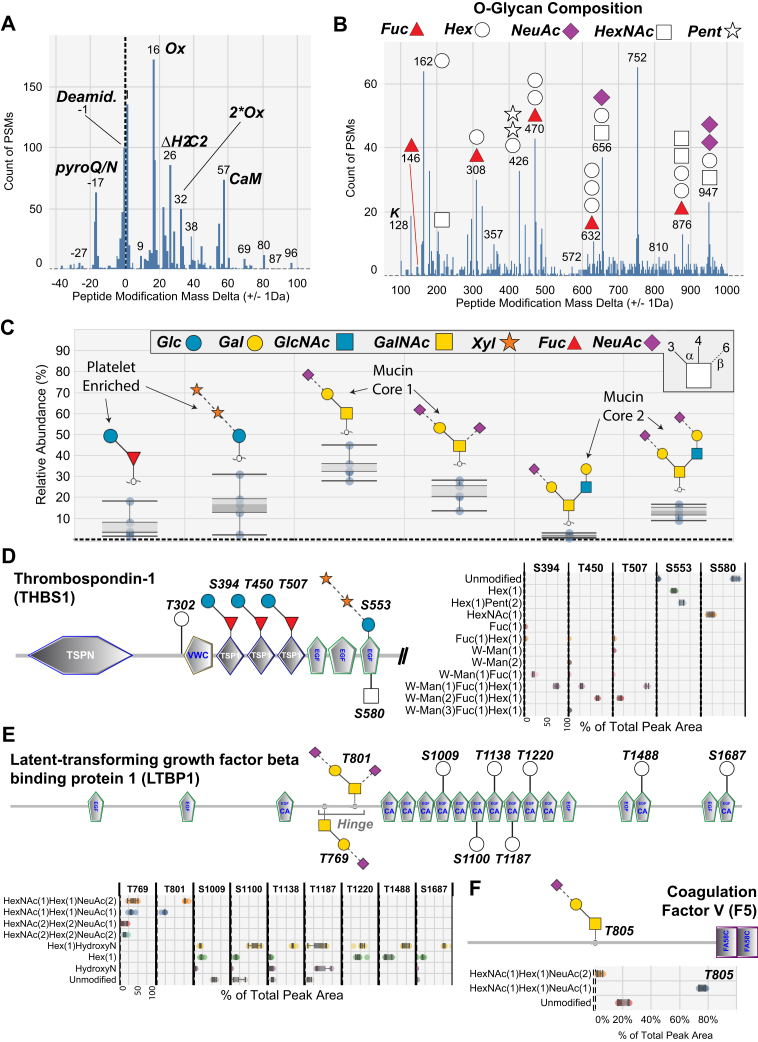
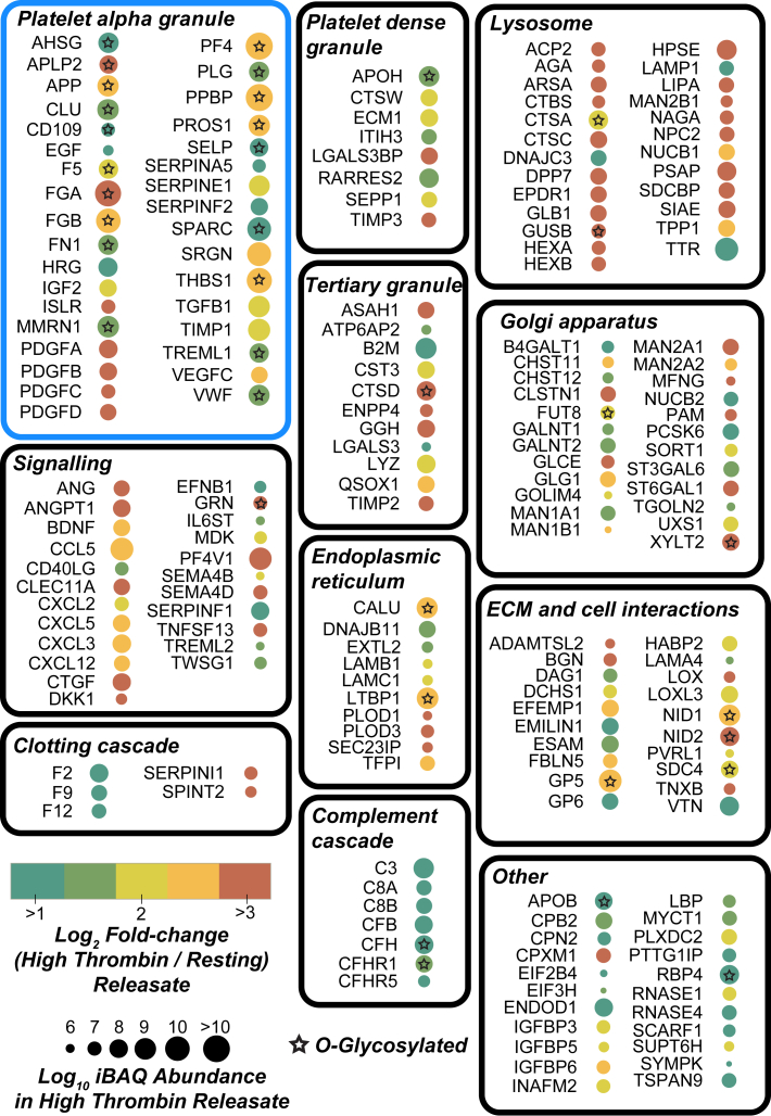
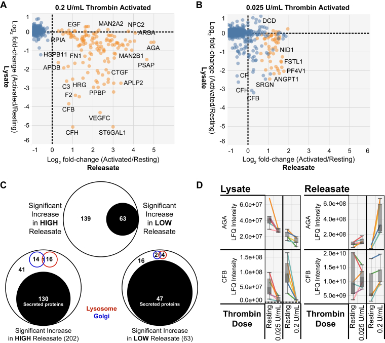
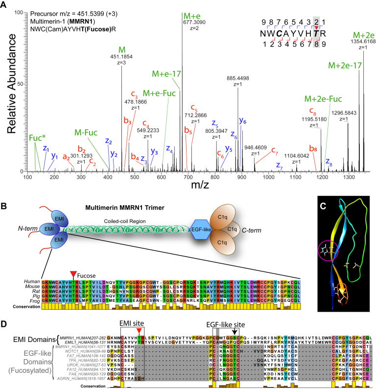
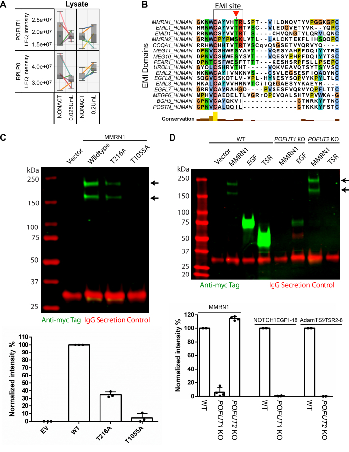
Platelet activation induces the secretion of proteins that promote platelet aggregation and inflammation. However, detailed analysis of the released platelet proteome is hampered by platelets' tendency to preactivate during their isolation and a lack of sensitive protocols for low abundance releasate analysis. Here, we detail the most sensitive analysis to date of the platelet releasate proteome with the detection of >1300 proteins. Unbiased scanning for posttranslational modifications within releasate proteins highlighted O-glycosylation as being a major component. For the first time, we detected O-fucosylation on previously uncharacterized sites including multimerin-1 (MMRN1), a major alpha granule protein that supports platelet adhesion to collagen and is a carrier for platelet factor V. The N-terminal elastin microfibril interface (EMI) domain of MMRN1, a key site for protein-protein interaction, was O-fucosylated at a conserved threonine within a new domain context. Our data suggest that either protein O-fucosyltransferase 1, or a novel protein O-fucosyltransferase, may be responsible for this modification. Mutating this O-fucose site on the EMI domain led to a >50% reduction of MMRN1 secretion, supporting a key role of EMI O-fucosylation in MMRN1 secretion. By comparing releasates from resting and thrombin-treated platelets, 202 proteins were found to be significantly released after high-dose thrombin stimulation. Complementary quantification of the platelet lysates identified >3800 proteins, which confirmed the platelet origin of releasate proteins by anticorrelation analysis. Low-dose thrombin treatment yielded a smaller subset of significantly regulated proteins with fewer secretory pathway enzymes. The extensive platelet proteome resource provided here (larancelab.com/platelet-proteome) allows identification of novel regulatory mechanisms for drug targeting to address platelet dysfunction and thrombosis.
Keywords: EMI domain; fucose; glycosylation; human; platelet; secretion; secretome.
Copyright © 2024 The Authors. Published by Elsevier Inc. All rights reserved.
Conflict of interest statement
Conflict of interest The authors declare no competing interests.
Figures
Similar articles
-
FUT10 and FUT11 are protein O-fucosyltransferases that modify protein EMI domains.Nat Chem Biol. 2025 Apr;21(4):598-610. doi: 10.1038/s41589-024-01815-x. Epub 2025 Jan 7. Nat Chem Biol. 2025. PMID: 39775168 Free PMC article.
-
A 2D-DIGE-based proteomic analysis reveals differences in the platelet releasate composition when comparing thrombin and collagen stimulations.Sci Rep. 2015 Feb 3;5:8198. doi: 10.1038/srep08198. Sci Rep. 2015. PMID: 25645904 Free PMC article.
-
Proteomics of the TRAP-induced platelet releasate.J Proteomics. 2009 Feb 15;72(1):91-109. doi: 10.1016/j.jprot.2008.10.009. Epub 2008 Nov 8. J Proteomics. 2009. PMID: 19049909
-
Taking the stock of granule cargo: Platelet releasate proteomics.Platelets. 2017 Mar;28(2):119-128. doi: 10.1080/09537104.2016.1254762. Epub 2016 Dec 8. Platelets. 2017. PMID: 27928935 Review.
-
Platelets in Healthy and Disease States: From Biomarkers Discovery to Drug Targets Identification by Proteomics.Int J Mol Sci. 2020 Jun 25;21(12):4541. doi: 10.3390/ijms21124541. Int J Mol Sci. 2020. PMID: 32630608 Free PMC article. Review.
Cited by
-
The Proteome Content of Blood Clots Observed Under Different Conditions: Successful Role in Predicting Clot Amyloid(ogenicity).Molecules. 2025 Feb 3;30(3):668. doi: 10.3390/molecules30030668. Molecules. 2025. PMID: 39942772 Free PMC article. Review.
-
Protein O-Fucosyltransferases: Biological Functions and Molecular Mechanisms in Mammals.Molecules. 2025 Mar 26;30(7):1470. doi: 10.3390/molecules30071470. Molecules. 2025. PMID: 40286076 Free PMC article. Review.
-
Novel antibodies detect nucleocytoplasmic O-fucose in protist pathogens, cellular slime molds, and plants.bioRxiv [Preprint]. 2024 Oct 22:2024.10.15.618526. doi: 10.1101/2024.10.15.618526. bioRxiv. 2024. Update in: mSphere. 2025 Feb 25;10(2):e0094524. doi: 10.1128/msphere.00945-24. PMID: 39464065 Free PMC article. Updated. Preprint.
-
Novel antibodies detect nucleocytoplasmic O-fucose in protist pathogens, cellular slime molds, and plants.mSphere. 2025 Feb 25;10(2):e0094524. doi: 10.1128/msphere.00945-24. Epub 2025 Feb 6. mSphere. 2025. PMID: 39912628 Free PMC article.
-
GlycoMaple: recent updates and applications in visualization and analysis of glycosylation pathways.Anal Bioanal Chem. 2025 Feb;417(5):885-894. doi: 10.1007/s00216-024-05594-1. Epub 2024 Oct 17. Anal Bioanal Chem. 2025. PMID: 39414644 Free PMC article. Review.
References
-
- Pagel O., Walter E., Jurk K., Zahedi R.P. Taking the stock of granule cargo: platelet releasate proteomics. Platelets. 2017;28:119–128. - PubMed
-
- Harrison P., Martin Cramer E. Platelet α-granules. Blood Rev. 1993;7:52–62. - PubMed
-
- Kieffer N., Guichard J., Farcet J.-P., Vainchenker W., Breton-Gorius J. Biosynthesis of major platelet proteins in human blood platelets. Eur. J. Biochem. 1987;164:189–195. - PubMed
-
- Ossovskaya V.S., Bunnett N.W. Protease-activated receptors: contribution to physiology and disease. Physiol. Rev. 2004;84:579–621. - PubMed
MeSH terms
Substances
Grants and funding
LinkOut - more resources
Full Text Sources
Molecular Biology Databases
