. 2025 Jan:67:289-299.
doi: 10.1016/j.jare.2024.01.017. Epub 2024 Jan 17.

RvD1 improves resident alveolar macrophage self-renewal via the ALX/MAPK14/S100A8/A9 pathway in acute respiratory distress syndrome

Affiliations

RvD1 improves resident alveolar macrophage self-renewal via the ALX/MAPK14/S100A8/A9 pathway in acute respiratory distress syndrome

Yang Ye et al. J Adv Res. 2025 Jan.

Abstract

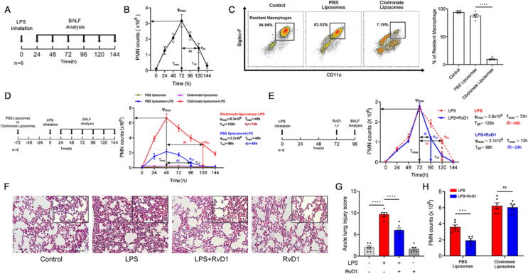
Introduction: Acute respiratory distress syndrome (ARDS) is a pulmonary inflammatory process primarily caused by sepsis. The resolution of inflammation is an active process involving the endogenous biosynthesis of specialized pro-resolving mediators, including resolvin D1 (RvD1). Resident alveolar macrophages (RAMs) maintain pulmonary homeostasis and play a key role in the resolution phase. However, the role of RAMs in promoting the resolution of inflammation by RvD1 is unclear.

Objectives: Here, we investigated the mechanisms of RvD1 on regulating RAMs to promote the resolution of ARDS.

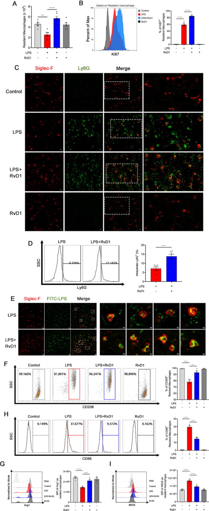
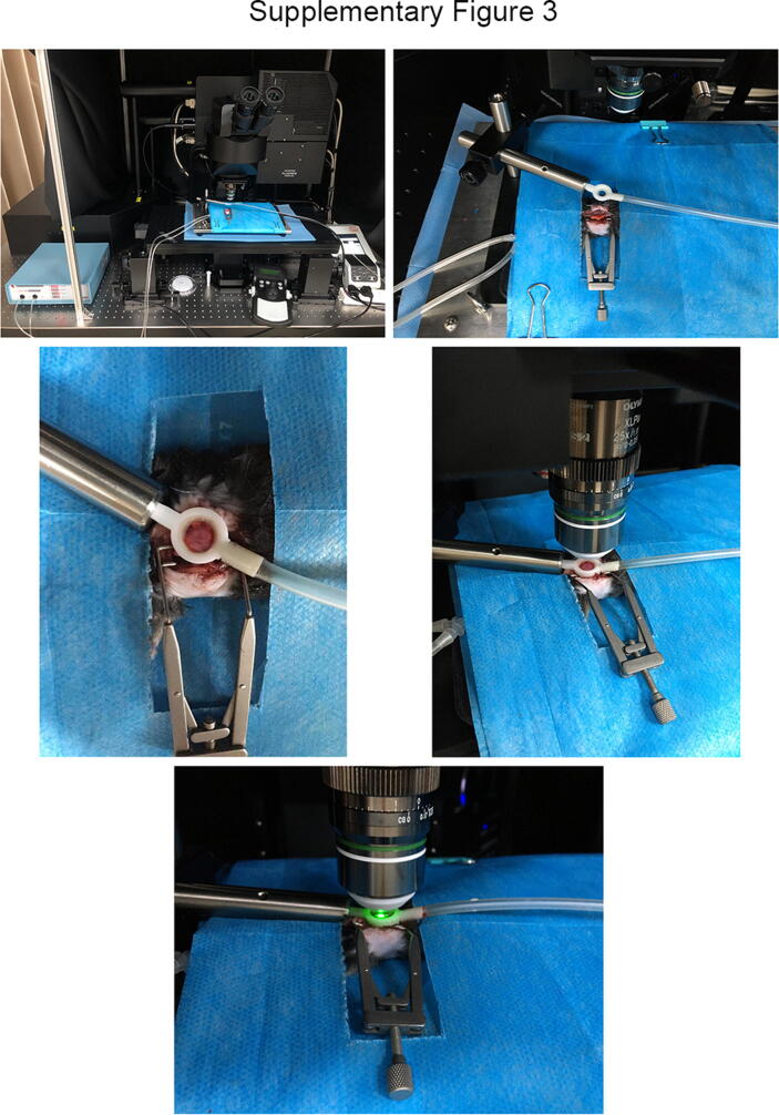
Methods: Mice were administered lipopolysaccharide and/or Escherichia coli via aerosol inhalation to establish a self-limited ARDS model. Then, RvD1 was administered at the peak inflammatory response. RAMs self-renewal was measured by flow cytometry, RAM phagocytosis was measured by two-photon fluorescence imaging. In addition, plasma was collected from intensive care unit patients on days 0-2, 3-5, and 6-9 to measure RvD1 and S100A8/A9 levels using triple quadrupole/linear ion trap mass spectrometry.

Results: RAMs were found to play a pivotal role in resolving inflammation during ARDS, and RvD1 enhanced RAM proliferation and phagocytosis, which was abrogated by a lipoxin A4 receptor (ALX, RvD1 receptor) inhibitor. Both primary RAMs transfected with rS100A8/A9 and/or S100A8/A9 siRNA and S100A9-/- mice (also deficient in S100A8 function) showed higher turnover and phagocytic function, indicating that RvD1 exerted its effects on RAMs by inhibiting S100A8/A9 production in the resolution phase. RvD1 reduced S100A8/A9 and its upstream MAPK14 levels in vivo and in vitro. Finally, in the patients, RvD1 levels were lower, but S100A8/A9 levels were higher.

Conclusions: We propose that RvD1 improved RAM self-renewal and phagocytosis via the ALX/MAPK14/S100A8/A9 signaling pathway. Plasma RvD1 and S100A8/A9 levels were negatively correlated, and associated with the outcome of sepsis-induced ARDS.

Keywords: Acute respiratory distress syndrome; Resident alveolar macrophages; Resolution of inflammation; Resolvin D1; S100A8/A9.


Conflict of interest statement

Declaration of competing interest The authors declare that they have no known competing financial interests or personal relationships that could have appeared to influence the work reported in this paper.

Figures

None
Graphical abstract
Fig. 1
Fig. 1
RvD1 promotes the inflammatory resolution of LPS-induced ARDS dependent on RAMs. Mice were treated with LPS (1 mg/kg) by aerosol inhalation, and BALF was collected from 0 h to 144 h (A) to establish a self-limited murine ARDS model (B). Neutrophil counts were determined by flow cytometry, and resolution indices (Ri) were calculated (B). RAM depletion was achieved by the intratracheal instillation of 50 μL of liposome clodronate (5 mg/mL). Phosphate-buffered saline liposomes were used as controls (C). Three days later, mice were administered LPS by aerosol inhalation, and Ψmax, Tmax, R50, T50, and Ri were calculated (D). Then, RvD1 was administered to mice 72 h after LPS inhalation. Twenty-four hours later, BALF was collected. Infiltrated neutrophils were enumerated to calculate the Ri (E). Lung tissues were harvested and stained with H&E to evaluate the effects of RvD1 on lung histology (F). Original magnification × 200 and × 400. Acute lung injury scores were determined based on the standards mentioned in the Materials and Methods (G). After RAM depletion, RvD1 was administered to mice 72 h after LPS inhalation. Twenty-four hours later, neutrophil counts in the BALF were determined by flow cytometry (H). Ψmax:neutrophil numbers at maximum; Tmax: the time point when neutrophil numbers reached Ψmax; R50: decrease in neutrophil numbers to 50 % of the Ψmax; T50: the time point when the neutrophil numbers decreased to R50; Ri, the time interval from Ψmax to R50 (T50-Tmax). The data are presented as the mean ± SEM, n = 6–8. **p < 0.01, ****p < 0.0001, ns: not significant.
Fig. 2
Fig. 2
RvD1 increases RAM proliferation in LPS-induced ARDS. RvD1 was administered to mice 72 h after LPS inhalation. Twenty-four hours later, RAM numbers (A) and proliferation (Ki67+ cells) (B) were measured by flow cytometry. PE anti-mouse CD170 (Siglec-F) antibody (red) and FITC anti-mouse Ly6G antibody (green) were given by aerosol inhalation to visualize pulmonary RAMs (red) and neutrophils (green), respectively. Scale bar, 20 µm. Bar graph of intracellular Ly6G staining gated on RAM subsets in collected BALF (C). RAM phagocytosis was measured by two-photon fluorescence imaging. PE anti-mouse CD170 (Siglec-F) antibody (red) and FITC-LPS (green) were given by aerosol inhalation into the pulmonary tract (D). Red indicates RAMs. Green indicates LPS. Scale bar, 20 µm (left) and 10 µm (right). CD206, Arg1 (M2) and CD86, iNOS (M1) RAMs were also detected by flow cytometry. The data are presented as the mean ± SEM, n = 6–8. *p < 0.05, **p < 0.01, ***p < 0.001, ****p < 0.0001. (For interpretation of the references to colour in this figure legend, the reader is referred to the web version of this article.)
Fig. 3
Fig. 3
RvD1 inhibits RAM-derived S100A8/A9 expression in LPS-induced ARDS. RvD1 was administered to mice 72 h after LPS inhalation. Twenty-four hours later, RAMs were sorted to perform transcriptional profiling experiments. RNA-seq was performed to identify genes differentially expressed by ≥ 2.0-fold (A). Genes upregulated in the LPS group compared to the controls and genes downregulated in the RvD1 treatment group compared to the LPS group were analyzed (B). Sample correlation or variance of logarithm-transformed counts from RNA-seq datasets of control, LPS- and RvD1-treated RAMs were analyzed by PCA (C). Pyramid plot with bidirectional log10 (p-value) demonstrated the involved gene ontology (GO) terms and pathways from submitted up- or downregulated gene lists (D). mRNA expression of selected genes detected by qRT-PCR in the sorted RAMs (E). S100A8/A9+ was mainly expressed on RAMs (F). The S100A8/A9 expression on RAMs was also measured by flow cytometry (G). The data are presented as the mean ± SEM, n = 3–6. **p < 0.01, ***p < 0.001, ****p < 0.0001.
Fig. 4
Fig. 4
RvD1 promotes primary RAM self-renewal and phagocytic function dependent on S100A8/A9 in vitro. Primary RAMs were isolated from mice and co-cultured with 50 nM S100A8 or S100A9 siRNA for 48 h before LPS/IFN-γ stimulation. Twenty-four hours later, the percentage of Ki67+ cells was measured by flow cytometry (A). Primary RAMs were cultured with S100A8 or S100A9 plasmid for 48 h to calculate the percentage of Ki67+ RAMs (B). Cell cycle was detected after co-culturing with S100A8 or S100A9 siRNA (C). Primary RAMs were co-cultured with 50 nM S100A8 or S100A9 siRNA for 48 h before FITC-LPS/IFN-γ stimulation. Twenty-four hours later, the percentage of intracellular FITC-LPS in RAMs was measured (D). FITC-LPS in primary RAMs was determined by immunofluorescent images (E). The percentage of CD206 RAMs was detected after treatment with S100A8 or S100A9 siRNA (F). Primary RAMs were co-cultured with LPS/IFN-γ in the presence or absence of RvD1 (100 nM) for 24 h to measure S100A8 and S100A9 mRNA expression (G, H), then proliferation (I) and cell cycle (J) were analyzed. RAMs were co-cultured with 50 nM S100A8 or S100A9 siRNA for 48 h before LPS/IFN-γ treatment with or without RvD1 (100 nM) stimulation. Twenty-four hours later, proliferation was measured by CCK8 (K). siNeg: negative control siRNA. The data are presented as the mean ± SEM, n = 6–8. *p < 0.05, **p < 0.01, ***p < 0.001, ****p < 0.0001, ns: not significant.
Fig. 5
Fig. 5
RvD1 promotes RAM self-renewal and phagocytic function dependent on S100A8/A9 in LPS-stimulated S100A9-/- mice. S100A9-/- mice were given LPS (1 mg/kg) by aerosol inhalation, and BALF was collected from 0 h to 120 h, and the infiltrated neutrophils were enumerated to calculate the Ri (A). RAM numbers were counted at each time point by flow cytometry (B), and the mean fluorescence intensity (MFI) of Ki67+ RAMs was measured (C). PE anti-mouse CD170 (Siglec-F) antibody (red) and FITC anti-mouse Ly6G antibody (green) were given by aerosol inhalation to visualize pulmonary RAMs (red) and neutrophils (green), respectively. Scale bar, 20 µm. rS100A8 or rS100A9 (1 μg and 10 μg) was administered to S100A9-/- mice 48 h after LPS inhalation (E) to observe the MFI of Ki67+ RAMs and intracellular Ly6G staining in RAMs (G). RvD1 was administrated to S100A9-/- mice 48 h after LPS inhalation (E) to determine PMN numbers (H), RAM counts (I), the MFI of Ki67+ RAMs (J), and intracellular Ly6G+ cells in RAMs (K). The data are presented as the mean ± SEM, n = 6–8. *p < 0.05, **p < 0.01, ***p < 0.001, ****p < 0.0001. (For interpretation of the references to colour in this figure legend, the reader is referred to the web version of this article.)
Fig. 6
Fig. 6
Negative correlation between plasma RvD1 and S100A8/A9 levels is associated with the outcomes of sepsis-induced ARDS patients. Thirty-five sepsis-induced ARDS patients admitted to the ICU and eight healthy volunteers were enrolled. Plasma was collected at different times. RvD1-d5 was used as the internal standard (A). Plasma RvD1 levels were measured by liquid chromatography-tandem mass spectrometry (LC-MS/MS) (B, C). RvD1 levels were analyzed in survivors and non-survivors (D). Plasma S100A8/A9 levels were also measured by enzyme-linked immunosorbent assays, and (E) the concentration in survivors and non-survivors was also analyzed (F). Finally, the correlation between plasma RvD1 and S100A8/A9 levels was assessed (G). The data are presented as the mean ± SEM, n = 4–6. *p < 0.05, **p < 0.01, ***p < 0.001, ****p < 0.0001.
Supplementary figure 1
Supplementary figure 2
Supplementary figure 3
Supplementary figure 4
Supplementary figure 5
Supplementary figure 6
Supplementary figure 7
Supplementary figure 8
Supplementary figure 9
Supplementary figure 10

References

    1. Guillen-Guio B., Lorenzo-Salazar J.M., Ma S.F., Hou P.C., Hernandez-Beeftink T., Corrales A., et al. Sepsis-associated acute respiratory distress syndrome in individuals of European ancestry: a genome-wide association study. Lancet Respir Med. 2020;8(3):258–266. - PMC - PubMed
    1. Ware L.B., Matthay M.A. The acute respiratory distress syndrome. N Engl J Med. 2000;342(18):1334–1349. - PubMed
    1. Chiang N., Fredman G., Bäckhed F., Oh S.F., Vickery T., Schmidt B.A., et al. Infection regulates pro-resolving mediators that lower antibiotic requirements. Nature. 2012;484(7395):524–528. - PMC - PubMed
    1. Serhan C.N., Levy B.D. Resolvins in inflammation: emergence of the pro-resolving superfamily of mediator. J Clin Invest. 2018;128(7):2657–2669. - PMC - PubMed
    1. Krishnamoorthy S., Recchiuti A., Chiang N., Yacoubian S., Lee C.H., Yang R., et al. Resolvin D1 binds human phagocytes with evidence for proresolving receptors. Proc Natl Acad Sci USA. 2010;107(4):1660–1665. - PMC - PubMed

LinkOut - more resources