. 2024 Mar 18;30(4):367-380.
doi: 10.1261/rna.079911.123.

Characterization of tRNA splicing enzymes RNA ligase and tRNA 2'-phosphotransferase from the pathogenic fungi Mucorales

Affiliations

Characterization of tRNA splicing enzymes RNA ligase and tRNA 2'-phosphotransferase from the pathogenic fungi Mucorales

Shreya Ghosh et al. RNA. .

Abstract

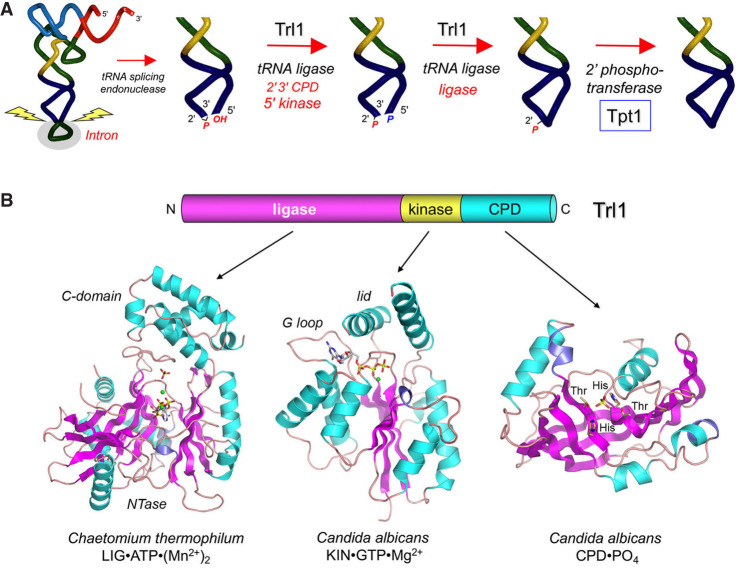
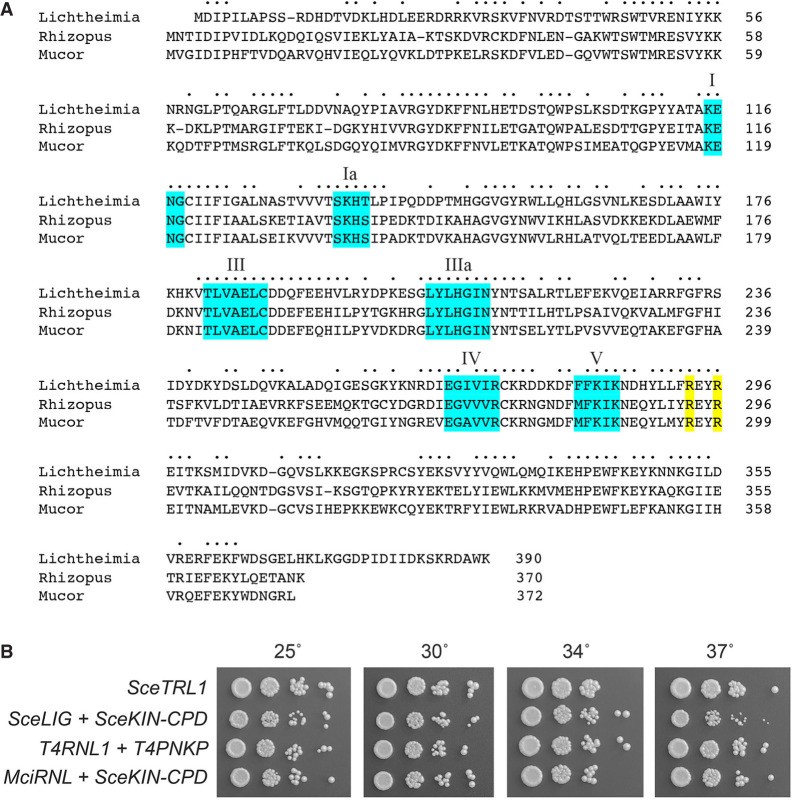
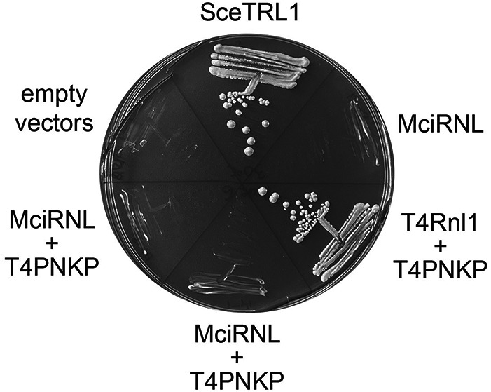
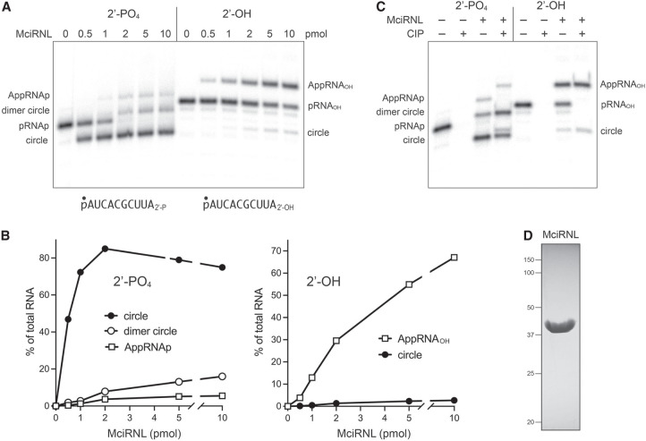
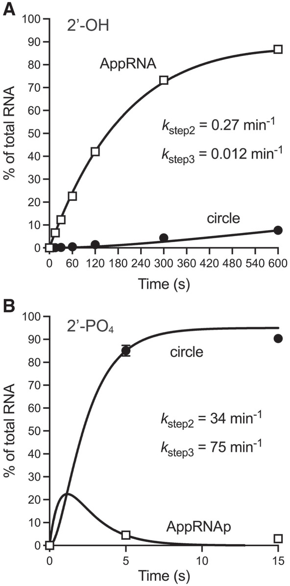
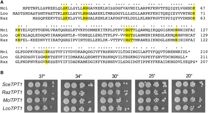
Fungal Trl1 is an essential trifunctional tRNA splicing enzyme that heals and seals tRNA exons with 2',3'-cyclic-PO4 and 5'-OH ends. Trl1 is composed of C-terminal cyclic phosphodiesterase and central polynucleotide kinase end-healing domains that generate the 3'-OH,2'-PO4 and 5'-PO4 termini required for sealing by an N-terminal ATP-dependent ligase domain. Trl1 enzymes are present in many human fungal pathogens and are promising targets for antifungal drug discovery because their domain structures and biochemical mechanisms are unique compared to the mammalian RtcB-type tRNA splicing enzyme. Here we report that Mucorales species (deemed high-priority human pathogens by WHO) elaborate a noncanonical tRNA splicing apparatus in which a monofunctional RNA ligase enzyme is encoded separately from any end-healing enzymes. We show that Mucor circinelloides RNA ligase (MciRNL) is active in tRNA splicing in vivo in budding yeast in lieu of the Trl1 ligase domain. Biochemical and kinetic characterization of recombinant MciRNL underscores its requirement for a 2'-PO4 terminus in the end-joining reaction, whereby the 2'-PO4 enhances the rates of RNA 5'-adenylylation (step 2) and phosphodiester synthesis (step 3) by ∼125-fold and ∼6200-fold, respectively. In the canonical fungal tRNA splicing pathway, the splice junction 2'-PO4 installed by RNA ligase is removed by a dedicated NAD+-dependent RNA 2'-phosphotransferase Tpt1. Here we identify and affirm by genetic complementation in yeast the biological activity of Tpt1 orthologs from three Mucorales species. Recombinant M. circinelloides Tpt1 has vigorous NAD+-dependent RNA 2'-phosphotransferase activity in vitro.

Keywords: fungal pathogen; tRNA 2′-phosphotransferase; tRNA ligase; tRNA splicing.


Figures

FIGURE 1.
FIGURE 1.
Canonical pathway of fungal tRNA splicing and structural organization of the trifunctional fungal tRNA ligase Trl1. (A) Intron removal by tRNA splicing endonuclease leaves 2′,3′-cyclic phosphate and 5′-OH ends on the broken tRNA halves. The tRNA exons are then joined by Trl1—a trifunctional tRNA ligase. Trl1 catalyzes two end-healing reactions, performed by a 5′-OH polynucleotide kinase domain and a polynucleotide 2′,3′-cyclic phosphodiesterase (CPD) domain, to generate the 5′-PO4 and 3′-OH,2′-PO4 termini required for sealing by an ATP-dependent RNA ligase domain. The 2′-PO4 at the resulting splice junction is removed by the NAD+-dependent RNA 2′-phosphotransferase enzyme Tpt1. (B) Fungal Trl1 consists of N-terminal ligase, central kinase, and C-terminal CPD catalytic modules. The structures of the ligase (pdb 6N0T), kinase (pdb 6U03), and CPD (pdb 6U05) domains from the indicated fungi are shown in complexes with active site ligands.
FIGURE 2.
FIGURE 2.
Mucorales encode a stand-alone RNA ligase enzyme active in tRNA splicing in vivo. (A) Alignment of the primary structures of monofunctional Trl1 LIG domain homologs from M. circinelloides (Genbank EPB83728.1), R. azygosporus (Genbank RCH90547.1), and L. corymbifera (Genbank CDH55495.1). Positions of side chain identity/similarity are indicated by dots. Six signature ligase adenylyltransferase motifs I, Ia, III, IIIa, IV, and V are shaded in cyan. Two conserved arginine residues implicated in the recognition of an RNA 2′-PO4 terminus are shaded in yellow. (B) Complementation of S. cerevisiae trl1Δ was assayed by plasmid shuffle. Aliquots (3 µL) of serial tenfold dilutions of FOA-resistant trl1Δ strains expressing the indicated genes were spotted on YPD agar plates and incubated at 25°C, 30°C, 34°C, and 37°C. Photographs of the plates are shown.
FIGURE 3.
FIGURE 3.
Mucor RNL is inactive in vivo when paired with the T4 end-healing enzyme Pnkp. The S. cerevisiae trl1Δ p(CEN URA3 TRL1) “shuffle” strain was transformed with marked plasmids expressing the indicated genes and with empty vector controls. Individual transformants were streaked on agar medium containing FOA, which was photographed after incubation for 10 d at 30°C. Two independent trl1Δ p(CEN URA3 TRL1) MciRNL + T4PNKP isolates were tested for growth on FOA.
FIGURE 4.
FIGURE 4.
RNA ligase activity of recombinant MciRNL. (A) Reaction mixtures (10 µL) containing 50 mM Tris-acetate, pH 6.5, 5 mM MgCl2, 0.1 µM (1 pmol) 5′ 32P-labeled 10-mer RNA with either a 3′-OH,2′-PO4 end (left side) or a 3′-OH,2′-OH end (right side) and increasing amounts of MciRNL as specified were incubated at 37°C for 5 min. The reactions were quenched with an equal volume of 95% formamide/50 mM EDTA, and the products were analyzed by electrophoresis (at 58 W constant power) through a 40-cm 20% polyacrylamide gel containing 7 M urea in 45 mM Tris-borate, 1 mM EDTA. The 5′-radiolabeled RNAs were visualized by scanning the gel with a Fujifilm FLA-7000 imaging device. The positions and identities of the RNA substrates, RNA-adenylate intermediates, and ligated products are indicated on the left and right. (B) The distributions of radiolabeled RNAs in each lane in panel A were quantified by analysis of the gel scans in ImageQuant and are plotted as a function of input MciRNL for the ligation reactions with the 2′-PO4 (left panel) and 2′-OH (right panel) RNA substrates. (C) Product analysis via treatment with CIP (calf intestine alkaline phosphatase). Ligation reactions containing 1 pmol 5′ 32P-labeled pRNA2′p and 10 pmol MciRNL (where indicated by +) were incubated at 37°C for 5 min. The mixtures were then incubated for 10 min at 37°C with 5 U CIP (purchased from NEB) where indicated by + above the lanes. The mixtures were quenched and then analyzed by urea-PAGE as described in A. (D) An aliquot (10 µg) of the recombinant MciRNL preparation was analyzed by SDS-PAGE. The Coomassie blue-stained gel is shown. The positions and sizes (kDa) of marker proteins are indicated on the left. The expected molecular weight of MciRNL is 43 kDa.
FIGURE 5.
FIGURE 5.
Kinetics of single-turnover ligation by MciRNL–AMP. (A) Ligation of 2′-OH RNA. Reaction mixtures (80 µL) containing 50 mM Tris-acetate, pH 6.5, 5 mM MgCl2, 0.1 µM 5′ 32P-labeled 10-mer RNA with a 3′-OH,2′-OH end, and 1 µM MciRNL were incubated at 37°C. The reactions were initiated by adding MciRNL to a prewarmed reaction mixture. Aliquots (10 µL) were withdrawn before adding enzyme (time 0) or at the times specified after enzyme addition, and quenched immediately with an equal volume of 95% formamide/50 mM EDTA. The products were analyzed by urea-PAGE. The distribution of radiolabeled RNAs is plotted as a function of reaction time. Each datum in the graph is an average of three independent experiments ± SEM, except for the 10-min time point, which is an average of two independent experiments. (B) Ligation of 2′-PO4 RNA. Individual replicate reaction mixtures (10 µL) containing 50 mM Tris-acetate, pH 6.5, 5 mM MgCl2, 0.1 µM 5′ 32P-labeled 10-mer RNA with a 3′-OH,2′-PO4 end, and 1 µM MciRNL were incubated at 37°C. The reactions were initiated by adding MciRNL to a prewarmed reaction mixture. The reactions were quenched after 5 or 15 sec with an equal volume of 95% formamide/50 mM EDTA. The products were analyzed by urea-PAGE. The distribution of radiolabeled RNAs is plotted as a function of reaction time. Each datum in the graph is an average of nine replicate reactions ± SEM. The data in panels A and B were fit by nonlinear regression in Prism to a unidirectional two-step kinetic mechanism.
FIGURE 6.
FIGURE 6.
Effect of pH and magnesium on single-turnover pRNA2′p ligation. (A) pH dependence. Reaction mixtures (10 µL) containing either 50 mM Tris-acetate (pH 4.5, 5.0, 5.5, 6.0, or 6.5) or 50 mM Tris-HCl (pH 7.0, 7.5, 8.0, 8.5, 9.0, or 9.5), 5 mM MgCl2, 1 pmol 5′ 32P-labeled pRNA2′p, and 10 pmol MciRNL were incubated at 37°C for 1 min. (B) Magnesium dependence. Reaction mixtures (10 µL) containing 50 mM Tris-acetate, pH 6.5, 1 pmol 5′ 32P-labeled pRNA2′p, 10 pmol MciRNL, and MgCl2 as specified or 5 mM EDTA were incubated at 37°C for 1 min. The reaction products were analyzed by urea-PAGE and visualized by scanning the gel with a Fujifilm FLA-7000 imaging device.
FIGURE 7.
FIGURE 7.
MciRNL adenylylation. (A) pH dependence. Reaction mixtures (10 µL) containing either 50 mM Tris-acetate (pH 4.5, 5.0, 5.5, 6.0, or 6.5) or 50 mM Tris-HCl (pH 7.0, 7.5, 8.0, 8.5, 9.0, or 9.5), 5 mM MgCl2, 50 µM [α32P]ATP, and 10 µM (100 pmol) MciRNL were incubated at 37°C for 5 min, then quenched with SDS, and analyzed by SDS-PAGE. A scan of the gel is shown. The positions and sizes (kDa) of marker proteins are indicated on the left. (B) ATP concentration-dependence. Reaction mixtures (10 µL) containing 50 mM Tris-HCl, pH 9.0, 5 mM MgCl2, 10 µM (100 pmol) MciRNL, and 50, 100, 200, 300, 400, or 500 µM [α32P]ATP were incubated at 37°C for 5 min, then quenched with SDS, and analyzed by SDS-PAGE. A scan of the gel is shown. The extents of MciRNL–[32P]AMP formation are indicated below the lanes.
FIGURE 8.
FIGURE 8.
Mucorales Tpt1 proteins have tRNA 2′-phosphotransferase activity in vivo. (A) Alignment of the amino acid sequences of Tpt1 homologs from M. circinelloides (Genbank EPB89638.1), R. azygosporus (Genbank RCI00563.1), and L. corymbifera (Genbank CDH53741.1). Positions of side chain identity/similarity are indicated by dots above the alignment. The conserved amino acids that make atomic contacts with Tpt1 reaction substrates and products are highlighted in yellow shading. (B) Complementation of S. cerevisiae tpt1Δ was assayed by plasmid shuffle. Aliquots (3 µL) of serial tenfold dilutions of FOA-resistant tpt1Δ strains expressing the indicated TPT1 genes were spotted on YPD agar plates and incubated at 20°C, 25°C, 30°C, 34°C, and 37°C. Photographs of the plates are shown.
FIGURE 9.
FIGURE 9.
2′-phosphotransferase activity of recombinant Mucor Tpt1. (A) An aliquot (7.5 µg) of the recombinant MciTpt1 preparation was analyzed by SDS-PAGE. The Coomassie blue-stained gel is shown. The positions and sizes (kDa) of marker proteins are indicated on the left. The expected molecular weight of MciTpt1 is 24 kDa. (B) Reaction mixtures (10 µL) containing 100 mM Tris-HCl, pH 7.5, 1 mM NAD+, 0.2 µM (2 pmol) 5′ 32P-labeled 2′-PO4 branchpoint-containing 6-mer RNA oligonucleotide (as shown), and 0, 1, 2.5, 5, 10, or 20 pmol MciTpt1 were incubated at 37°C for 30 min. The reactions were quenched by the addition of three volumes of cold 90% formamide, 50 mM EDTA. The samples were analyzed by electrophoresis (at 55 W constant power) through a 40-cm 20% polyacrylamide gel containing 7 M urea in 45 mM Tris-borate, 1 mM EDTA. The 5′-radiolabeled 2′-PO4 substrate RNA and the 2′-OH product RNA were visualized by scanning the gel with a Fujifilm FLA-7000 imaging device. The extents of product formation were quantified by analysis of the gel scans in ImageQuant and are plotted as a function of input MciTpt1. Each datum is the average of four independent titration experiments ± SEM.
Shreya Ghosh
Shreya Ghosh

Similar articles

Cited by

References

    1. Amitsur M, Levitz R, Kaufmann G. 1987. Bacteriophage T4 anticodon nuclease, polynucleotide kinase and RNA ligase reprocess the host lysine tRNA. EMBO J 6: 2499–2503. 10.1002/j.1460-2075.1987.tb02532.x - DOI - PMC - PubMed
    1. Banerjee A, Ghosh S, Goldgur Y, Shuman S. 2019a. Structure and two-metal mechanism of fungal tRNA ligase. Nucleic Acids Res 47: 1428–1439. 10.1093/nar/gky1275 - DOI - PMC - PubMed
    1. Banerjee A, Goldgur Y, Schwer B, Shuman S. 2019b. Atomic structures of the RNA end-healing 5′-OH kinase and 2′,3′-cyclic phosphodiesterase domains of fungal tRNA ligase: conformational switches in the kinase upon binding of the GTP phosphate donor. Nucleic Acids Res 47: 11826–11838. 10.1093/nar/gkz1049 - DOI - PMC - PubMed
    1. Banerjee A, Munir A, Abdullahu L, Damha MJ, Goldgur Y, Shuman S. 2019c. Structure of tRNA splicing enzyme Tpt1 illuminates the mechanism of RNA 2′-PO4 recognition and ADP-ribosylation. Nat Commun 10: 218. 10.1038/s41467-018-08211-9 - DOI - PMC - PubMed
    1. Culver GM, McCraith SM, Zillmann M, Kierzek R, Michaud N, LaReau RD, Turner DH, Phizicky EM. 1993. An NAD derivative produced during transfer RNA splicing: ADP-ribose 1″-2″ cyclic phosphate. Science 261: 206–208. 10.1126/science.8392224 - DOI - PubMed

LinkOut - more resources