

Incidental Thalamic Lesions Identified on Brain MRI in Pediatric and Young Adult Patients: Imaging Features and Natural History
- PMID: 38238093
- PMCID: PMC11285995
- DOI: 10.3174/ajnr.A8090
Incidental Thalamic Lesions Identified on Brain MRI in Pediatric and Young Adult Patients: Imaging Features and Natural History
Abstract
Background and purpose: Nonspecific, localized thalamic signal abnormalities of uncertain significance are occasionally found on pediatric brain MR imaging. The goal of this study is to describe the MR imaging appearance and natural history of these lesions in children and young adults.
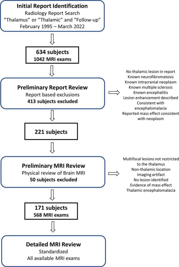
Materials and methods: This retrospective study evaluated clinically acquired brain MR imaging examinations obtained from February 1995 to March 2022 at a large, tertiary care pediatric hospital. Examinations with non-mass-like and nonenhancing thalamic lesions were identified based on term search of MR imaging reports. A total of 221 patients formed the initial group for imaging assessment. Additional exclusions during imaging review resulted in 171 patients. Imaging appearance and size changes were assessed at baseline and at follow-up examinations.
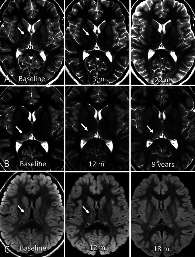
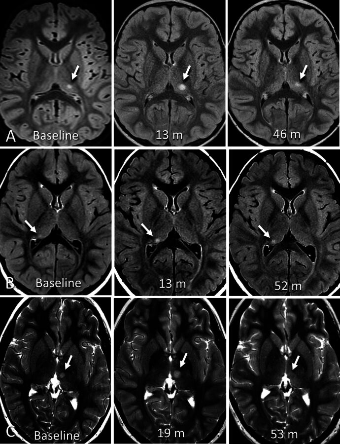
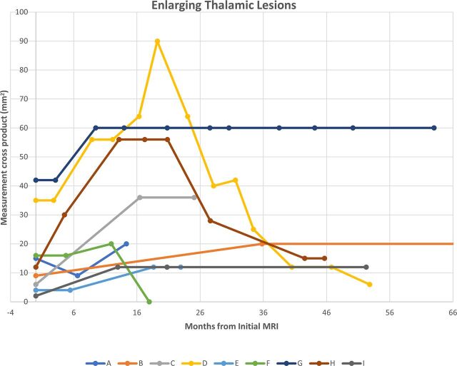
Results: A total of 171 patients (102 male) at a median age of 11 years (range: 1-23 years), 568 MR imaging examinations, and 180 thalamic lesions were included. Median time from baseline to the last follow-up MR imaging was 542 days (range: 46-5730 days). No lesion enhanced at any time point. On imaging follow-up, 11% of lesions (18/161) became smaller, 10% (16/161) resolved, 73% (118/161) remained stable, and 6% (9/161) increased in size at some point during evaluation. Median time interval from baseline to enlargement was 430 days (range: 136-1074 days).
Conclusions: Most incidental, non-mass-like thalamic signal abnormalities were stable, decreased in size, or resolved on follow-up imaging and are likely of no clinical significance. Surveillance strategies with longer follow-up intervals may be adequate in the management of such findings.
© 2024 by American Journal of Neuroradiology.
Figures
Similar articles
-
Prevalence and MRI findings of incidentally detected pituitary non-enhancing lesion on brain MRI in children.J Pediatr Endocrinol Metab. 2021 Apr 5;34(5):591-598. doi: 10.1515/jpem-2020-0518. Print 2021 May 26. J Pediatr Endocrinol Metab. 2021. PMID: 33818038
-
Natural History and Management of Incidentally Discovered Focal Brain Lesions Indeterminate for Tumor in Children.Neurosurgery. 2020 Mar 1;86(3):357-365. doi: 10.1093/neuros/nyz078. Neurosurgery. 2020. PMID: 30989228
-
Prevalence and natural history of arachnoid cysts in children.J Neurosurg Pediatr. 2010 Jun;5(6):578-85. doi: 10.3171/2010.2.PEDS09464. J Neurosurg Pediatr. 2010. PMID: 20515330
-
Thalamic lesions: a radiological review.Behav Neurol. 2014;2014:154631. doi: 10.1155/2014/154631. Epub 2014 Jul 2. Behav Neurol. 2014. PMID: 25100900 Free PMC article. Review.
-
The management of brainstem gliomas in patients with neurofibromatosis 1.Neurology. 1996 Jun;46(6):1652-60. doi: 10.1212/wnl.46.6.1652. Neurology. 1996. PMID: 8649565 Review.
Cited by
-
Incidental brain tumor findings in children: prevalence, natural history, management, controversies, challenges, and dilemmas.Childs Nerv Syst. 2024 Oct;40(10):3179-3187. doi: 10.1007/s00381-024-06598-z. Epub 2024 Aug 31. Childs Nerv Syst. 2024. PMID: 39215810 Free PMC article. Review.
-
Pediatric thalamic incidentalomas: a retrospective analysis of their characteristics, evolution, management, and prognostic factors for progression.Acta Neurochir (Wien). 2025 Aug 8;167(1):218. doi: 10.1007/s00701-025-06632-2. Acta Neurochir (Wien). 2025. PMID: 40779174 Free PMC article.
References
MeSH terms
LinkOut - more resources
Full Text Sources
Medical