Review
. 2024 Feb 20;137(4):394-407.
doi: 10.1097/CM9.0000000000002976. Epub 2024 Jan 18.

Chronic stress as an emerging risk factor for the development and progression of glioma

Affiliations
Review

Chronic stress as an emerging risk factor for the development and progression of glioma

Lan Yi et al. Chin Med J (Engl). .

Abstract

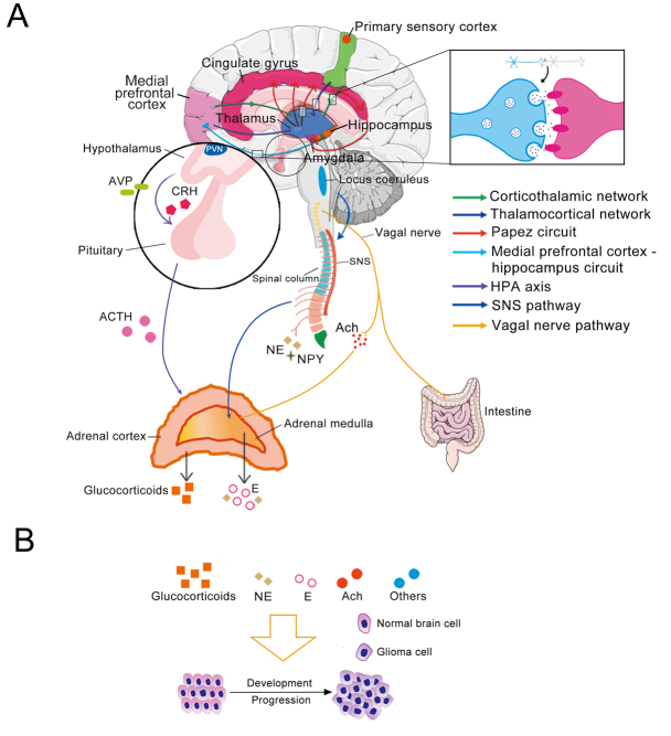
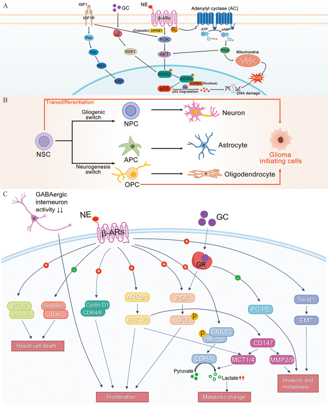
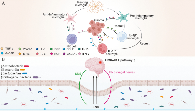
Gliomas tend to have a poor prognosis and are the most common primary malignant tumors of the central nervous system. Compared with patients with other cancers, glioma patients often suffer from increased levels of psychological stress, such as anxiety and fear. Chronic stress (CS) is thought to impact glioma profoundly. However, because of the complex mechanisms underlying CS and variability in individual tolerance, the role of CS in glioma remains unclear. This review suggests a new proposal to redivide the stress system into two parts. Neuronal activity is dominant upstream. Stress-signaling molecules produced by the neuroendocrine system are dominant downstream. We discuss the underlying molecular mechanisms by which CS impacts glioma. Potential pharmacological treatments are also summarized from the therapeutic perspective of CS.


Conflict of interest statement

None.

Figures

Figure 1
Figure 1
Processing of the stress system and its associated media. (A) The upstream signals of the stress system are mainly received and processed by different cortical areas and limbic systems through neural circuits for various types of stressors. The cortex communicates bidirectionally with the thalamus through a cortico-thalamo-cortical loop to make real-time judgments about information. The components of the limbic system, such as the thalamus, cingulate gyrus and hippocampus, exchange information through the Papez circuit, which plays an important role in the cortical control of emotion and memory storage. The hippocampus stores information transmitted multiple times by the Papez circuit and forms long-term memories. The medial prefrontal cortex–hippocampal circuit transmits information between the medial prefrontal cortex and the hippocampus. A neural circuit consists mainly of a population of neurons connected by synapses which perform specific function when activated. The CNS regulates the activity of the HPA axis by controlling the secretion of CRH and AVP in the PVN. As a result, the pituitary gland releases ACTH, which causes the adrenal cortex to release glucocorticoids. Simultaneously, the CNS activates the SNS, resulting in the secretion of E from the adrenal medulla. The secretion of acetylcholine by the vagal nerve binds to muscarinic receptors on chromaffin cells in the adrenal medulla, promoting the secretion of CAs. Sympathetic nerve endings also secrete small amounts of NPY. The vagal pathway acts as a bridge between the brain and the gut. (B) Several stress system molecules have been implicated in promoting glioma development and progression, including glucocorticoids, NE, E, Ach and other molecules secreted by the CNS. ACTH: Adrenocorticotropic hormone; Ach: Acetylcholine; AVP: Arginine vasopressin; CAs: Catecholamines; CNS: Central nervous system; CRH: Corticotropin-releasing hormone; CS: Chronic stress; E: Epinephrine; HPA axis: Hypothalamic-pituitary-adrenal axis; NE: Norepinephrine; NPY: Neuropeptide Y; PVN: Paraventricular nucleus; SNS: Sympathetic nervous system. The Figure was partly generated using Servier medical art repository (https://smart.servier.com), provided by Servier and licensed under a Creative Commons Attribution 3.0 unported license, and partly adapted from BioRender.com with permission.
Figure 2
Figure 2
Mechanisms by which CS promotes malignant progression in glioma cell. (A) CS stimulates olfactory neurons to secrete IGF1, which binds to IGF1R on OPCs and activates classical RTK pathways. CS triggers the neuroendocrine system to secrete NE and GCs, which activate the PI3K-AKT and SGK1-MDM2 pathways to promote the p53 degradation. Meanwhile, NE also activates PKA to promote mitochondrial generation of ROS, which in turn promotes DNA damage. (B) NSCs give rise to NPCs, astrocyte progenitor cells and OPCs, which in turn each differentiate into neurons, astrocytes and oligodendrocytes. Two solid red lines (transdifferentiation) indicate cell types that have been recognized as cells of malignant glioma origin. (C) CS promotes abnormal signaling pathways in glioma cells, mainly through NE binding to β-ARs on the surface of glioma cells. GCs combined with intracellular GRs and a decrease in GABAergic interneuron activity could act synergistically. AC: Adenylyl cyclase; AKT: Protein kinase B; APC: Astrocyte precursor cell; ARRB1: β-arrestin 1; ATP: Adenosine triphosphate; cAMP: Cyclic adenosine monophosphate; β-AR: β adrenoceptor; Bax: B-cell lymphoma 2-associated X protein; BCL-2: B-cell lymphoma 2; BCL-XL: B-cell lymphoma-extra large; CD147: Cluster of differentiation 147; CDK4/6: Cyclin-dependent kinase 4/6; CS: Chronic stress; DNA: Deoxyribonucleic acid; EMT: Epithelial to mesenchymal transition; ERK: Extracellular signal-regulated kinase; GABAergic: Gamma aminobutyric acidergic; GC: Glucocorticoid; GR: Glucocorticoid receptor; Gs: G protein; HIF-1α: Hypoxia-inducible factor 1α; IGF1: Insulin-like growth factor 1; IGF1R: Insulin-like growth factor 1 receptor; LDHA: Lactate dehydrogenase A; MCT1/4: Monocarboxylate transporters 1/4; MDM2: Murine double minute 2; MEK: Mitogen-activated protein kinase kinase; MMP2/9: Matrix metallopeptidase 2/9; NE: Norepinephrine; NK cell: Natural killer cell; NPC: Neural progenitor cells; NSC: Neural stem cell; OPC: Oligodendrocyte precursor cell; P: Phosphorylation; PC/PE: Phosphatidylcholine to phosphatidylethanolamine ratio; PI3K: Phosphoinositide 3-kinase; PKA: Protein kinase A; p53: Tumor protein 53; Raf: Rapidly accelerated fibrosarcoma; Ras: Rat sarcoma virus; ROS: Reactive oxygen species; RTK: Receptor tyrosine kinase; SGK1: Serum and glucocorticoid-regulated protein kinase 1; Ubi: Ubiquitin; Twist1: Twist family basic helix-loop-helix transcription factor 1; +: Increase; -: Decrease. The Figure was partly adapted from BioRender.com with permission.
Figure 3
Figure 3
Effects of chronic stress on immune cells and gut microbiota. (A) CS activates resting microglia to become either anti-inflammatory or pro-inflammatory microglia, predominantly pro-inflammatory. Pro-inflammatory microglia recruit IL-1β+ immune cells such as monocytes and neutrophils into the brain. Recruited monocytes and neutrophils secrete pro-inflammatory factors that promote glioma development, such as TNF-α, EGF, HGF, IL-1β and IL-8. At the same time, glioma cells secrete VCAM-1 to promote monocyte adhesion to glioma cells. Neutrophils bind to G-CSF secreted by glioma cells to promote their further proliferation. Anti-inflammatory microglia block communication between pro-inflammatory microglia and natural killer cells, inhibiting natural killer cells from eliminating glioma cells. Dashed black lines represent processes suppressed by CS and solid black lines represent processes facilitated by CS. (B) The gut-brain-microbiota axis and CS. CS has been shown to alter the species and abundance of the gut microbiota. Specifically, Lactobacillus, Bacteroidia and Actinobacteria have been found to decrease in number, while pathogenic bacteria increase in the intestines. The brain receives information from the gut through the ENS via the SNS and PNS. Reductions in Lactobacillus have been linked to increased PI3K-AKT signaling in gliomas, which can stimulate glioma cell proliferation. CCL2: C-C motif chemokine ligand 2; CXCL10: C-X-C motif chemokine ligand 10; EGF: Epidermal growth factor; ENS: Enteric nervous system; G-CSF: Granulocyte colony stimulating factor; HGF: Hepatocyte growth factor; IFN-γ: Interferon-gamma; IL-1β: Interleukin-1β; IL-6: Interleukin-6; IL-8: Interleukin-8; IL-15: Interleukin-15; NK cell: Natural killer cell; PNS: Parasympathetic nervous system; SNS: Sympathetic nervous system; TNF-α: Tumor necrosis factor α; VCAM-1: Vascular cell adhesion molecule-1. The Figure was partly generated using Servier medical art repository (https://smart.servier.com), provided by Servier, licensed under a Creative Commons Attribution 3.0 unported license. The Figure was partly generated using Servier medical art repository (https://smart.servier.com), provided by Servier and licensed under a Creative Commons Attribution 3.0 unported license, and partly adapted from BioRender.com with permission.

Similar articles

Cited by

References

    1. Sung H Ferlay J Siegel RL Laversanne M Soerjomataram I Jemal A, et al. . Global Cancer Statistics 2020: GLOBOCAN Estimates of Incidence and Mortality Worldwide for 36 Cancers in 185 Countries. CA Cancer J Clin 2021;71:209–249. doi: 10.3322/caac.21660. - PubMed
    1. Lv K Cao X Wang R Du P Fu J Geng D, et al. . Neuroplasticity of Glioma Patients: Brain Structure and Topological Network. Front Neurol 2022;13:871613. doi: 10.3389/fneur.2022.871613. - PMC - PubMed
    1. Weller M Wick W Aldape K Brada M Berger M Pfister SM, et al. . Glioma. Nat Rev Dis Primers 2015;1:15017. doi: 10.1038/nrdp.2015.17. - PubMed
    1. Lin D Wang M Chen Y Gong J Chen L Shi X, et al. . Trends in Intracranial Glioma Incidence and Mortality in the United States, 1975-2018. Front Oncol 2021;11:748061. doi: 10.3389/fonc.2021.748061. - PMC - PubMed
    1. Zhao Z Zhang KN Wang Q Li G Zeng F Zhang Y, et al. . Chinese Glioma Genome Atlas (CGGA): A Comprehensive Resource with Functional Genomic Data from Chinese Glioma Patients. Genomics Proteomics Bioinformatics 2021;19:1–12. doi: 10.1016/j.gpb.2020.10.005. - PMC - PubMed