

Morphometric network-based abnormalities correlate with psychiatric comorbidities and gene expression in PCDH19-related developmental and epileptic encephalopathy
- PMID: 38238304
- PMCID: PMC10796344
- DOI: 10.1038/s41398-024-02753-x
Morphometric network-based abnormalities correlate with psychiatric comorbidities and gene expression in PCDH19-related developmental and epileptic encephalopathy
Abstract
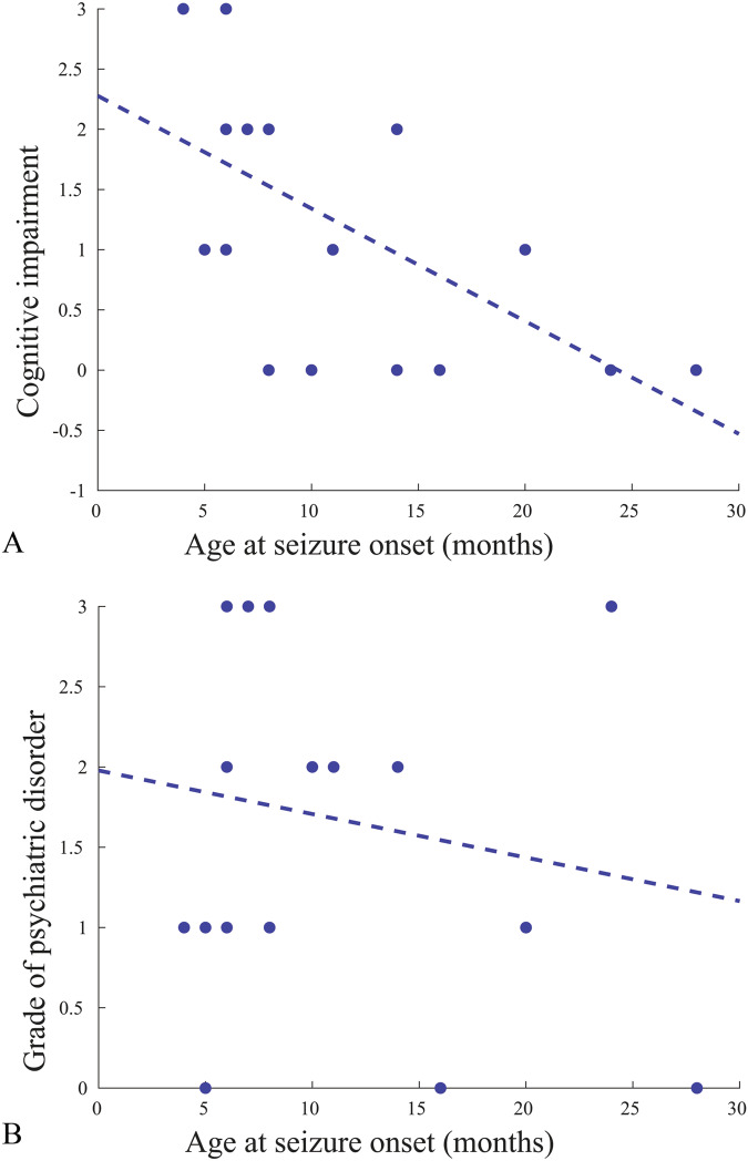
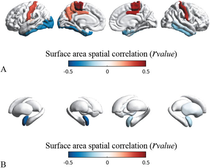
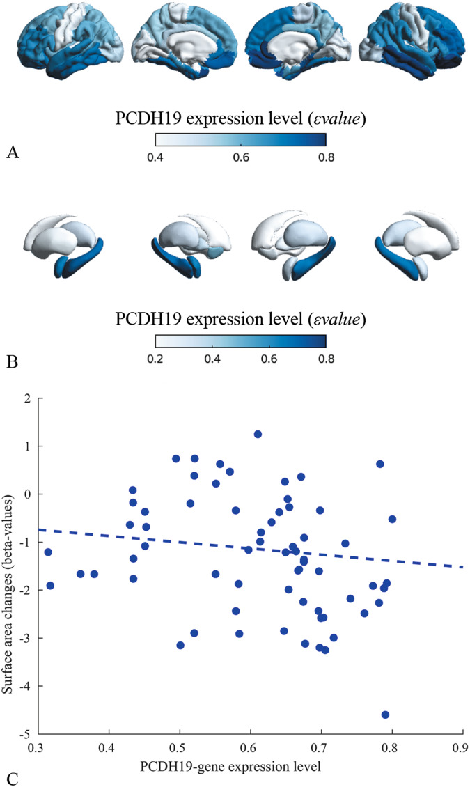
Protocadherin-19 (PCDH19) developmental and epileptic encephalopathy causes an early-onset epilepsy syndrome with limbic seizures, typically occurring in clusters and variably associated with intellectual disability and a range of psychiatric disorders including hyperactive, obsessive-compulsive and autistic features. Previous quantitative neuroimaging studies revealed abnormal cortical areas in the limbic formation (parahippocampal and fusiform gyri) and underlying white-matter fibers. In this study, we adopted morphometric, network-based and multivariate statistical methods to examine the cortex and substructure of the hippocampus and amygdala in a cohort of 20 PCDH19-mutated patients and evaluated the relation between structural patterns and clinical variables at individual level. We also correlated morphometric alterations with known patterns of PCDH19 expression levels. We found patients to exhibit high-significant reductions of cortical surface area at a whole-brain level (left/right pvalue = 0.045/0.084), and particularly in the regions of the limbic network (left/right parahippocampal gyri pvalue = 0.230/0.016; left/right entorhinal gyri pvalue = 0.002/0.327), and bilateral atrophy of several subunits of the amygdala and hippocampus, particularly in the CA regions (head of the left CA3 pvalue = 0.002; body of the right CA3 pvalue = 0.004), and differences in the shape of hippocampal structures. More severe psychiatric comorbidities correlated with more significant altered patterns, with the entorhinal gyrus (pvalue = 0.013) and body of hippocampus (pvalue = 0.048) being more severely affected. Morphometric alterations correlated significantly with the known expression patterns of PCDH19 (rvalue = -0.26, pspin = 0.092). PCDH19 encephalopathy represents a model of genetically determined neural network based neuropsychiatric disease in which quantitative MRI-based findings correlate with the severity of clinical manifestations and had have a potential predictive value if analyzed early.
© 2024. The Author(s).
Conflict of interest statement
The authors declare no competing interests.
Figures


Similar articles
-
Quantitative MRI-Based Analysis Identifies Developmental Limbic Abnormalities in PCDH19 Encephalopathy.Cereb Cortex. 2020 Oct 1;30(11):6039-6050. doi: 10.1093/cercor/bhaa177. Cereb Cortex. 2020. PMID: 32582916
-
Multicenter retrospective study of patients with PCDH19-related epilepsy: The first Hungarian cohort.Epileptic Disord. 2024 Oct;26(5):685-693. doi: 10.1002/epd2.20264. Epub 2024 Jul 17. Epileptic Disord. 2024. PMID: 39017914
-
Neuronal network activity and connectivity are impaired in a conditional knockout mouse model with PCDH19 mosaic expression.Mol Psychiatry. 2024 Jun;29(6):1710-1725. doi: 10.1038/s41380-023-02022-1. Epub 2023 Mar 30. Mol Psychiatry. 2024. PMID: 36997609 Free PMC article.
-
A systematic review and meta-analysis of 271 PCDH19-variant individuals identifies psychiatric comorbidities, and association of seizure onset and disease severity.Mol Psychiatry. 2019 Feb;24(2):241-251. doi: 10.1038/s41380-018-0066-9. Epub 2018 Jun 11. Mol Psychiatry. 2019. PMID: 29892053 Free PMC article.
-
PCDH19-related infantile epileptic encephalopathy: an unusual X-linked inheritance disorder.Hum Mutat. 2012 Apr;33(4):627-34. doi: 10.1002/humu.22029. Epub 2012 Feb 14. Hum Mutat. 2012. PMID: 22267240 Review.
Cited by
-
Bayesian Rare Variant Analysis Identifies Novel Schizophrenia Putative Risk Genes.J Pers Med. 2024 Aug 2;14(8):822. doi: 10.3390/jpm14080822. J Pers Med. 2024. PMID: 39202013 Free PMC article.
-
Expanding the Mutational Landscape and Clinical Phenotype of CHD2-Related Encephalopathy.Neurol Genet. 2024 Jul 11;10(4):e200168. doi: 10.1212/NXG.0000000000200168. eCollection 2024 Aug. Neurol Genet. 2024. PMID: 39035822 Free PMC article.
References
-
- Kolc KL, Sadleir LG, Scheffer IE, Ivancevic A, Roberts R, Pham DH, et al. A systematic review and meta-analysis of 271 PCDH19-variant individuals identifies psychiatric comorbidities, and association of seizure onset and disease severity. Mol Psychiatry. 2019;24:241–51. doi: 10.1038/s41380-018-0066-9. - DOI - PMC - PubMed
MeSH terms
Substances
Grants and funding
LinkOut - more resources
Full Text Sources
Medical
Molecular Biology Databases
Miscellaneous
