

Microglia-mediated drug substance transfer promotes chemoresistance in brain tumors: insights from an in vitro co-culture model using GCV/Tk prodrug system
- PMID: 38238749
- PMCID: PMC10795391
- DOI: 10.1186/s12935-024-03213-8
Microglia-mediated drug substance transfer promotes chemoresistance in brain tumors: insights from an in vitro co-culture model using GCV/Tk prodrug system
Abstract
Background: It is well known that tumor-associated macrophages (TAMs) play essential roles in brain tumor resistance to chemotherapy. However, the detailed mechanisms of how TAMs are involved in brain tumor resistance are still unclear and lack a suitable analysis model.
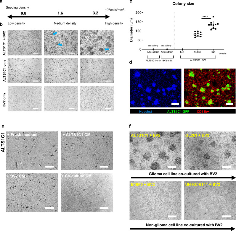
Methods: A BV2 microglial cells with ALTS1C1 astrocytoma cells in vitro co-culture system was used to mimic the microglia dominating tumor stroma in the tumor invasion microenvironment and explore the interaction between microglia and brain tumor cells.
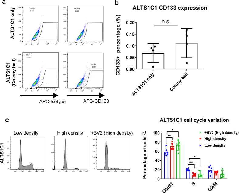
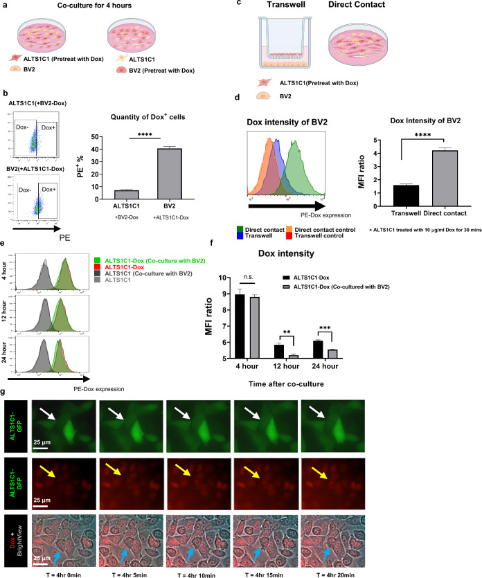
Results: Our result suggested that microglia could form colonies with glioma cells under high-density culturing conditions and protect glioma cells from apoptosis induced by chemotherapeutic drugs. Moreover, this study demonstrates that microglia could hijack drug substances from the glioma cells and reduce the drug intensity of ALTS1C1 via direct contact. Inhibition of gap junction protein prevented microglial-glioma colony formation and microglia-mediated chemoresistance.
Conclusions: This study provides novel insights into how glioma cells acquire chemoresistance via microglia-mediated drug substance transferring, providing a new option for treating chemo-resistant brain tumors.
Keywords: Chemoresistance; Glioma; Microglia; Tumor microenvironment.
© 2024. The Author(s).
Conflict of interest statement
The authors declare no competing interests.
Figures


Similar articles
-
Distinct roles of small extracellular vesicles from resident and infiltrating macrophages on glioma growth and mobility.J Cancer. 2025 Jan 1;16(3):969-981. doi: 10.7150/jca.103595. eCollection 2025. J Cancer. 2025. PMID: 39781357 Free PMC article.
-
Herpes simplex virus thymidine kinase gene therapy in experimental rat BT4C glioma model: effect of the percentage of thymidine kinase-positive glioma cells on treatment effect, survival time, and tissue reactions.Cancer Gene Ther. 2000 Mar;7(3):413-21. doi: 10.1038/sj.cgt.7700132. Cancer Gene Ther. 2000. PMID: 10766347
-
Tumor-secreted SDF-1 promotes glioma invasiveness and TAM tropism toward hypoxia in a murine astrocytoma model.Lab Invest. 2012 Jan;92(1):151-62. doi: 10.1038/labinvest.2011.128. Epub 2011 Sep 5. Lab Invest. 2012. PMID: 21894147
-
Tumor-Associated Macrophages in Gliomas-Basic Insights and Treatment Opportunities.Cancers (Basel). 2022 Mar 4;14(5):1319. doi: 10.3390/cancers14051319. Cancers (Basel). 2022. PMID: 35267626 Free PMC article. Review.
-
Microglia/Brain Macrophages as Central Drivers of Brain Tumor Pathobiology.Neuron. 2019 Nov 6;104(3):442-449. doi: 10.1016/j.neuron.2019.08.028. Neuron. 2019. PMID: 31697921 Free PMC article. Review.
Cited by
-
Distinct roles of small extracellular vesicles from resident and infiltrating macrophages on glioma growth and mobility.J Cancer. 2025 Jan 1;16(3):969-981. doi: 10.7150/jca.103595. eCollection 2025. J Cancer. 2025. PMID: 39781357 Free PMC article.
References
-
- Brandes AA, Basso U, Reni M, Vastola F, Tosoni A, Cavallo G, et al. First-line chemotherapy with cisplatin plus fractionated temozolomide in recurrent glioblastoma multiforme: a phase II study of the Gruppo Italiano Cooperativo di Neuro-Oncologia. J Clin Oncol. 2004;22(9):1598–1604. doi: 10.1200/jco.2004.11.019. - DOI - PubMed
Grants and funding
LinkOut - more resources
Full Text Sources
Miscellaneous
