Review
. 2024 Jan 18;25(1):37.
doi: 10.1186/s12931-023-02615-y.

Time-Controlled Adaptive Ventilation (TCAV): a personalized strategy for lung protection

Affiliations
Review

Time-Controlled Adaptive Ventilation (TCAV): a personalized strategy for lung protection

Hassan Al-Khalisy et al. Respir Res. .

Abstract

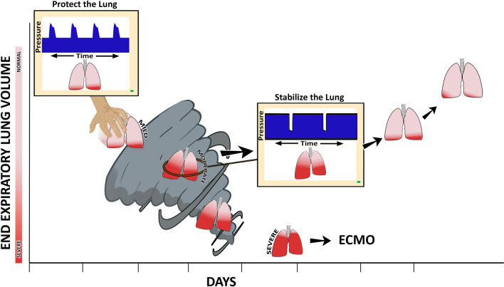
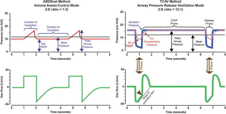
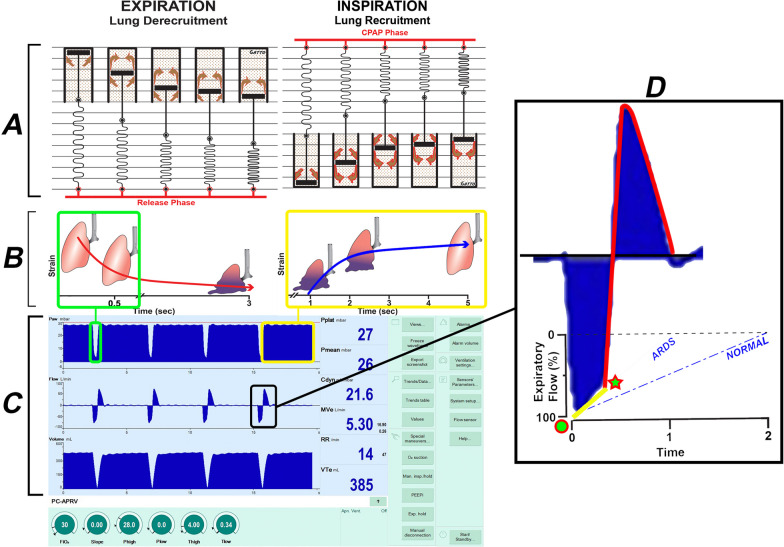
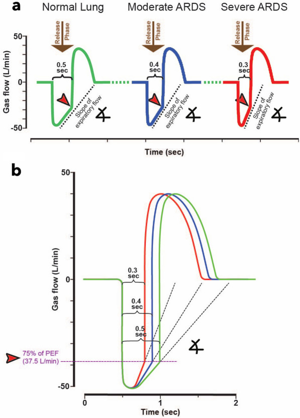
Acute respiratory distress syndrome (ARDS) alters the dynamics of lung inflation during mechanical ventilation. Repetitive alveolar collapse and expansion (RACE) predisposes the lung to ventilator-induced lung injury (VILI). Two broad approaches are currently used to minimize VILI: (1) low tidal volume (LVT) with low-moderate positive end-expiratory pressure (PEEP); and (2) open lung approach (OLA). The LVT approach attempts to protect already open lung tissue from overdistension, while simultaneously resting collapsed tissue by excluding it from the cycle of mechanical ventilation. By contrast, the OLA attempts to reinflate potentially recruitable lung, usually over a period of seconds to minutes using higher PEEP used to prevent progressive loss of end-expiratory lung volume (EELV) and RACE. However, even with these protective strategies, clinical studies have shown that ARDS-related mortality remains unacceptably high with a scarcity of effective interventions over the last two decades. One of the main limitations these varied interventions demonstrate to benefit is the observed clinical and pathologic heterogeneity in ARDS. We have developed an alternative ventilation strategy known as the Time Controlled Adaptive Ventilation (TCAV) method of applying the Airway Pressure Release Ventilation (APRV) mode, which takes advantage of the heterogeneous time- and pressure-dependent collapse and reopening of lung units. The TCAV method is a closed-loop system where the expiratory duration personalizes VT and EELV. Personalization of TCAV is informed and tuned with changes in respiratory system compliance (CRS) measured by the slope of the expiratory flow curve during passive exhalation. Two potentially beneficial features of TCAV are: (i) the expiratory duration is personalized to a given patient's lung physiology, which promotes alveolar stabilization by halting the progressive collapse of alveoli, thereby minimizing the time for the reopened lung to collapse again in the next expiration, and (ii) an extended inspiratory phase at a fixed inflation pressure after alveolar stabilization gradually reopens a small amount of tissue with each breath. Subsequently, densely collapsed regions are slowly ratcheted open over a period of hours, or even days. Thus, TCAV has the potential to minimize VILI, reducing ARDS-related morbidity and mortality.

Keywords: APRV; ARDS; ARMA; Acute respiratory distress syndrome; Alveolar opening and collapse time constants; Driving pressure; Dynamic alveolar mechanics; Open lung approach; Regional alveolar instability; Stress-multipliers; TCAV; Tidal volume; VILI; Ventilator-induced lung injury; Viscoelastic.


Conflict of interest statement

MKS has received a Research Grant and GFN an Unrestricted Educational Grant from Dräger Medical Systems, Inc. GFN, MKS have presented and received honoraria and/or travel reimbursement at event(s) sponsored by Dräger Medical Systems, Inc., outside of the published work. GFN, MKS, have lectured for Intensive Care On-line Network, Inc. (ICON). NMH is the founder of ICON, of which PLA is an employee. NMH holds patents on a method of initiating, managing and/or weaning airway pressure release ventilation, as well as controlling a ventilator in accordance with the same. DWK and JH are co-founders and shareholders of OscillaVent, Inc., and are co-inventors on a patent involving multi-frequency oscillatory ventilation. DWK and JH also receive research support from ZOLL Medical Corporation, and DWK is a consultant for Lungpacer Medical, Inc. JHTB is a consultant to and shareholder of OscillaVent, Inc., and has two patents pending in the field of MV. The authors maintain that industry had no role in the design and conduct of the study; the collection, management, analysis, or interpretation of the data; nor the preparation, review, or approval of the manuscript.

Figures

Fig. 1
Fig. 1
An ever-shrinking, baby lung, known as a VILI Vortex has been used to describe the evolution of ventilator-induced lung injury (VILI) [28]. Upper left: The ‘patient’ with mostly open lung tissue (pink) and a lesser amount of collapsed tissue (red) defined as Mild ARDS is placed on ARDSnet LVT ventilation. The LVT and low airway pressures strategy is designed to ‘rest’ the ‘baby lung’, however, this ventilation method allows the acutely injured tissue to continually collapse pushing it into the VILI Vortex. Lung pathogenesis moves from Mild to Moderate ARDS as normal tissue progressively shrinks (pink → red), Disease progression into Severe ARDS is inevitable if unchecked at which point rescue methods such as extracorporeal membrane oxygenation (ECMO) may be necessary. ARDS causes the lung to become time and pressure dependent. This means that it will take more time for the alveoli to open and less time for them to collapse at any given airway pressure. Thus, the alveolar opening can be accelerated by an extended inspiratory time, and alveolar collapse minimized by a short expiratory time. The brief time at inspiration is not adequate to open collapsed alveoli while the extended time at expiration will not prevent alveolar collapse using the ARDSnet approach (Upper left Protect the Lung, Ventilator Monitor blue Pressure/Time curve). The open lung approach to rapidly reopen the lung (seconds or minutes) using recruitment maneuvers and higher PEEP has not been successful at reducing ARDS-related mortality. Using inspiratory and expiratory duration in addition to pressure to open and stabilize alveoli has been shown very effective and lung protective by our group and others [, , –91]. An extended inspiratory time will progressively recruit alveoli and a brief expiratory time will prevent re-collapse. A correctly set time-controlled ventilator method will stabilize alveoli (Center, Stabilize the Lung, Ventilator Monitor blue Pressure/Time curve) using a short expiratory time pulling the lung from the Vortex. Once progressive lung collapse is halted, collapsed tissue can be reopened slowly (Red lung tissue turning pink) over hours or days depending on the level of lung pathophysiology [, , –91]. This figure depicts the ability of TCAV to be used after ARDS has developed or as a rescue mode but if applied early during mild ARDS movement of the lung into the Vortex could be prevented. Reproduced from Reference [29], under terms of the Creative Commons Attribution 4.0 International License
Fig. 2
Fig. 2
A Pressure/Time and Flow/Time curves = generated by the ARDSnet method to set and adjust the Volume Assist-Control mode. Key features include an inspiratory: expiratory ratio of 1:3 where the peak/plateau inspiratory pressure is brief. A set positive end-expiratory pressure (Set-PEEP) and FiO2 are adjusted using oxygenation as the trigger for change [20]. B Pressure/Time and Flow/Time curves for the Time Controlled Adaptive Ventilation (TCAV) method to set and adjust the Airway Pressure Release Ventilation (APRV) mode. Key features include an inspiratory: expiratory ratio of ~ 12:1, where. the continuous positive airway pressure (CPAP) Phase is ~ 90% of each breath. A tidal volume (VT), which is measured as the volume of gas released during the Release Phase (brown arrow), is not set but is influenced by changes in (i) respiratory system compliance (CRS), (ii) the CPAP Phase pressure, and (iii) the duration of the Release Phase. The Release Phase is determined by the Slope of the Expiratory Flow Curve (red arrowhead), which is a breath-to-breath measure of CRS. The lower the CRS, the faster the lung recoil, the steeper the slope, and the shorter the Release Phase, further reducing VT. Thus, the VT will be low in a non-compliant, injured lung and will increase in size only when the lung recruits and CRS increases. Since a change in CRS directs the Release Phase duration, which in turn adjusts the VT and the time-controlled PEEP (TC-PEEP) the TCAV method is both personalized and adaptive as the patient’s lung gets better or worse [104]. Reproduced from Reference [104], under terms of the Creative Commons Attribution 4.0 International License
Fig. 3
Fig. 3
The ability of the TCAV method to stabilize and then open the lung is based on opening and collapse time constants, which are greatly altered if pulmonary surfactant is deactivated, and the viscoelastic system by which the lung changes volume. A Viscoelastic lung volume change. EXPIRATION (Lung Derecruitment): Viscoelastic volume change can be modeled using the spring connected in parallel with a dashpot. When airway pressure begins to fall during the Release Phase (Fig. 2B), there is a very short time delay before alveolar collapse begins, followed by rapid collapse (spring), and then a gradual, continual collapse over time (dash moving slowing through the pot). INSPIRATION (Lung Recruitment): When airway pressure is reapplied during the CPAP Phase (Fig. 2B, CPAP Phase), the reverse sequence of events occurs during lung opening: slight delay → rapid recruitment → gradual progressive recruitment. B Diagram of viscoelastic lung opening and collapse over time. If the expiratory time is very brief (≤ 0.5 s), lung tissue will not have time to collapse (green box). Lung tissue will continue to recruit without a change in airway pressure for as long as the CPAP Phase is applied. (yellow boxes). C A ventilator monitor showing typical TCAV method Pressure/ Flow/ Volume/ Time curves. Using the TCAV method the extended CPAP Phase continually 'nudges' the lung open over time (yellow boxes) (Additional file 3, Additional file 4) and establishes durable lung recruitment by not giving the lung sufficient time to collapse during the brief Release Phase (green boxes). D A blow-up of the expiratory and inspiratory flow curves is seen on the ventilator monitor (black box). The animal being ventilated had ARDS so the slope of the expiratory flow curve (SlopeEF) was very steep (ARDS, yellow line) as compared to the SlopeEF in a healthy lung (NORMAL, blue dashed line). At the termination of the brief Release Phase (green star) the lung is rapidly reinflated to the set CPAP Pressure. Panel A Adapted from Reference [29], under terms of the Creative Commons Attribution 4.0 International License
Fig. 4
Fig. 4
Expiratory Gas Flow/Time curve using the TCAV method to set TLow, the duration of the Release Phase (Fig. 2B, Release Phase). As lung injury increases from Normal Lung to Moderate and Severe ARDS, the respiratory system compliance (CRS) decreases, increasing the collapse recoil of the lung. The increased lung recoil causes faster gas flow during expiration resulting in a steeper slope of the expiratory flow curve (SlopeEF). a Using the SlopeEF to set the Release Phase duration (Fig. 2B, Release Phase), the Normal Lung has a release time of 0.5 s, Moderate ARDS 0.4 s, and Severe ARDS 0.3 s. Expiratory flow is terminated (red arrowhead) by the clinician by adjusting the TLow, following which the lung is rapidly reinflated to the CPAP Phase (Fig. 2B, CPAP Phase). Thus, using the TCAV method personalizes and adapts the Release Phase (TLow) according to the patient’s lung physiology. b Calculation of the termination point on the expiratory flow curve using the TCAV method. Termination of expiratory flow (TEF) is calculated as 75% of the peak expiratory flow (PEF) (PEF 50L/min × 0.75 = TEF 37.5L/min). Adapted from Reference [104], under terms of the Creative Commons Attribution 4.0 International License
Fig. 5
Fig. 5
Closed-loop systems for both the VILI Vortex and TCAV personalized lung protection (Fig. 1). VILI Vortex—injury collapses lung tissue and reduces respiratory system compliance (CRS) → redistributing a fixed tidal volume into a heterogeneously damaged lung → leads to maldistribution of gas within the lung damaging alveoli by both atelectrauma and volutrauma → causing progressive lung collapse (VILI Vortex) → further reducing CRS. TCAV—injury collapses lung tissue and reduces CRS → changes in CRS are manifest as a change in the slope of the expiratory flow curve (SlopeEF) → the SlopeEF is used to set the duration of the Release Phase and is thus directed by changes in the patient’s CRS (Fig. 2B, Release Phase) → directed by changes in the patient’s CRS the Release Phase is set sufficiently short to prevent alveolar collapse resulting in a gradual lung recruitment → lung recruitment increases CRS. The slope of the expiratory flow curve (SlopeEF) can be used as a dynamic feedback signal to adaptively change the expiratory duration necessary to maintain lung stability. Changes in the SlopeEF will identify if CRS is low or high and used to personalize and adaptively adjust the Expiratory Duration (TLow) necessary to maintain an open and stable lung, regardless of lung injury severity. The left side of the figure does not have this feedback mechanism which may lead to further alveolar collapse. On the right side of the figure the change in SlopeEF allows a stop and brake and adjustments made to T-low to halt the VILI Vortex

Similar articles

Cited by

References

    1. Matthay MA, Zemans RL, Zimmerman GA, Arabi YM, Beitler JR, Mercat A, Herridge M, Randolph AG, Calfee CS. Acute respiratory distress syndrome. Nat Rev Dis Primers. 2019;5:18. doi: 10.1038/s41572-019-0069-0. - DOI - PMC - PubMed
    1. Slutsky AS, Ranieri VM. Ventilator-induced lung injury. N Engl J Med. 2013;369:2126–2136. doi: 10.1056/NEJMra1208707. - DOI - PubMed
    1. Caser EB, Zandonade E, Pereira E, Gama AM, Barbas CS. Impact of distinct definitions of acute lung injury on its incidence and outcomes in Brazilian ICUs: prospective evaluation of 7,133 patients*. Crit Care Med. 2014;42:574–582. doi: 10.1097/01.ccm.0000435676.68435.56. - DOI - PubMed
    1. Laffey JG, Bellani G, Pham T, Fan E, Madotto F, Bajwa EK, Brochard L, Clarkson K, Esteban A, Gattinoni L, et al. Potentially modifiable factors contributing to outcome from acute respiratory distress syndrome: the LUNG SAFE study. Intensive Care Med. 2016;42:1865–1876. doi: 10.1007/s00134-016-4571-5. - DOI - PubMed
    1. Fan E, Brodie D, Slutsky AS. Acute respiratory distress syndrome: advances in diagnosis and treatment. JAMA. 2018;319:698–710. doi: 10.1001/jama.2017.21907. - DOI - PubMed

MeSH terms

LinkOut - more resources