

Age-dependent change and intraskeletal variability in secondary osteons of elderly Australians
- PMID: 38238907
- PMCID: PMC11095313
- DOI: 10.1111/joa.14010
Age-dependent change and intraskeletal variability in secondary osteons of elderly Australians
Abstract
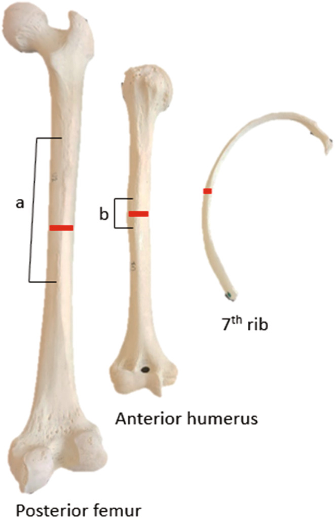
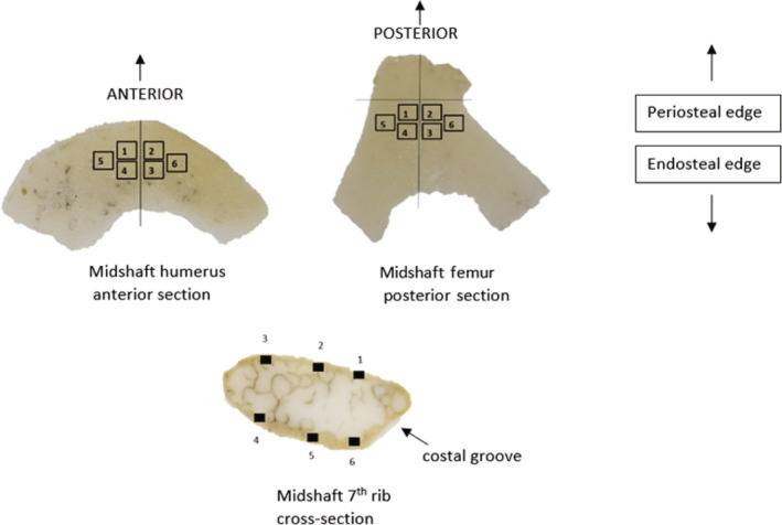
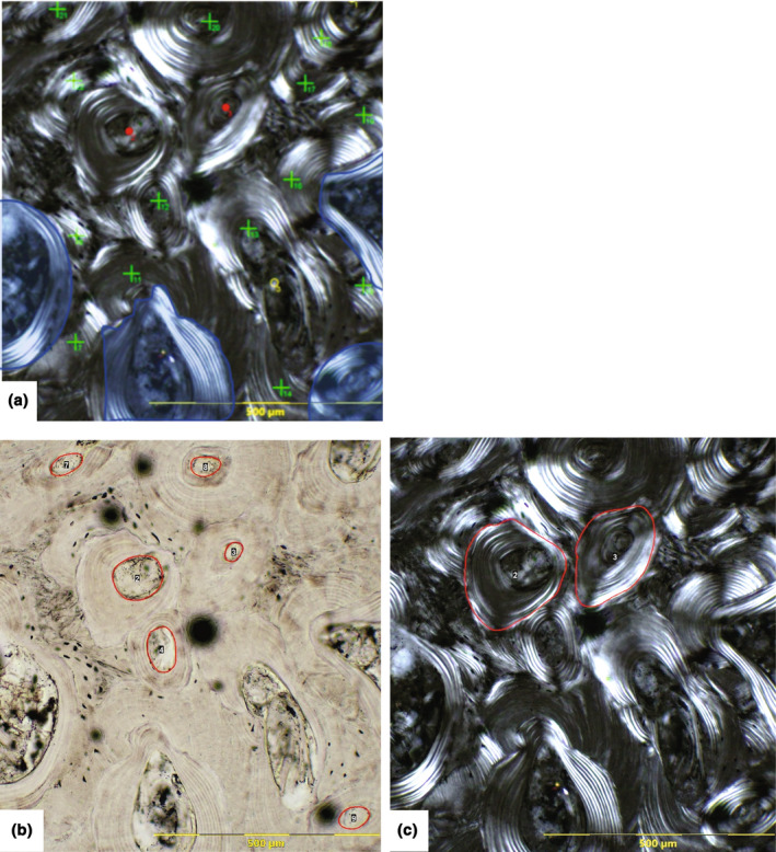
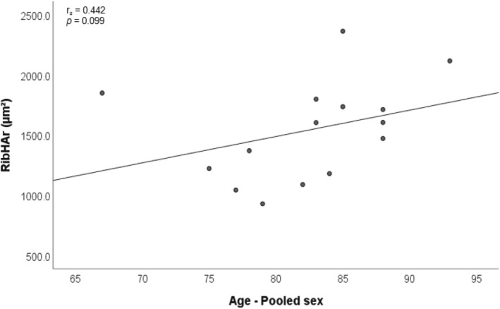
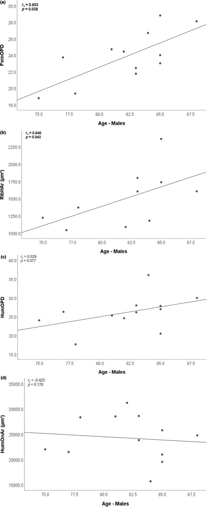
There is a need to fully understand intra-skeletal variability within different populations to develop and improve age-at-death estimation methods. This study evaluates age-related histomorphometric changes in three different bones intra-individually in a modern Australian sample. Four female and 13 male elderly Australian adult donors (67-93 years) were examined for osteon population density (OPD), osteon area (On.Ar), and Haversian canal area (H.Ar) of secondary osteons to compare between femora, ribs, and humeri and assess against age. In the pooled sex sample, no statistically significant correlations were observed between age and each histological variable. In the males, OPD of the femur increased significantly with age, as did porosity in the rib. In the male humeri, OPD increased moderately with age, while H.Ar was decreased moderately with age. Intra-bone comparisons showed that males had significantly higher osteon counts in their ribs compared to their femora, while their ribs showed statistically significantly less porosity than their humeri. When bone size was accounted for, by adjusting the femur and humerus histology data by robusticity indices, histology values were found to be similar between bones within the same individual. This is despite the upper and lower limbs receiving different ranges and types of biomechanical load. Our findings demonstrate that bone size influences histomorphometry, and this could confound age-at-death estimations that have not been adjusted for robusticity. Future studies would benefit from examining bone histomorphometry within a larger sample size and incorporating bone robusticity measures into histology analyses.
Keywords: age‐related histomorphometry; intra‐skeletal variation; osteon; remodelling; robusticity index.
© 2024 The Authors. Journal of Anatomy published by John Wiley & Sons Ltd on behalf of Anatomical Society.
Conflict of interest statement
The authors have no conflicts of interest.
Figures
Similar articles
-
Relation between cross-sectional bone geometry and double zonal osteon frequency and morphology.Am J Phys Anthropol. 2020 Apr;171(4):598-612. doi: 10.1002/ajpa.23954. Epub 2019 Nov 1. Am J Phys Anthropol. 2020. PMID: 31675105
-
Secondary osteon and Haversian canal dimensions as behavioral indicators.Am J Phys Anthropol. 2006 Dec;131(4):460-8. doi: 10.1002/ajpa.20454. Am J Phys Anthropol. 2006. PMID: 16685724
-
Variability in osteon size in recent human populations.Am J Phys Anthropol. 1998 Jun;106(2):219-27. doi: 10.1002/(SICI)1096-8644(199806)106:2<219::AID-AJPA8>3.0.CO;2-K. Am J Phys Anthropol. 1998. PMID: 9637185
-
Age estimation in the combined long bones and ribs by histomorphometry: Past, present, and future.Med Sci Law. 2024 Jan;64(1):52-71. doi: 10.1177/00258024231208280. Epub 2023 Oct 25. Med Sci Law. 2024. PMID: 37876174 Review.
-
Histomorphometry specific to anthropological studies concerning the human condition.J Anthropol Sci. 2024 Dec 26;102:69-85. doi: 10.4436/JASS.10203. J Anthropol Sci. 2024. PMID: 39003728 Review.
References
-
- Abdullah, H. , Jamil, M.M.A. , Ambar, R. & Nor, F.M. (2018) Bone histology: a key for human sex determination after death. Journal of Physics: Conference Series, 1019, 012010.
-
- Ahlqvist, J. & Damsten, O. (1969) A modification of Kerley's method for the microscopic determination of age in human bone. Journal of Forensic Science, 14, 205–212. - PubMed
-
- An, Y.H. , Moreira, P.L. , Kang, Q.K. & Gruber, H.E. (2003) Principles of embedding and common protocols. In: An, Y. & Martin, K.L. (Eds.) Handbook of histology methods for bone and cartilage. New York: Springer.
-
- Andreasen, C.M. , Bakalova, L.P. , Brüel, A. , Hauge, E.M. , Kiil, B.J. , Delaisse, J.M. et al. (2020) The generation of enlarged eroded pores upon existing intracortical canals is a major contributor to endocortical trabecularization. Bone, 130, 115–127. - PubMed
-
- Andronowski, J.M. & Crowder, C. (2019) Bone area histomorphometry. Journal of Forensic Sciences, 64, 486–493. - PubMed
Publication types
MeSH terms
LinkOut - more resources
Full Text Sources
Research Materials
