Review
. 2024 Feb 27;24(5):996-1029.
doi: 10.1039/d3lc00860f.

Lipid vesicle-based molecular robots

Affiliations
Review

Lipid vesicle-based molecular robots

Zugui Peng et al. Lab Chip. .

Abstract

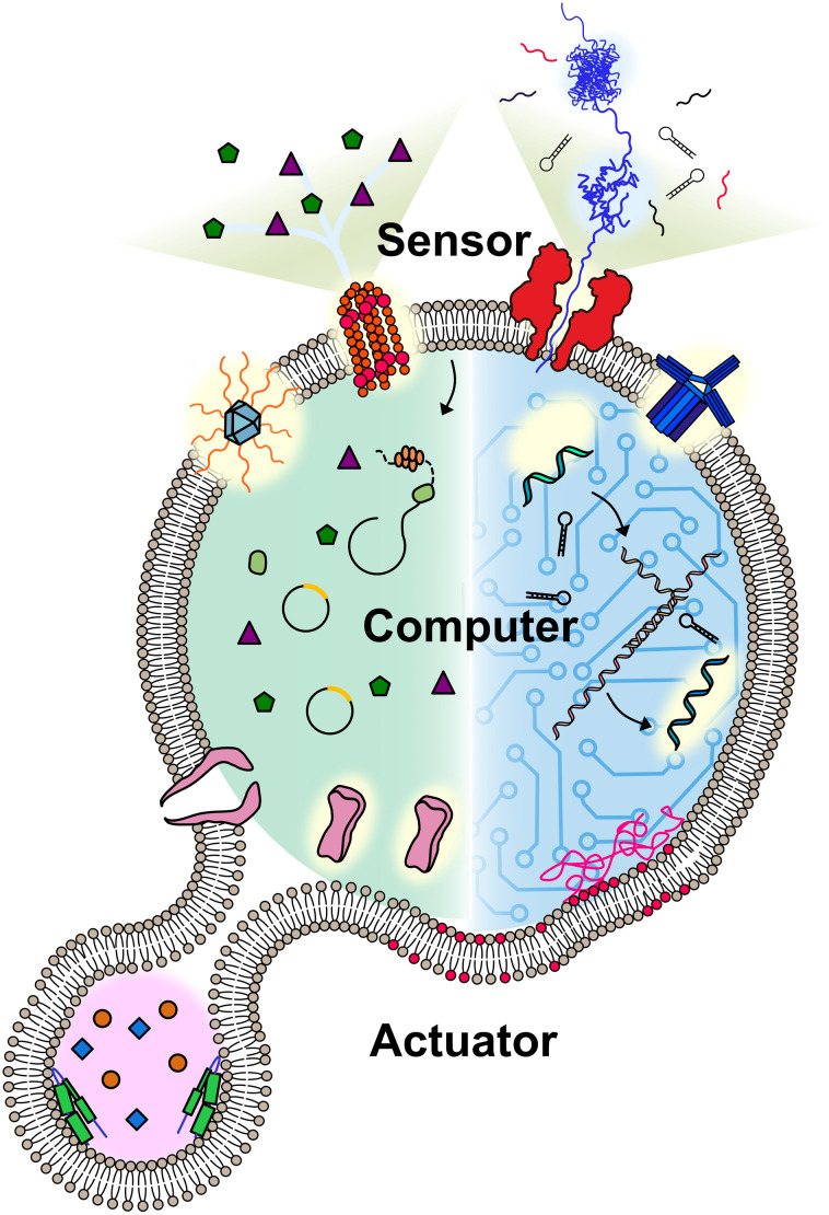
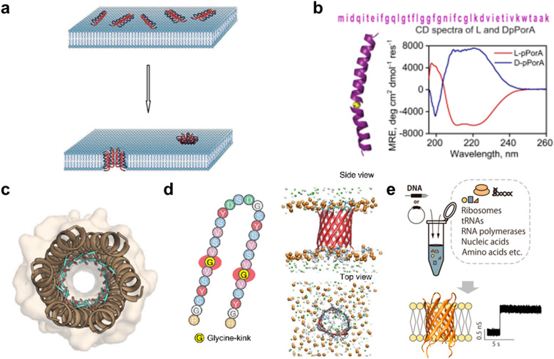
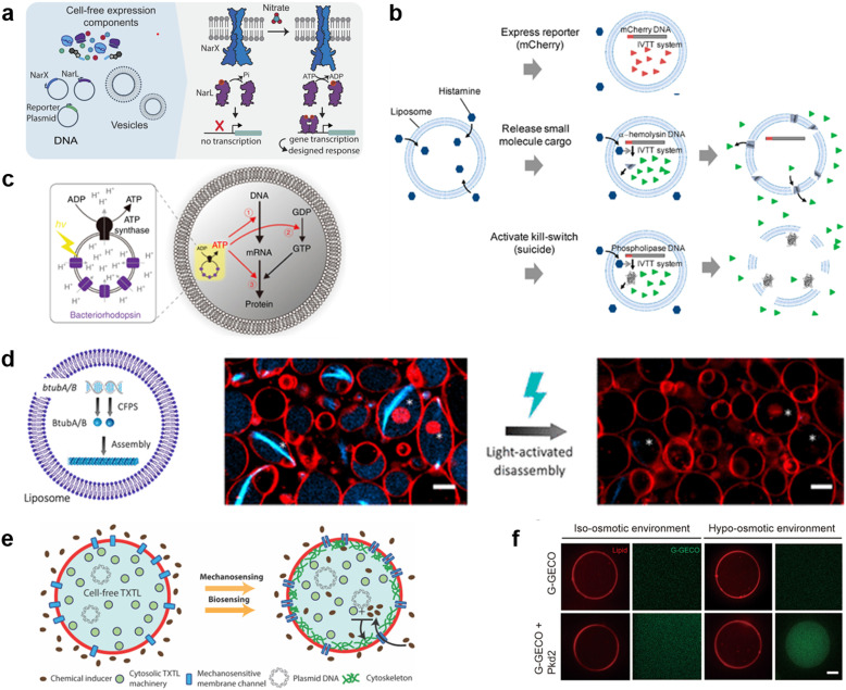
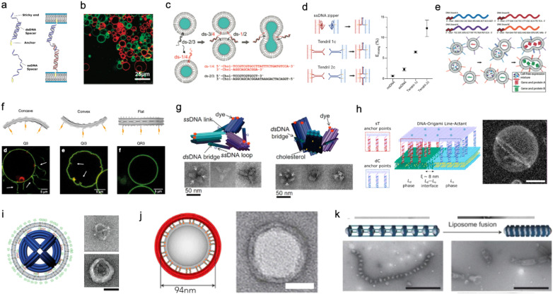
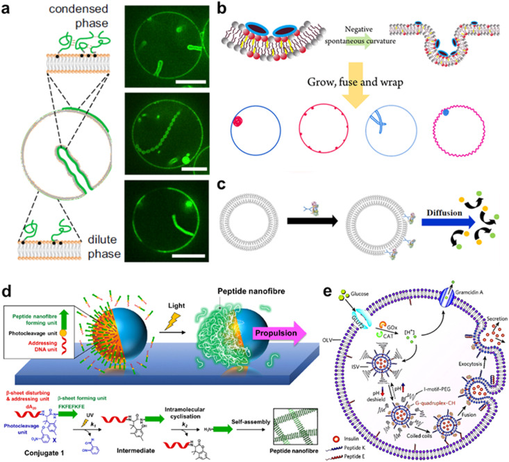
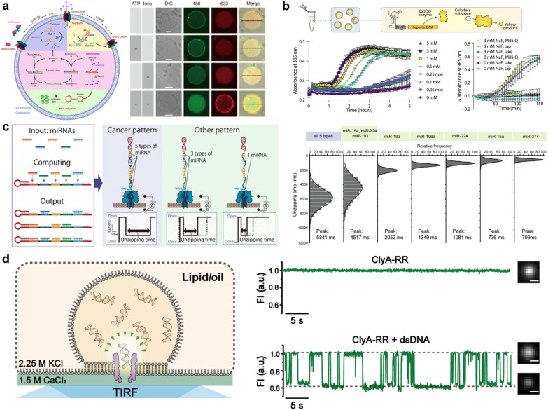
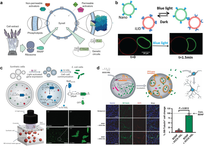
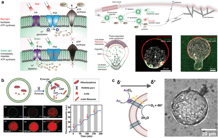
A molecular robot, which is a system comprised of one or more molecular machines and computers, can execute sophisticated tasks in many fields that span from nanomedicine to green nanotechnology. The core parts of molecular robots are fairly consistent from system to system and always include (i) a body to encapsulate molecular machines, (ii) sensors to capture signals, (iii) computers to make decisions, and (iv) actuators to perform tasks. This review aims to provide an overview of approaches and considerations to develop molecular robots. We first introduce the basic technologies required for constructing the core parts of molecular robots, describe the recent progress towards achieving higher functionality, and subsequently discuss the current challenges and outlook. We also highlight the applications of molecular robots in sensing biomarkers, signal communications with living cells, and conversion of energy. Although molecular robots are still in their infancy, they will unquestionably initiate massive change in biomedical and environmental technology in the not too distant future.


Conflict of interest statement

There are no conflicts to declare.

Figures

Fig. 1
Fig. 1. Conceptual illustration of a molecular robot including: a body to encapsulate molecular machinery, sensors to collect signals, computers to make decisions, and actuators to perform the tasks.
Fig. 2
Fig. 2. Schematic showing examples of different vesicle architectures that can be controllably manufactured using microfluidic techniques.
Fig. 3
Fig. 3. Examples of different techniques used to generate Giant Unilamellar Vesicles (GUVs). a, Emulsion phase transfer, where lipid stabilised w/o droplets are taken through a w/o column through a centrifugal force, resulting in the formation of a second monolayer, and hence the establishment of a lipid bilayer vesicle. b, Continuous droplet interface crossing encapsulation (cDICE) involving instrumentation which houses concentric layers of fluids with different densities, where aqueous droplets that emerge from a capillary are driven by centrifugal force through multiple lipid interfaces. c, Octanol-assisted liposome assembly (OLA) method of generating lipid vesicles on-chip. This method involves generating a lipid stabilised w/o/w double emulsion followed by extraction of the intermediate oil phase, leaving behind a lipid membrane. Figures reproduced with permission from: a, ref. , copyright 2023, MDPI; b, ref. , copyright 2016, the Royal Society of Chemistry; c, ref. , copyright 2016, Nature Publishing Group.
Fig. 4
Fig. 4. Protein nanopores for transmitting information. a, Illustration of the mechanism of nanopore DNA sequencing. b, Transport of T7 RNA polymerase by the SLO nanopore in a lipid vesicle system. c, Molecular surface illustration of the YaxAB nanopore. d, Protein detection with a monobody containing tFhuA nanopores. e, A comparison of the crystal structure (pink) and the designed structure (gray) of a de novo-designed transmembrane eight-strand β-barrel. Figures reproduced with permission from: a, ref. , copyright 2021, Elsevier; b, ref. , copyright 2021, Wiley-VCH; c, ref. , copyright 2023, American Chemical Society; d, ref. , copyright 2023, Nature Publishing Group; e, ref. , copyright 2021, AAAS.
Fig. 5
Fig. 5. Peptide nanopores for transmitting information. a, Formation of nanopores in lipid membranes by alamethicin. b, DpPorA peptide sequence and CD spectra for DpPorA (blue) and LpPorA (red). c, X-ray crystal structure of the nanopore formed by the de novo-designed α-helix peptide. d, SVG28 sequence and simulated pore structure. e, Utilizing CFPS to synthesize variants of SVG28. Figures reproduced with permission from: a, ref. , copyright 2014, Hindawi Publishing Corporation; b, ref. , copyright 2022, Nature Publishing Group; c, ref. , copyright 2022, American Chemical Society; d, ref. , copyright 2023, Nature Publishing Group; e, ref. , copyright 2023, American Chemical Society.
Fig. 6
Fig. 6. DNA nanopores and receptors for transmitting information. a, First reported DNA origami nanopore that transported dye molecules across the lipid membranes of GUVs. b, DNA origami nanopore with the largest inner diameter. c, DNA origami nanopore for size-selective transport, controlled by changing the number of lid-like DNA strands on top of the pore. d, DNA origami nanopore that reversibly opens and closes upon addition of key DNA strands. e, DNA nanopore composed of six DNA strands. f, Charge selective transport of dye molecules by a six-helix DNA nanopore. g, Receptor-mimicking DNA nanostructures capable of detecting ATP and lysozyme. h, Receptor-mimicking DNA nanostructures capable of detecting DNA strands. Figures reproduced with permission from: a, ref. , copyright 2016, Nature Publishing Group; b, ref. , copyright 2021, American Chemical Society; c, ref. , copyright 2021, Royal Chemical Society; d, ref. , copyright 2023, Nature Publishing Group; e, ref. , copyright 2023, Wiley-VCH; f, ref. , copyright 2021, American Chemical Society; g, ref. , copyright 2021, Royal Chemical Society; h, ref. , copyright 2021, American Chemical Society.
Fig. 7
Fig. 7. Synthetic channels for transmitting information. a, Chemical structures of aromatic molecules (left). Single crystal structure of aromatic molecules and its linearly self-assembling channel structure (central). Illustration of selective transmembrane transport of Li+ (right). b, Chemical structure of foldamer (left). Side and top view of the foldamer-based synthetic channel. Illustration of selective transmembrane transport of I (right). c, Schematic showing light-driven transport of metal ions and an enlarged view of the molecular motor. d, Chemical structure of amphiphilic cyclophanes with perfluorinated aromatic units (left) and its response to mechanical stress with selective transport of K+. e, Illustration of benzoimidazole (Bzim)-modified 5hmC-containing DNA2 translocation through a SWCNT. f, Illustration and single-crystal X-ray structure of the tetrahedral MOCs (left). Transport process of amino acids through the tetrahedral MOCs (right). Figures reproduced with permission from: a, ref. , copyright 2023, Wiley-VCH; b, ref. , copyright 2020, Wiley-VCH; c, ref. , copyright 2021, American Chemical Society; d, ref. , copyright 2022, American Chemical Society; e, ref. , copyright 2013, Nature Publishing Group; f, ref. , copyright 2021, American Chemical Society.
Fig. 8
Fig. 8. DNA computing for signal processing. a, Suggested mechanism of operation of the automaton by DNA-based state transition using the restriction nuclease FokI and ligase (right). b, “Seesawing”, “thresholding”, and “reporting” DNA reactions. c, The decision architecture (left) of the neural network. Fluorescence levels of α, β and γ, measured in approximately 25 000 droplets (right). d, Illustration of the AND gate operation using DNA droplets (left). CLSM images for droplet phase separation corresponding to four input patterns (right). Scale bars: 10 μm. Figures reproduced with permission from: a, ref. , copyright 2003, National Academy of Sciences; b, ref. , copyright 2012, Elsevier; c, ref. , copyright 2023, Royal Chemical Society; d, ref. , copyright 2022, Wiley-VCH.
Fig. 9
Fig. 9. Cell-free protein synthesis system (CFPS) DNA for signal processing. a, Using a two-component sensing system (NarX–NarL) to sense various substances. b, Controlling the fate of GUVs with a histamine-responsive riboswitch. c, Synthesis of a reporter protein using light irradiation of ATP synthase and bacteriorhodopsin-containing GUVs. d, Deforming GUVs from elliptical to spherical shape by degrading CFPS synthesized BtubA/B formed microtubes into monomers using light irradiation. Scale bars: 5 μm. e, Expression of a bacterial cytoskeletal protein with GUVs loaded with CFPS and the mechanosensitive protein MscL by exposing the GUVs to hypo-osmotic solution. f, Activating the CFPS-synthesized ion channel Pkd2 with osmotic stress on the membrane to enhance the influx of calcium ions. Scale bars: 10 μm. Figures reproduced with permission from: a, ref. , copyright 2023, National Academy of Sciences; b, ref. , copyright 2019, American Chemical Society; c, ref. , copyright 2020, Nature Publishing Group; d, ref. , copyright 2021, American Chemical Society; e, ref. , copyright 2019, American Chemical Society; f, ref. , copyright 2022, American Society for Cell Biology.
Fig. 10
Fig. 10. DNA nanostructures for actuating lipid membranes. a, Schematic of DNA linkers anchored to membranes and mediating adhesion between lipid vesicles. Linkers may feature a double-stranded DNA spacer (top) or be fully single-stranded (bottom). b, Confocal micrograph of GUVs adhering due the action of single-stranded DNA linkers (right), adapted from ref. . c, DNA “zipper” constructs mediating lipid vesicle fusion. d, Comparison of fusion efficiency (right) from various DNA zipper and “tendril” constructs (left). e, Cascades of lipid vesicle fusion reactions mediated by DNA zippers trigger CFPS. f, Curved DNA origami (top) influence the morphology of GUVs as determined with confocal microscopy (bottom). g, Clathrin-like DNA origami triskelia of controllable curvature (top) and their TEM images (bottom). h, DNA origami lineactants (left) accumulate at the interface between liquid ordered and liquid disordered domains in phase-separated GUVs (confocal projection, right) thanks to the phase-selectivity of double cholesterol (dC) and single tocopherol (sT) anchors. Scale bar: 10 μm. i, Virus-like DNA particle obtained by templating the formation of a lipid vesicle around a spherical DNA origami decorated with lipids (left) and TEM images of a bare (top right) and lipid-enveloped (bottom right) origami. Scale bar: 50 nm. j, Lipid vesicles of controlled size templated by lipid-modified DNA-origami rings. Images are TEM micrographs of the constructs shown on the immediate left. Scale bars: 50 nm. k, Liposomes captured by dynamic DNA origami arrays. Scale bars: 100 nm. Figures reproduced with permission from: a, ref. , copyright 2019, IOP Publishing; b, ref. , copyright 2007, American Chemical Society; c, ref. , copyright 2008, American Chemical Society; d, ref. , copyright 2022, Royal Chemical Society; e, ref. , copyright 2019, Wiley-VCH; f, ref. , copyright 2018, Nature Publishing Group; g, ref. , copyright 2019, American Chemical Society; h, ref. , copyright 2023, American Chemical Society; i, ref. , copyright 2014, American Chemical Society; j, ref. , copyright 2017, Elsevier; k, ref. , copyright 2020, Wiley-VCH.
Fig. 11
Fig. 11. Proteins and peptides for actuating lipid membranes. a, GUV deformation triggered by phase separation of FUS LC adhered on the lipid membrane. Scale bars: 10 μm. b, GUV deformation triggered by de novo peptides. c, Propulsion of ATPase-coated GUVs. d, Light-induced propulsion of a phase-separated GUVs driven by local peptide nanofibre growth (top) and the photocleavage reaction of a DNA-peptide conjugate (bottom). e, Illustration of the biochemical process inside OLVs. Vesicle fusion is triggered by the interaction between peptide K and peptide E. Figures reproduced with permission from: a, ref. , copyright 2021, National Academy of Sciences; b, ref. , copyright 2018, American Chemical Society; c, ref. , copyright 2019, American Chemical Society; d, ref. , copyright 2018, Nature Publishing Group; e, ref. , copyright 2021, Royal Chemical Society.
Fig. 12
Fig. 12. Molecular robots for detection of molecules. a, Detection of Sr2+ by GUVs with a DNA nanopore and circuit (left), as observed by the increase of 488 fluorescent signals from SG-I in the presence of ATP and Sr2+ (right). b, Illustration of the GUVs designed to detect sodium fluoride (top) and its absorbance over time in response to in vitro samples (bottom left) and real-world samples (bottom right). c, Using nanopores and DNA computing to detect five types of miRNAs (left) from histograms of the unzipping time of each miRNA pattern (right). d, Illustration of DiffusiOptoPhysiology (DOP) method (left). Comparison of the signal of Fluo-8 with/without dsDNA in the solution (right). Figures reproduced with permission from: a, ref. , copyright 2020, Nature Publishing Group; b, ref. , copyright 2023, AAAS; c, ref. , copyright 2022, American Chemical Society; d, ref. , copyright 2019, AAAS.
Fig. 13
Fig. 13. Molecular robots for communication with living cells. a, Modular design of genetic circuit interactions within and between GUVs. b, Controlling the distance between GUVs with light-oxygen-voltage 2 protein. Scale bar = 25 μm. c, Schematic representation of communication between GUVs with bacteria (top). GFP is expressed only when the GUVs are exposed to UV light (bottom). Scale bar = 200 μm. Scale bar (bottom right) = 20 μm. d, Schematic representation of communication between GUVs with neural stem cells (top). GUVs function to enhance stem cell differentiation, as revealed by the increase in the percentage of bIII-tubulin-overexpressing neurons (bottom). Scale bar = 50 μm. Figures reproduced with permission from: a, ref. , copyright 2020, Royal Chemical Society; b, ref. , copyright 2019, Wiley-VCH; c, ref. , copyright 2023, Nature Publishing Group; d, ref. , copyright 2020, AAAS.
Fig. 14
Fig. 14. Molecular robots for conversion of energy. a, Facilitating or impeding the ATP synthesis with different light color exposure. (left) Optical stimulation couples ATP synthesis with ATP-dependent actin polymerization and morphological change of the GUVs (top right). Confirmation of the formed cytoskeletal proteins and their impact on membrane deformation with microscopy (bottom right). Scale bar = 20 μm. b, Mitochondrion-containing GUVs' response to pyruvate addition (top). Time-dependent process of cytoskeletal proteins polymerization triggered by the addition of pyruvate (bottom). Scale bar = 10 μm. c, Schematic showing polarization of carbon nanotubes within lipid membranes, triggering the reduction of gold chloride to solid gold deposits (left). Observed deposition of gold on GUVs (right). Scale bar = 20 μm. a, ref. , copyright 2023, Frontiers Media and ref. , copyright 2020, Wiley-VCH; b, ref. , copyright 2022, Wiley-VCH; c, ref. , copyright 2022, Wiley-VCH.

Similar articles

Cited by

References

    1. Dietrich-Buchecker C. O. Sauvage J. P. Kintzinger J. P. Une nouvelle famille de molecules : les metallo-catenanes. Tetrahedron Lett. 1983;24:5095–5098. doi: 10.1016/S0040-4039(00)94050-4. - DOI
    1. Anelli P. L. Spencer N. Fraser Stoddart J. A Molecular Shuttle. J. Am. Chem. Soc. 1991;113:5131–5133. doi: 10.1021/ja00013a096. - DOI - PubMed
    1. Koumura N. Zijlstra R. W. J. van Delden R. A. Harada N. Feringa B. L. Light-driven monodirectionalmolecular rotor. Nature. 1999;401:152–154. doi: 10.1038/43646. - DOI - PubMed
    1. Kudernac T. et al., Electrically driven directional motion of a four-wheeled molecule on a metal surface. Nature. 2011;479:208–211. doi: 10.1038/nature10587. - DOI - PubMed
    1. Hagiya M. Konagaya A. Kobayashi S. Saito H. Murata S. Molecular robots with sensors and intelligence. Acc. Chem. Res. 2014;47:1681–1690. doi: 10.1021/ar400318d. - DOI - PubMed

Publication types