Review
. 2024 Jan;14(1):133-154.
doi: 10.1016/j.apsb.2023.08.033. Epub 2023 Sep 3.

Plant-derived nanovesicles as an emerging platform for cancer therapy

Affiliations
Review

Plant-derived nanovesicles as an emerging platform for cancer therapy

Hanzhe Liu et al. Acta Pharm Sin B. 2024 Jan.

Abstract

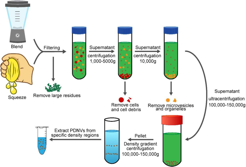
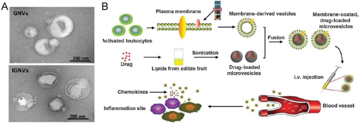
Plant-derived nanovesicles (PDNVs) derived from natural green products have emerged as an attractive nanoplatform in biomedical application. They are usually characterized by unique structural and biological functions, such as the bioactive lipids/proteins/nucleic acids as therapeutics and targeting groups, immune-modulation, and long-term circulation. With the rapid development of nanotechnology, materials, and synthetic chemistry, PDNVs can be engineered with multiple functions for efficient drug delivery and specific killing of diseased cells, which represent an innovative biomaterial with high biocompatibility for fighting against cancer. In this review, we provide an overview of the state-of-the-art studies concerning the development of PDNVs for cancer therapy. The original sources, methods for obtaining PDNVs, composition and structure are introduced systematically. With an emphasis on the featured application, the inherent anticancer properties of PDNVs as well as the strategies in constructing multifunctional PDNVs-based nanomaterials will be discussed in detail. Finally, some scientific issues and technical challenges of PDNVs as promising options in improving anticancer therapy will be discussed, which are expected to promote the further development of PDNVs in clinical translation.


Conflict of interest statement

The authors declare no conflicts of interest.

Figures

Image 1
Graphical abstract
Figure 1
Figure 1
Bioactive components contained in PNDVs and their biological properties to take an effect on the occurrence, development and metastasis of tumors by acting on tumor cells or cells in the tumor microenvironment (TME), or even by modulating the TME.
Figure 2
Figure 2
Technique flow for the isolation of plant-derived nanovesicles (PDNVs) by different times of differential centrifugation and ultracentrifugation.
Figure 3
Figure 3
(A) Schematic illustration of the isolation and therapeutic functions of tea flower-derived nanovesicles via inducing oxidative stress, mitochondrial damage, cell cycle arrest and apoptosis. Adapted with permission from Ref. 46. Copyright © 2022 Elsevier. (B) Illustration of the preventive and therapeutic effects of tea leaf-derived nanovesicles on IBD and CAC through the ability to target macrophages via the galactose on the surface, the modulation of microbiota, protection of epithelial barrier, antioxidation, and anti-inflammation. Adapted with permission from Ref. 47. Copyright © 2021 Elsevier.
Figure 4
Figure 4
(A) Schematic of EVs isolation using ELD. (B) Cell viability of AGS, BGC-823 and SGC-7901 cells treated with different concentrations of LDEVs. (C) Tumor growth curve of the control and LDEVs treatment groups. Adapted with permission from Ref. 55. Copyright 2020 BioMed Central.
Figure 5
Figure 5
(A) Schematic of the isolation, selective cytotoxicity and synergistic effect of plant sap-derived extracellular vesicles (PD-ENVs). Adapted with permission from Ref. 57. Copyright 2020 Multidisciplinary Digital Publishing Institute. (B) Schematic of the isolation, characterization and anti-metastatic effects of DM-derived nanovesicles (DMS-EVs) in a 3D microfluidic cancer metastasis model. Adapted with permission from Ref. 58. Copyright 2020 Multidisciplinary Digital Publishing Institute.
Figure 6
Figure 6
(A) Schematic diagram of the therapeutic mechanism of 5-Fu, bitter melon-derived nanovesicles (Group: BMEVs), and 5-FU-loaded bitter melon-derived nanovesicles (Group: BMNVs + 5-Fu). (B) Fluorescence images of the intracellular ROS levels after BMNVs treatment, scale bar = 200 μm. (C) Tumor growth curve of the control, 5-FU, BMNVs and BMNVs + 5-FU groups. (D) Representative images of tumors. Adapted with permission from Ref. 71. Copyright 2021 BioMed Central.
Figure 7
Figure 7
(A) Schematic diagram of nanovesicles isolation from cabbage and the functional analysis and drug delivery of Cabex and Rabex. (B) Fluorescence microscopy image of Cabex-delivered DNA oligonucleotides (red). Hoechst 33342 was used for nuclei staining (Blue). Scale bar indicates 50 μm. (C) Cell viability of SW480 cells after treatment with Cabex loaded with DOX. Adapted with permission. Adapted with permission from Ref. 73. Copyright © 2021 Elsevier. (D) Schematic of the preparation of HSP 70-loaded grapefruit-derived nanovesicles (GF-EVs). (E) Fluorescence micrograph of DLD1 cells co-cultured with GF-EVs loaded with HSP70-AF647. Adapted with permission from Ref. 75. Copyright 2021 Nature Publishing Group.
Figure 8
Figure 8
Several ways of engineering PDNVs.
Figure 9
Figure 9
(A) The morphology and size of sucrose-gradient band 2 was characterized by NTA and TEM. (B) Ex vivo fluorescence imaging and quantification of tumors from mice treated with ACNVs (I) and PEG-ACNVs (II). (C) Ex vivo fluorescence imaging and quantification of blood samples from mice treated with ACNVs (I) and PEG-ACNVs (II). (D) Representative images of tumors in mice after different treatments. Adapted with permission from Ref. 91. Copyright 2021 Dove Medical Press.
Figure 10
Figure 10
(A) Representative images of cells transfected with biotinylated eYFP using GNVs or Lipofectamine 2000. (B) Activity of luciferase expressed in T cells transfected with biotin-labeled anti-CD4 or CD8 antibodies loaded with GNVs encapsulated with psiCHECK2. (C) Representative images of each group of tumors after treatment (left) and mean intensity of fluorescence signals (right). (D) Changes of tumor volume in different groups after subcutaneous injection of tumor cells. Adapted with permission from Ref. 93. Copyright 2013 Nature Publishing Group.
Figure 11
Figure 11
(A) Scanning electron microscopy image of GNVs and IGNVs. Adapted with permission. (B) Schematic of the preparation of the IGNVs and GNVs for targeted delivery of drugs to the sites of inflammation. Adapted with permission from Ref. 94. Copyright © 2015 American Association for Cancer Research.
Figure 12
Figure 12
(A) Schematic of intravenous injection of GDNVs for targeted delivery of chemotherapeutic agents to tumors through blood vessels. Adapted with permission. Adapted with permission from Ref. 96. Copyright © 2016 Elsevier. (B) Process for isolating nanovesicles from ginger roots (GDENs). (C) Concept of ligand displaying by arrowtail and partially loading by arrowhead due to different orientation. (D) Fluorescence microscope image of Alexa647-labeled arrowtail and arrowhead RNA nanoparticle (red) incubated with KB cell (green). (E) Size of KB cell-derived xenograft tumors in nude mice treated intravenously every two days for two weeks. Adapted with permission from Ref. 97. Copyright 2018 Nature Publishing Group.

Cited by

References

    1. Printezi M.I., Kilgallen A.B., Bond M.J.G., Štibler U., Putker M., Teske A.J., et al. Toxicity and efficacy of chronomodulated chemotherapy: a systematic review. Lancet Oncol. 2022;23:e129–e143. - PubMed
    1. Qu Y., Chu B., Wei X., Chen Y., Yang Y., Hu D., et al. Cancer-cell-biomimetic nanoparticles for targeted therapy of multiple myeloma based on bone marrow homing. Adv Mater. 2022;34 - PubMed
    1. Gong L., Zhang Y., Zhao J., Zhang Y., Tu K., Jiao L., et al. All-in-one biomimetic nanoplatform based on hollow polydopamine nanoparticles for synergistically enhanced radiotherapy of colon cancer. Small. 2022;18 - PubMed
    1. Zhang S., Zhang Y., Feng Y., Wu J., Hu Y., Lin L., et al. Biomineralized two-enzyme nanoparticles regulated tumor glycometabolism inducing tumor cell pyroptosis and robust anti-tumor immunotherapy. Adv Mater. 2022;34 - PubMed
    1. Agarwalla P., Ogunnaike E.A., Ahn S., Ligler F.S., Dotti G., Brudno Y. Scaffold-mediated static transduction of T cells for CAR-T cell therapy. Adv Healthcare Mater. 2020;9 - PMC - PubMed

LinkOut - more resources