. 2024 Jan 4:17:1295805.
doi: 10.3389/fncel.2023.1295805. eCollection 2023.

Analysis of α-syn and parkin interaction in mediating neuronal death in Drosophila model of Parkinson's disease

Affiliations

Analysis of α-syn and parkin interaction in mediating neuronal death in Drosophila model of Parkinson's disease

Sonia Narwal et al. Front Cell Neurosci. .

Abstract

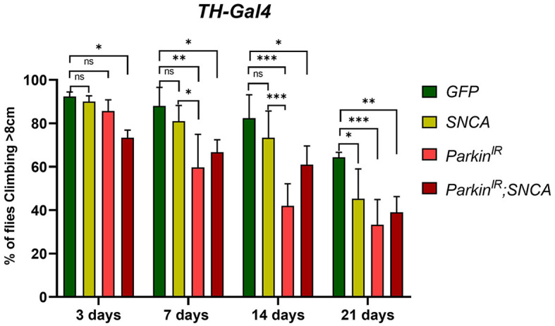
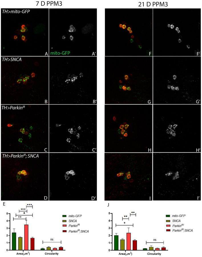
One of the hallmarks of Parkinson's Disease (PD) is aggregation of incorrectly folded α-synuclein (SNCA) protein resulting in selective death of dopaminergic neurons. Another form of PD is characterized by the loss-of-function of an E3-ubiquitin ligase, parkin. Mutations in SNCA and parkin result in impaired mitochondrial morphology, causing loss of dopaminergic neurons. Despite extensive research on the individual effects of SNCA and parkin, their interactions in dopaminergic neurons remain understudied. Here we employ Drosophila model to study the effect of collective overexpression of SNCA along with the downregulation of parkin in the dopaminergic neurons of the posterior brain. We found that overexpression of SNCA along with downregulation of parkin causes a reduction in the number of dopaminergic neuronal clusters in the posterior region of the adult brain, which is manifested as progressive locomotor dysfunction. Overexpression of SNCA and downregulation of parkin collectively results in altered mitochondrial morphology in a cluster-specific manner, only in a subset of dopaminergic neurons of the brain. Further, we found that SNCA overexpression causes transcriptional downregulation of parkin. However, this downregulation is not further enhanced upon collective SNCA overexpression and parkin downregulation. This suggests that the interactions of SNCA and parkin may not be additive. Our study thus provides insights into a potential link between α-synuclein and parkin interactions. These interactions result in altered mitochondrial morphology in a cluster-specific manner for dopaminergic neurons over a time, thus unraveling the molecular interactions involved in the etiology of Parkinson's Disease.

Keywords: Drosophila melanogaster; Parkinson's disease; dopaminergic neurons; mitochondrial morphology; parkin; tyrosine hydroxylase; α-synuclein.


Conflict of interest statement

The authors declare that the research was conducted in the absence of any commercial or financial relationships that could be construed as a potential conflict of interest.

Figures

Figure 1
Figure 1
SNCA overexpression and parkin downregulation (parkinIR) independently, and together (parkinIR; SNCA) in dopaminergic neurons (DA), exhibit locomotor dysfunctions: Climbing assay indicates loss of locomotor function with age in flies. A total of 30 (N = 30) flies were used per genotype and 10 flies (n = 10) were used for the climbing assay. Data is represented as mean with SEM. Statistical analysis was performed using Two-way ANOVA followed by Tukey's multiple comparison test. P value: *(0.033), **(0.002), ***(< 0.001), ns-not significant (0.12).
Figure 2
Figure 2
SNCA and parkinIR expression independently and together (parkinIR; SNCA) cause DA cluster specific neuronal loss (A) Schematic representation of DA neuronal clusters (PPL1, PPM1&2, PPM3, PPL2) in the posterior region of adult brain. (B) Representative confocal Maximum Intensity Projection (MIP) of WT adult brain stained with GFP (green) and Tyrosine Hydroxylase (TH) (Red) to reveal DA neurons in the posterior region. An adult brain of the desired genotype was dissected and stained for TH. (C–G) 7-day-old adult fly brains show TH stain. (C, C') Control flies, (D, D') SNCA overexpression, (E, E') ParkinIR expression, (F, F') ParkinIR; SNCA expression, show the TH-stain which is (G) quantified. (H-L) 21-day-old adult fly brains showing TH stain. (H, H') Control flies, (I, I') SNCA overexpression, (J, J') ParkinIR expression, (K, K') ParkinIR; SNCA expression shows the TH-stain and is (L) quantified. Scale bar 50μm. A total of four adult brains were used (n = 4) per genotype. Data is represented as mean with SEM. Statistical analysis was performed using Two-way ANOVA followed by Tukey's multiple comparison test. P value: *(0.033), **(0.002), ***(< 0.001), ns-not significant (0.12).
Figure 3
Figure 3
SNCA affects the expression of parkin at the transcriptional level but not the translational level. (A) 7-day old parkin mRNA level (B) 21-day-old parkin mRNA level. (C) Parkin immunoblot and (E) quantification of 7-day-old adult brains normalized to beta-Tubulin. (D) Parkin immunoblot and (F) quantification of 21-day old adult brains normalized to beta-Tubulin. Data is represented as mean with SEM. Statistical analysis was performed using One-way ANOVA followed by Tukey's multiple comparison test. P value: *(0.033), ** (0.002), ns-not significant (0.12).
Figure 4
Figure 4
SNCA overexpression results in swollen mitochondria, parkinIR expression has shown elongated whereas together (parkinIR; SNCA) shows fragmented mitochondria in PPL1 DA clusters. Adult brains of the desired genotype expressing the mitochondria-targeted green fluorescent protein (mitoGFP) in TH- TH-positive (red) cells. (A–J) 7-day & 21-day old adult fly brains showing mitoGFP in PPL1 cluster. Control brains showing mitoGFP at (A, A') 7-day and (F, F') 21-day. SNCA overexpressing flies show mitoGFP (B, B') in 7-day and in (G, G') 21-day. ParkinIR-expressing flies show mitoGFP in (C, C') 7-day and in (H, H') 21-day. ParkinIR; SNCA expressing flies show mitoGFP in (D, D') 7-day and further enhanced in (I, I') 21-day. (E) Quantification of mitochondria morphology (area and circularity) was done using ImageJ Mito-Morphology Macro. Scale bar 10μm. A total of four adult brains were used (n = 4) per genotype. Data is represented as mean with SEM. Statistical analysis was performed using Two-way ANOVA followed by Tukey's multiple comparison test. P value: *(0.033), ns-not significant (0.12).
Figure 5
Figure 5
SNCA overexpression and parkinIR; SNCA have shown fragmented mitochondria, whereas parkinIR expression has shown elongated mitochondria in PPM3 DA clusters. Adult brains of the desired genotype expressing the mitochondria-targeted green fluorescent protein (mitoGFP) in TH- TH-positive (red) cells. (A–J) 7-day & 21-day old adult fly brains showing mitoGFP in PPM3 cluster. Control brains showing mitoGFP at (A, A') 7-day and (F, F') 21-day. SNCA overexpressing flies show mitoGFP (B, B') in 7-day and in (G, G') 21-day. ParkinIR expressing flies show mitoGFP in (C, C') 7-day and in (H, H') 21-day. ParkinIR; SNCA expressing flies show mitoGFP in (D, D') 7-day and in (I, I') 21-day also. (E) Quantification of mitochondria morphology (area and circularity) was done using ImageJ Mito-Morphology Macro. Scale bar 10 μm. A total of four adult brains were used (n = 4) per genotype. Data is represented as mean with SEM. Statistical analysis was performed using Two-way ANOVA followed by Tukey's multiple comparison test. P value: *(0.033), **(0.002), ***(< 0.001), ns-not significant (0.12).
Figure 6
Figure 6
Schematic representation of the effect of α-syn and parkin on specific DA neuronal clusters in the adult fly brain.

References

    1. Agostini F., Bubacco L., Chakrabarti S., Bisaglia M. (2023). α-synuclein toxicity in drosophila melanogaster is enhanced by the presence of iron: implications for Parkinson's disease. Antioxidants 12, 261. 10.3390/antiox12020261 - DOI - PMC - PubMed
    1. Appel-Cresswell S., Vilarino-Guell C., Encarnacion M., Sherman H., Yu I., Shah B., et al. . (2013). Alpha-synuclein p.H50Q, a novel pathogenic mutation for Parkinson's disease. Mov. Disord. 28, 811–813. 10.1002/mds.25421 - DOI - PubMed
    1. Aryal B., Lee Y. (2019). Disease model organism for Parkinson disease: Drosophila melanogaster. BMB Rep. 52, 250–258. 10.5483/BMBRep.2019.52.4.204 - DOI - PMC - PubMed
    1. Barone M. C., Sykiotis G. P., Bohmann D. (2011). Genetic activation of Nrf2 signaling is sufficient to ameliorate neurodegenerative phenotypes in a Drosophila model of Parkinson's disease. DMM Dis. Model. Mech. 4, 701–707. 10.1242/dmm.007575 - DOI - PMC - PubMed
    1. Bier E. (2005). Drosophila, the golden bug, emerges as a tool for human genetics. Nat. Rev. Genet. 6, 9–23. 10.1038/nrg1503 - DOI - PubMed

LinkOut - more resources