. 2024 Jan 4:14:1294565.
doi: 10.3389/fimmu.2023.1294565. eCollection 2023.

MediMer: a versatile do-it-yourself peptide-receptive MHC class I multimer platform for tumor neoantigen-specific T cell detection

Affiliations

MediMer: a versatile do-it-yourself peptide-receptive MHC class I multimer platform for tumor neoantigen-specific T cell detection

Marten Meyer et al. Front Immunol. .

Abstract

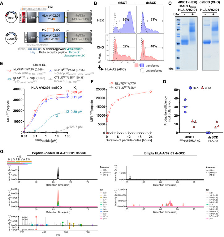
Peptide-loaded MHC class I (pMHC-I) multimers have revolutionized our capabilities to monitor disease-associated T cell responses with high sensitivity and specificity. To improve the discovery of T cell receptors (TCR) targeting neoantigens of individual tumor patients with recombinant MHC molecules, we developed a peptide-loadable MHC class I platform termed MediMer. MediMers are based on soluble disulfide-stabilized β2-microglobulin/heavy chain ectodomain single-chain dimers (dsSCD) that can be easily produced in large quantities in eukaryotic cells and tailored to individual patients' HLA allotypes with only little hands-on time. Upon transient expression in CHO-S cells together with ER-targeted BirA biotin ligase, biotinylated dsSCD are purified from the cell supernatant and are ready to use. We show that CHO-produced dsSCD are free of endogenous peptide ligands. Empty dsSCD from more than 30 different HLA-A,B,C allotypes, that were produced and validated so far, can be loaded with synthetic peptides matching the known binding criteria of the respective allotypes, and stored at low temperature without loss of binding activity. We demonstrate the usability of peptide-loaded dsSCD multimers for the detection of human antigen-specific T cells with comparable sensitivities as multimers generated with peptide-tethered β2m-HLA heavy chain single-chain trimers (SCT) and wild-type peptide-MHC-I complexes prior formed in small-scale refolding reactions. Using allotype-specific, fluorophore-labeled competitor peptides, we present a novel dsSCD-based peptide binding assay capable of interrogating large libraries of in silico predicted neoepitope peptides by flow cytometry in a high-throughput and rapid format. We discovered rare T cell populations with specificity for tumor neoepitopes and epitopes from shared tumor-associated antigens in peripheral blood of a melanoma patient including a so far unreported HLA-C*08:02-restricted NY-ESO-1-specific CD8+ T cell population. Two representative TCR of this T cell population, which could be of potential value for a broader spectrum of patients, were identified by dsSCD-guided single-cell sequencing and were validated by cognate pMHC-I multimer staining and functional responses to autologous peptide-pulsed antigen presenting cells. By deploying the technically accessible dsSCD MHC-I MediMer platform, we hope to significantly improve success rates for the discovery of personalized neoepitope-specific TCR in the future by being able to also cover rare HLA allotypes.

Keywords: T cell receptor discovery; T cells; neoepitope screening; peptide-MHC class I multimer; personalized medicine; tumor immunotherapy; tumor neoantigen.


Conflict of interest statement

The authors declare that the research was conducted in the absence of any commercial or financial relationships that could be construed as a potential conflict of interest. The author(s) declared that they were an editorial board member of Frontiers, at the time of submission. This had no impact on the peer review process and the final decision.

Figures

Figure 1
Figure 1
Successful CHO cell-based production of in vivo biotinylated, empty, peptide-receptive, disulfide-stabilized β2m-HLA-A*02:01 single-chain dimers (dsSCD). (A) Schematic representation of soluble disulfide-trapped peptide-β2m-HLA-A*02:01 single-chain trimer (dtSCT) (upper panel) and dsSCD (lower panel). dtSCT consist of a single polypeptide chain comprising a peptide ligand, human β2-microglobulin (β2m) and MHC-I ectodomain that are covalently linked via flexible glycine-serine-rich linkers (GSL). The SCT intramolecular disulfide trap is generated between a cysteine [C] in the first GSL and a C residue replacing a conserved MHC-I tyrosine [Y] residue at position 84. In dsSCD, β2m is fused via a GSL to the MHC-I ectodomain that harbors an additional alanine [A] A139C, Y84C disulfide bridge that stabilizes the β2m-MHC-I complex also in the absence of a peptide ligand. dtSCT and dsSCD are C-terminally fused to an octa-histidine tag (His8), a biotin acceptor peptide that is followed by two consecutive (2x) thrombin cleavage sites fused to a murine IgG2a Fc part. All constructs are designed for mammalian cell expression and were always transiently co-transfected with ER-retained IgκSP-BirAKDEL biotin ligase to allow for site-specific biotinylation during dsSCD/dtSCT expression. (B) Representative intracellular HLA-A*02:01 dtSCT and dsSCD expression analysis by flow cytometry 48 hours post plasmid (filled lines) and mock (dotted line) transfection of HEK293-F (HEK) and CHO-S (CHO) cells using anti-HLA-A2 antibody BB7.2. (C) SDS-PAGE analysis of affinity chromatography-purified monomeric dtSCT and dsSCD from HEK and CHO cell supernatants, respectively, that have been treated with thrombin to cleave the Fc portion. DtSCT and dsSCD biotinylation is confirmed by equimolar addition of streptavidin prior to SDS-PAGE analysis leading to a gel shift. (D) Production efficiencies of dtSCT and dsSCD expressed in HEK or CHO. Values were calculated using the yield in mg of the final protein yield divided by the culture supernatant volume in liters used for affinity chromatography. Dots represent three independent cell transfections for the conditions shown. (E, F) Flow cytometry-based FITC-conjugated peptide (FITCpeptide) binding assay for bead-immobilized dsSCD. FITC median fluorescence intensities (MFI) of FITCpeptide-loaded dsSCD immobilized on streptavidin-conjugated beads in triplicates are shown. HLA-A*02:01 dsSCD beads were pulsed with the indicated concentrations of FITCpeptides (E), or by a FITCpeptide pulse at 1 µM for the indicated time periods starting with 5, 10, 30 and 60 minutes and ending with 24 hours (h) (F). HLA-A*02:01 %Rank EL prediction values are shown for the indicated peptides with K substitution and their binding levels as putative strong binders (SB, %Rank EL <0.5), weak binders (WB, %Rank EL <2) and non-binders (nonB, %Rank >2) according to the NetMHCpan 4.1 algorithm. The FITC conjugation of the peptide is neglected in this assessment. (E) Non-linear regression (one-site specific binding) of the FITC MFI values from means of triplicates (± SD) against the peptide concentration and calculated KD values in µM are shown. (F) Non-linear regression of the FITC MFI from means of triplicates (± SD) against the incubation time is shown. (G) Data-independent acquisition mass spectrometry (DIA-MS) of NLVPMVATV loaded (left panels) and empty (right panels) HLA-A*02:01 dsSCD. Left panels show the manual detection of the NLVPMVATV peptide eluted from the externally peptide-loaded HLA-A*02:01 dsSCD. Bars below the NLVPMVATV sequence indicate detected fragment ions in peptide-loaded sample. Top panels show extracted ion chromatograms (XICs) of the precursor and its isotopes, middle panels shows XICs of fragment ions. The top half of the bottom panel shows the MS2 spectra extracted at the highest point of the MS2 XIC, while the bottom half shows the mirrored spectrum as in silico predicted by PROSIT. The spectral angle (SA) was calculated and is indicated. Right panels show the lack of matching precursor and fragment ions for the empty dsSCD molecule within a 25 min window centered around the retention time of NLVPMVATV peptide. n.d, not detected.
Figure 2
Figure 2
Validation of 32 produced HLA-A, -B and -C dsSCD by FITCpeptide binding assay. Individual production yields of dsSCD allotypes and used FITCpeptides are listed in Supplementary Table 1 . For dsSCD testing, a set of peptides were rationally designed to have at a selected non-anchor residue a KFITC substitution (K*) based on known ligands, the NetMHCpan 4.1 binding motif viewer and %Rank EL values for the indicated HLA allotype. FACS histograms are shown for bead-immobilized dsSCD pulsed for 18 h with 1 µM of a binding FITCpeptide (peptide sequence and plot shown in teal) and non/poor binding FITCpeptide (black line) and bead background MFI (gray filled) for the indicated HLA allotypes.
Figure 3
Figure 3
Comparable antigen-specific labeling of TCR-transgenic CD8+ Jurkat and healthy donor-derived viral epitope-specific CD8+ T cell populations with peptide-loaded dsSCD and dtSCT multimers. (A) Schematic representation of dsSCD for the usage as antigen-specific labeling reagent for defined CD8+ T cell populations. Following an overnight peptide pulse, the biotinylated dsSCD are multimerized by the addition of a fluorochrome-labeled streptavidin. (B, C) Cognate dtSCD and dsSCD multimer labeling of CD8+ Jurkat 76 (J76CD8+) stably expressing various published TCR as recombinant chimeric TCR containing murine Cβ and Cα domains. (B) J76CD8+ cells stably expressing RA14 TCR recognizing HLA-A*02:01/HCMVpp65495-503 or DMF5 TCR recognizing HLA-A*02:01/MART-126-35 were labeled with HLA-A*02:01 dsSCD previously loaded at varying concentrations with peptide HCMVpp65495-503 (red symbols) or MART-126-35 (blue symbols), respectively, for 18 h followed by multimerization. The upper panel shows representative histograms of RA14 and DMF5 J76CD8+ labeled with dsSCD multimers loaded with 25 µM peptide (filled line, MFI values in plain font) or dtSCT multimers (dotted line, MFI values in italics). PE fluorescence-minus-one (FMO) controls of J76CD8+ are shown in filled gray. The lower panel shows RA14 (circles) and DMF5 (triangles) J76CD8+ staining efficiencies at varying peptide loading concentrations used for dsSCD loading in triplicates and fitting using non-linear regression. For control, RA14 and DMF5 J76CD8+ cells were stained with dsSCD loaded with the non-cognate peptide. (C) Antigen-specific staining of published neoepitope or tumor-associated antigen-specific HLA-A*11:01, HLA-C*03:04 and HLA-C*08:02-restricted TCR with multimerized dtSCT and dsSCD harboring the cognate peptide (teal) or a dsSCD/dtSCT-binding control peptide (black). (D, E) Detection of virus-specific human CD8+ T cell populations in healthy donors with dsSCD and dtSCT multimers using combinatorial dual-fluorochrome encoding. (D) Labeling of HLA-A*02:01+ healthy donor 01 (HD-01) CD8+ T cells from PBMC with HLA-A*02:01 multimers generated on the basis of dsSCD, dtSCT or the commercial easYmer. The upper panel shows a representative dot plot of a HCMVpp65495-503/HLA-A*02:01-specific CD8+ T cell population. The lower panel shows the identification of four different HLA-A*02:01-restricted multimer+ populations found in three independent experiments across all three tested pMHC-I multimer platforms. The data set was statistically analyzed by two-way ANOVA followed by Tukey’s multiple comparison test and no significant (ns) differences between the platforms were found. (E) Labeling of multiple HLA-typed healthy donors with pairs of dtSCT and dsSCD multimers representing 16 known viral and tumor-associated epitopes. Multimer+ populations using HLA-A*01:01, A*03:01, B*08:01 or C*06:02 dtSCT and dsSCD in HD-04 and HD-06 are exemplarily shown. (F) Correlation analysis of antigen-specific T cell frequencies detected by HLA-A*01:01, A*02:01, A*03:01 or C*06:02 dsSCD and dtSCT multimers in PBMC-derived CD8+ T cells of HD-01–07. The individual T cell frequencies and specificities are listed in Supplementary Table 2 .
Figure 4
Figure 4
Stability assessment of empty and peptide-loaded dsSCD. (A, B) Empty dsSCD were stored for up to one week at 4°C, 22°C or 37°C followed by a peptide pulse with MART-126-35 or InfV-AMP58-66 peptide and multimerization with streptavidin-PE. The shown durations and storage temperatures indicate conditions used for empty dsSCDs storage before dsSCD were finally loaded with 10 µM peptide and then kept at the indicated temperature peptide-loaded until 168 h were completed. To this add ~3 h incubation time until multimerization was accomplished and samples were analyzed by flow cytometry. Temperature-challenged dsSCD multimers, and dtSCT-based multimers serving as positive controls, were used to stain DMF5 TCR-transfected J76CD8+ (A) and healthy donor 07 (HD-07)-derived CD8+ T cells (B). (A) Representative histogram of DMF5 TCR J76CD8+ labeled with multimerized dsSCD that have been stored empty for 168 h at various temperatures (left panel) and MFI values of the entire experiment (right panel). (B) Staining of HD-07 CD8+ T cells with multimerized InfV-AMP58-66/HLA-A*02:01 dsSCD stored empty for 168 h at various temperatures (left panel) and MFI values of the entire experiment (right panel). Data represent single values. (C) Dissociation analysis of FITCpeptide-loaded dsSCD. Beads with immobilized HLA-A*02:01 dsSCD were pulsed overnight with 1 µM NLVPKFITCVATV at 4°C in PBS and were washed with PBS (dotted line) or left unwashed (solid line) at the indicated temperatures and incubation times followed by flow cytometric analysis of the remaining FITC MFI. The 0 h values represent the MFI baseline measured after the initial 24 h peptide pulse. (D) Analysis of the freezing compatibility of dsSCD. Empty HLA-A*02:01 dsSCD were stored in the presence of glycerol and BSA at -20°C (black dots) or left in PBS at 4°C (blue dots) for 3 weeks before usage as cognate or control peptide-loaded dsSCD multimers for the staining of HCMV pp65-reactive RA14 TCR-transfected J76CD8+ cells. In (A, C, D), the shown data represent the MFI values ± SD of the means of 3 technical replicates.
Figure 5
Figure 5
Analysis of HLA-A*02:01 dsSCD with cleavable β2m linker. (A) Schematic representation of a disulfide-stabilized β2m-HLA-A*02:01 single-chain dimer with an additional HRV 3C cleavage site at the C-terminal end of the glycine-serine linker between β2m and MHC-I ectodomain (dsSCD*). (B) SDS-PAGE analysis of affinity chromatography-purified monomeric HLA-A*02:01 with (dsSCD*) and without (dsSCD) HRV 3C cleavage site incubated overnight in the presence (+) or absence (–) of HRV 3C protease. After dsSCD* cleavage with HRV 3C, free linker-extended β2m is visible. For comparison human β2-microglobulin isolated from urine is shown. (C) Antigen-specific labeling of DMF5 and 1G4-TCR transgenic J76CD8+ cells with multimerized peptide-loaded dsSCD* with non-covalent β2m association (+ HRV 3C) and covalent β2m association (- HRV 3C). Here, dsSCD* were incubated with HRV 3C overnight followed by a consecutive peptide pulse (25 µM) and multimerization. A corresponding pMHC-I multimer staining using peptide-loaded dsSCD without HRV 3C cleavage site and dtSCT carrying the same peptides is additionally shown. (D) Analysis of the FITCpeptide-loading capacity of bead-immobilized dsSCD* and dsSCD treated with HRV 3C. HLA-A*02:01 dsSCD and dsSCD* were incubated overnight in the presence (+) or absence (–) of HRV 3C protease and additional supplementation with a 4.6-fold molar excess of free β2m (+) or no β2m (–) during this incubation step. Treated dsSCD and dsSCD* were immobilized on streptavidin beads and loaded with 1 µM NLVPKFITCVATV peptide overnight followed by flow cytometric analysis. Data represent mean values from 3 independent experiments in triplicates with statistical analysis by one-way ANOVA test followed by Tukey’s multiple comparison test. Error bars show the standard deviation. ns, not significant; *p< 0.05; **p< 0.01; ****p< 0.0001.
Figure 6
Figure 6
High-throughput dsSCD-based screening assay of peptide binding to HLA-I for in silico tumor neoepitope prediction validation. (A) Principle of a fast, flow cytometry-based assay for the reliable interrogation of large unlabeled peptide libraries for dsSCD binding exemplarily shown for HLA-A*02:01. As a first step, bead-immobilized dsSCD are loaded overnight with an unlabeled peptide at 10 µM followed by a 10 min pulse with a known FITC-labeled competitor peptide at 1 µM concentration. dsSCD occupancy by an unlabeled high-affinity target peptide, or high dsSCD receptivity to the competitor peptide due to prior loading with a very low-affinity peptide, is indicated by a low FITCpeptide signal (i.e. major reduction vs. +ve control signal) or a high FITCpeptide signal (i.e. minor reduction vs. +ve control signal) on the dsSCD-loaded bead, respectively. (B) Binding analysis of a panel of 20 known viral and tumor-associated (TAA) HLA-A*02:01 ligands and 7 predicted HLA-A*02:01 non-binding peptides by the dsSCD-based peptide screening assay. The MFI signal reduction in [%] relative to dsSCD-beads that have been loaded with FITCpeptide in the absence of a target peptide is shown. 100% MFI reduction indicates an approximation of the complete dsSCD occupancy by a target peptide. (C) Comparative peptide binding analysis by HLA-A*02:01 dsSCD peptide binding and easYmer complex formation (ECF) assay using known HLA-A*02:01 ligands. Shown is the dsSCD-bead MFI FITCpeptide signal reduction in [%] from Figure 6B in correlation with the bead-immobilized HLA-A*02:01 ECF displayed as MFI value of β2M in [%] of an ECF performed in the presence of a target peptide relative to an ECF using a designated positive (+ve) control peptide. (D) Comparative binding interrogation by ECF and dsSCD-binding assay covering all six HLA-I allotypes of the melanoma patient for in silico predicted tumor neoepitopes based on whole-exome sequencing and RNA-Seq data sets of the melanoma patient’s lymph node metastasis as well as selected TAA and viral epitopes. Individual peptides analyzed and peptide dsSCD-binding assay data are listed in Supplementary Table 3 and Supplementary Figure 3A , respectively. (C, D) Peptides that display a relative MFI lower than 25% (gray dotted lines) have been empirically determined as poor/non-binders. In all assays shown, the easYmers HLA-B*14:01 and HLA-C*05:01 were used as a surrogate for the patient’s HLA-B*14:02 and HLA-C*08:02 allotypes, respectively, displaying highly similar peptide binding motifs.
Figure 7
Figure 7
Detection of neoepitope- and TAA-specific CD8+ T cell populations in the peripheral blood of melanoma patient using multimerized easYmers, dtSCTs and dsSCD (MediMers) in a complementary manner. (A) Identified antigen-specific CD8+ T cell populations in the patient’s peripheral blood by dual color-encoded pMHC-I multimer staining covering all six HLA alleles of a melanoma patient. In total, 148 pMHC-I multimers were generated (107 predicted neoepitopes, 24 non-mutant tumor-associated antigens (TAA) and 17 viral epitopes) on the basis of a dsSCD for HLA-C*08:02 and easYmers covering the remaining five HLA-I allotypes. EasYmer HLA-B*14:01 was used as a surrogate for the patient’s HLA-B*14:02 allotype. For the initial cell staining up to 60 dual color-encoded pMHC-I multimer pairs were used in one single pMHC-I library and detected pMHC-I multimer+ populations were verified afterwards by at least two additional independent stainings with up to 10 dual color-encoded pMHC-I multimer pairs. Representative dot plots are shown in Supplementary Figure 3D . Zero values were converted to 0.0001 to allow for plotting on a log scale. (B, C) pMHC-I multimer-guided single-cell TCR repertoire and cell surface protein expression analysis. pMHC-I multimer+ CD8+ T cells were cell sorted using a pMHC-I multimer library comprising uniquely DNA-barcoded and population size-dependent dual color-encoded pMHC-I multimer pairs (see Supplementary Figure 2C ) combined with a panel of 30 DNA-barcoded cell phenotyping antibodies ( Supplementary Table 4 ). Multimerized dsSCD, easYmer and dtSCT were used in combination. An experimentally obtained 10x scSeq data set is shown as tSNE plot based on surface marker expression including the pMHC-I multimer labeling of a total of 3236 individual cells represented by dots. Cell clustering based on pMHC-I multimer barcode detection (B) and cell surface expression of selected T cell memory markers (C) is shown. (D, E) Validation of cloned MAGE-A3 and NY-ESO-1-specific TCR from the 10x scSEQ data set. TRBV/TRBJ and TRAV/TRAJ subtypes and respective CDR3 sequences are displayed in the table at the bottom. (D) Antigen-specific staining of a MAGE-A3168-176/A*01:01-specific TCR (MM-01) and two NY-ESO-1139-147/HLA-C*08:02-specific TCR (MM-02 and MM-03) expressed in J76CD8+ cells with multimerized dtSCT and dsSCD representing the cognate (teal lines) or a viral control (black lines) epitope. (E) Co-culture of TCR-expressing J76CD8+ cells with autologous peptide-pulsed B cells. The expression of early T cell activation marker CD69 ± SD in [%] of TCR+ J76CD8+ cells after 18h co-culture in triplicates in presence of cognate (teal) or a control (black) peptide at various concentrations. Two cloned dominant TCRs (MM-04 and MM-05) derived from the OSGEPV91D/HLA-C*08:02 multimer scSEQ cluster lacked expression in J76CD8+ cells or were not stained by corresponding the pMHC-I multimer, respectively. ND, not determined.

Similar articles

Cited by

References

    1. Altman JD, Moss PAH, Goulder PJR, Barouch DH, McHeyzer-Williams MG, Bell JI, et al. . Phenotypic analysis of antigen-specific T lymphocytes. Science (1996) 274:94–6. doi: 10.1126/science.274.5284.94 - DOI - PubMed
    1. Newell EW, Klein LO, Yu W, Davis MM. Simultaneous detection of many T-cell specificities using combinatorial tetramer staining. Nat Methods (2009) 6:497–9. doi: 10.1038/nmeth.1344 - DOI - PMC - PubMed
    1. Hadrup SR, Bakker AH, Shu CJ, Andersen RS, van Veluw J, Hombrink P, et al. . Parallel detection of antigen-specific T-cell responses by multidimensional encoding of MHC multimers. Nat Methods (2009) 6:520–6. doi: 10.1038/nmeth.1345 - DOI - PubMed
    1. Bentzen AK, Marquard AM, Lyngaa R, Saini SK, Ramskov S, Donia M, et al. . Large-scale detection of antigen-specific T cells using peptide-MHC-I multimers labeled with DNA barcodes. Nat Biotechnol (2016) 34:1037–45. doi: 10.1038/nbt.3662 - DOI - PubMed
    1. Dolton G, Lissina A, Skowera A, Ladell K, Tungatt K, Jones E, et al. . Comparison of peptide-major histocompatibility complex tetramers and dextramers for the identification of antigen-specific T cells. Clin Exp Immunol (2014) 177:47–63. doi: 10.1111/cei.12339 - DOI - PMC - PubMed

Publication types