. 2023 Nov 29:1:1.
doi: 10.1038/s44303-023-00001-3.

The chicken chorioallantoic membrane as a low-cost, high-throughput model for cancer imaging

Affiliations

The chicken chorioallantoic membrane as a low-cost, high-throughput model for cancer imaging

Lydia M Smith et al. Npj Imaging. .

Abstract

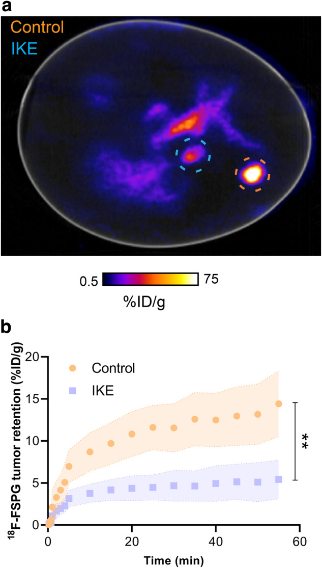
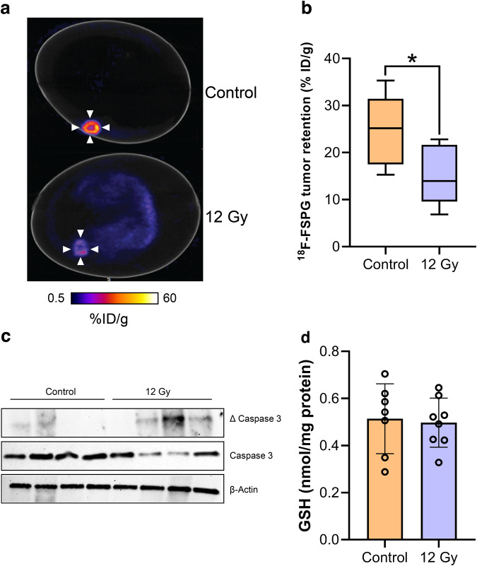
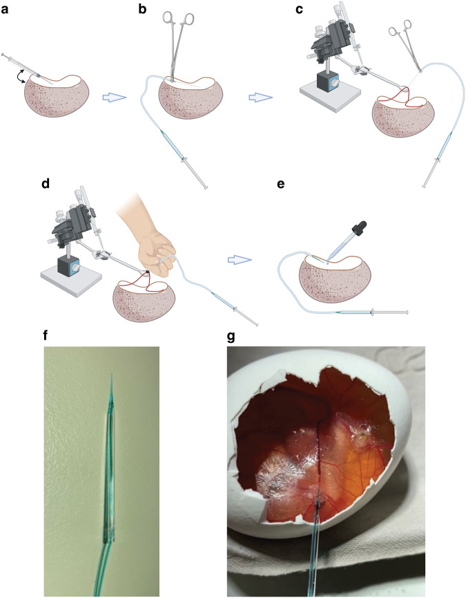
Mouse models are invaluable tools for radiotracer development and validation. They are, however, expensive, low throughput, and are constrained by animal welfare considerations. Here, we assessed the chicken chorioallantoic membrane (CAM) as an alternative to mice for preclinical cancer imaging studies. NCI-H460 FLuc cells grown in Matrigel on the CAM formed vascularized tumors of reproducible size without compromising embryo viability. By designing a simple method for vessel cannulation it was possible to perform dynamic PET imaging in ovo, producing high tumor-to-background signal for both 18F-2-fluoro-2-deoxy-D-glucose (18F-FDG) and (4S)-4-(3-18F-fluoropropyl)-L-glutamate (18F-FSPG). The pattern of 18F-FDG tumor uptake were similar in ovo and in vivo, although tumor-associated radioactivity was higher in the CAM-grown tumors over the 60 min imaging time course. Additionally, 18F-FSPG provided an early marker of both treatment response to external beam radiotherapy and target inhibition in ovo. Overall, the CAM provided a low-cost alternative to tumor xenograft mouse models which may broaden access to PET and SPECT imaging and have utility across multiple applications.


Conflict of interest statement

Competing Interests The authors declare no competing interests.

Figures

Fig. 1
Fig. 1. Optimization and characterization of in ovo tumor growth.
a Relative chick survival on E14 using a range of chemical and physical supports. b Percentage tumor take-rate of surviving embryos from each inoculation group. c Tumor weight at E14. d Representative BLI image of an in ovo NCI-H460 FLuc tumor. e Accompanying H&E section. f Separate and overlay images showing vasculature (lens culinaris agglutinin, 649 nm), tumor cell cytoskeleton (cytokeratin 18, 488 nm) and perfusion with nuclear stain Hoechst 33342 (350 nm). Error bars show standard deviation. *, p < 0.05; ***, p < 0.001.
Fig. 2
Fig. 2. Comparison of in ovo and in vivo 18F-FDG PET/CT imaging.
a Representative in ovo 18F-FDG PET/CT images 40–60 min p.i.. White arrows indicate the tumor. b Representative in vivo sagittal, coronal and axial (insert) 18F-FDG PET/CT images 40–60 min p.i.. White arrows indicate the tumor. Br, brain; H, heart; K, kidney. c Comparison of in ovo and in vivo 18F-FDG tumor pharmacokinetics. d In ovo and in vivo healthy and tumor tissue 18F-FDG uptake, expressed as the area under the TAC. Data is expressed as the mean plus standard deviation. n = 7 eggs, n = 9 mice. ***, p < 0.001.
Fig. 4
Fig. 4. Dynamic 18F-FSPG PET imaging in ovo.
a Representative in ovo 18F-FSPG PET/CT images 40–60 min p.i.. White arrows indicate the tumor. K, kidney. b TAC for tumor and yolk sac-associated 18F-FSPG retention in ovo. c AUC for major organs. Data is expressed as a mean plus standard deviation. n = 10. d xCT and NRF2 protein expression from NCI-H460 FLuc in ovo tumors. **, p < 0.01; ***, p < 0.001.
Fig. 5
Fig. 5. Ex vivo chicken embryo biodistribution with 18F-FSPG.
a Photo of the excised chick embryo. b Photos of key organs from the chick embryo at E14. c 18F-FSPG retention in key organs and in NCI-H460 FLuc tumors 60 min p.i. n = 3–6.
Fig. 6
Fig. 6. Inhibition of system xc- reduces 18F-FSPG uptake.
a Representative 18F-FSPG PET/CT image of an egg bearing both control and IKE-treated NCI-H460 FLuc in ovo tumors 40–60 min p.i. Orange circle shows the location of control tumor; blue circle shows the location of IKE-treated tumor. b 18F-FSPG TAC of control vs. IKE-treated tumors. Error bars represent one STD from the mean value. n = 6; **, p = 0.004.
Fig. 7
Fig. 7. External beam radiotherapy decreases 18F-FSPG retention.
a Representative 40-60 min 18F-FSPG PET/CT image of NCI-H460 FLuc-bearing eggs treated with 12 Gy radiotherapy or CT alone. b Quantification of 18F-FSPG tumor retention 40–60 min p.i.. n = 7; *, p = 0.017. c Western blot showing viability of control and radiation-treated NCI-H460 FLuc in ovo tumors. Total and cleaved (∆) caspase 3 were assessed, with actin used as a loading control (n = 4). d GSH concentrations for control and radiation-treated tumors. n = 8.
Fig. 8
Fig. 8. Evolution of Chick CAM cannulation methodology.
Various methods were trialed during cannulation optimization: a Direct injection with 30 g insulin syringe by hand. b Cut 30 g needle with needle holders. c Micromanipulators fixed with a hooked spatula attachment used to pull the vessel over and provide tension. d Glass needle and peristaltic pump tubing tied in with suture. e Glass needles injected by hand at branch points and secured in place using vetbond glue (optimized technique). f Photo of the final glass needle cannula used for all subsequent experiments. g Successful cannulation of a CAM vessel using (f). ae were created with BioRender.com.

Similar articles

Cited by

References

    1. Kerbel, R. S. What is the optimal rodent model for anti-tumor drug testing? Cancer Metastasis Rev.17, 301–304 (1998). - PubMed
    1. Richmond, A. & Su, Y. Mouse xenograft models vs GEM models for human cancer therapeutics. Dis. Model Mech.1, 78–82 (2008). - PMC - PubMed
    1. Lampreht Tratar, U., Horvat, S. & Cemazar, M. Transgenic mouse models in cancer research. Front. Oncol.8, 268 (2018). - PMC - PubMed
    1. Greenwood, H. E., Nyitrai, Z., Mocsai, G., Hobor, S. & Witney, T. H. High-throughput PET/CT imaging using a multiple-mouse imaging system. J. Nucl. Med.61, 292–297 (2020). - PMC - PubMed
    1. Martin, D. S. et al. Role of murine tumor models in cancer treatment research. Cancer Res.46, 2189–2192 (1986). - PubMed

LinkOut - more resources