

Liver sinusoidal endothelial cells as potential drivers of liver fibrosis (Review)
- PMID: 38240102
- PMCID: PMC10828992
- DOI: 10.3892/mmr.2024.13164
Liver sinusoidal endothelial cells as potential drivers of liver fibrosis (Review)
Abstract
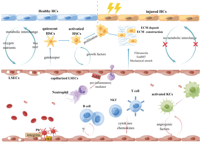
Liver fibrosis due to viral or metabolic chronic liver diseases is a major challenge of global health. It is a critical pre‑stage condition of severe hepatopathy, characterized by excessive accumulation of extracellular matrix components and ongoing chronic inflammation. To date, early prevention of liver fibrosis remains challenging. As the most abundant non‑parenchymal hepatic cell population, liver sinusoidal endothelial cells (LSECs) are stabilizers that maintain the intrahepatic environment. Notably, LSECs dysfunction appears to be implicated in the progression of liver fibrosis via numerous mechanisms. Following sustained liver injury, they lose their fenestrae (cytoplasmic pores) and change their crosstalk with other cellular interactions in the hepatic blood environment. LSEC‑targeted therapy has shown promising effects on fibrosis resolution, opening up new opportunities for anti‑fibrotic therapy. In light of this, the present study summarized changes in LSECs during liver fibrosis and their interactions with hepatic milieu, as well as possible therapeutic approaches that specially target LSECs.
Keywords: capillarization; crosstalk; endothelial dysfunction; liver fibrosis; liver sinusoidal endothelial cells.
Conflict of interest statement
The authors declare that they have no competing interests.
Figures
References
MeSH terms
LinkOut - more resources
Full Text Sources
Medical
Research Materials
