

Performance of a seed amplification assay for misfolded alpha-synuclein in cerebrospinal fluid and brain tissue in relation to Lewy body disease stage and pathology burden
- PMID: 38240849
- PMCID: PMC10799141
- DOI: 10.1007/s00401-023-02663-0
Performance of a seed amplification assay for misfolded alpha-synuclein in cerebrospinal fluid and brain tissue in relation to Lewy body disease stage and pathology burden
Abstract
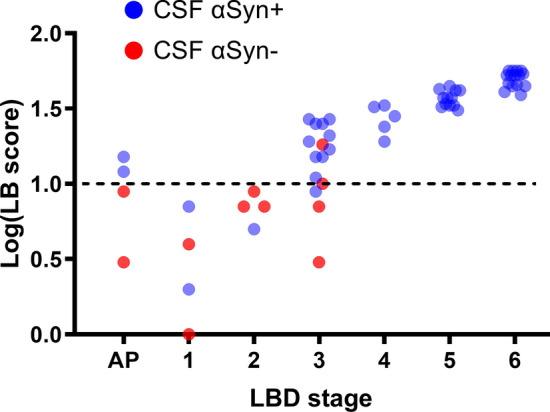
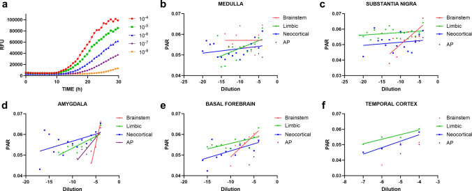
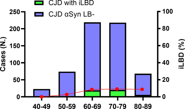
The development of in vitro seed amplification assays (SAA) detecting misfolded alpha-synuclein (αSyn) in cerebrospinal fluid (CSF) and other tissues has provided a pathology-specific biomarker for Lewy body disease (LBD). However, αSyn SAA diagnostic performance in early pathological stages or low Lewy body (LB) pathology load has only been assessed in small cohorts. Moreover, the relationship between SAA kinetic parameters, the number of αSyn brain seeds and the LB pathology burden assessed by immunohistochemistry has never been systematically investigated. We tested 269 antemortem CSF samples and 138 serially diluted brain homogenates from patients with and without neuropathological evidence of LBD in different stages by the αSyn Real-Time Quaking-Induced Conversion (RT-QuIC) SAA. Moreover, we looked for LB pathology by αSyn immunohistochemistry in a consecutive series of 604 Creutzfeldt-Jakob disease (CJD)-affected brains. αSyn CSF RT-QuIC showed 100% sensitivity in detecting LBD in limbic and neocortical stages. The assay sensitivity was significantly lower in patients in early stages (37.5% in Braak 1 and 2, 73.3% in Braak 3) or with focal pathology (50% in amygdala-predominant). The average number of CSF RT-QuIC positive replicates significantly correlated with LBD stage. Brain homogenate RT-QuIC showed higher sensitivity than immunohistochemistry for the detection of misfolded αSyn. In the latter, the kinetic parameter lag phase (time to reach the positive threshold) strongly correlated with the αSyn seed concentration in serial dilution experiments. Finally, incidental LBD prevalence was 8% in the CJD cohort. The present results indicate that (a) CSF RT-QuIC has high specificity and sufficient sensitivity to detect all patients with LB pathology at Braak stages > 3 and most of those at stage 3; (b) brain deposition of misfolded αSyn precedes the formation of LB and Lewy neurites; (c) αSyn SAA provides "quantitative" information regarding the LB pathology burden, with the lag phase and the number of positive replicates being the most promising variables to be used in the clinical setting.
Keywords: Amygdala; Diagnosis; Lewy bodies; Parkinson’s disease; RT-QuIC; α-Synuclein.
© 2024. The Author(s).
Conflict of interest statement
The authors declare there are no competing interests.
Figures

References
-
- Arnold MR, Coughlin DG, Brumbach BH, Smirnov DS, Concha-Marambio L, Farris CM, et al. α-Synuclein seed amplification in CSF and brain from patients with different brain distributions of pathological α-synuclein in the context of co-pathology and non-LBD diagnoses. Ann Neurol. 2022;92:650–662. doi: 10.1002/ana.26453. - DOI - PMC - PubMed
Publication types
MeSH terms
Substances
Grants and funding
LinkOut - more resources
Full Text Sources
Medical
