Review
. 2024 Jan;21(1):e00292.
doi: 10.1016/j.neurot.2023.10.002. Epub 2023 Dec 19.

Mitochondrial dysfunction in neurodegenerative disorders

Affiliations
Review

Mitochondrial dysfunction in neurodegenerative disorders

Madelyn M Klemmensen et al. Neurotherapeutics. 2024 Jan.

Abstract

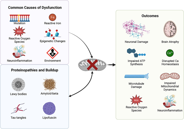
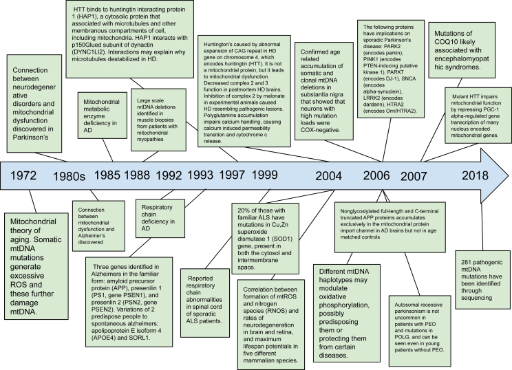
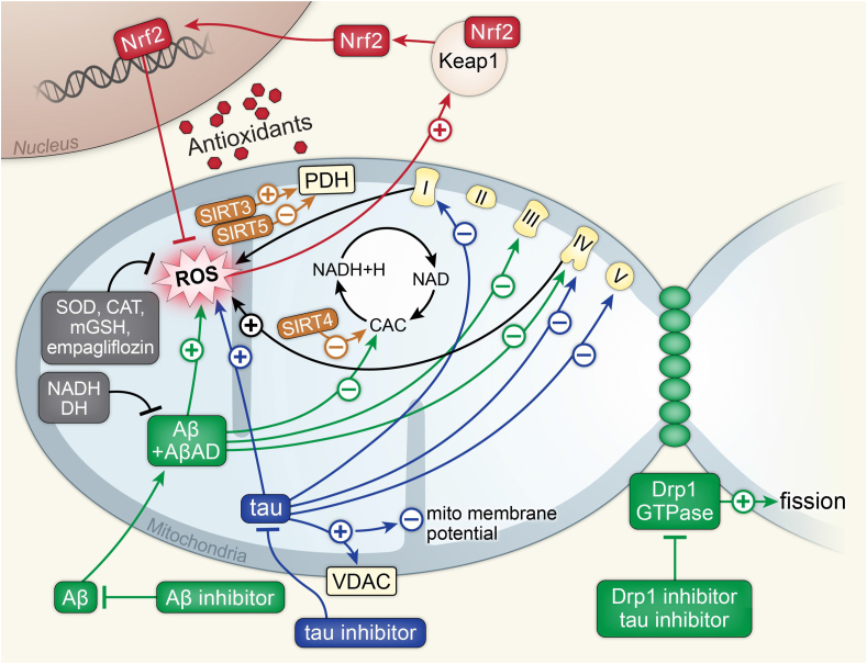
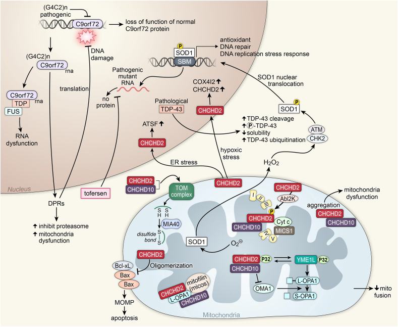
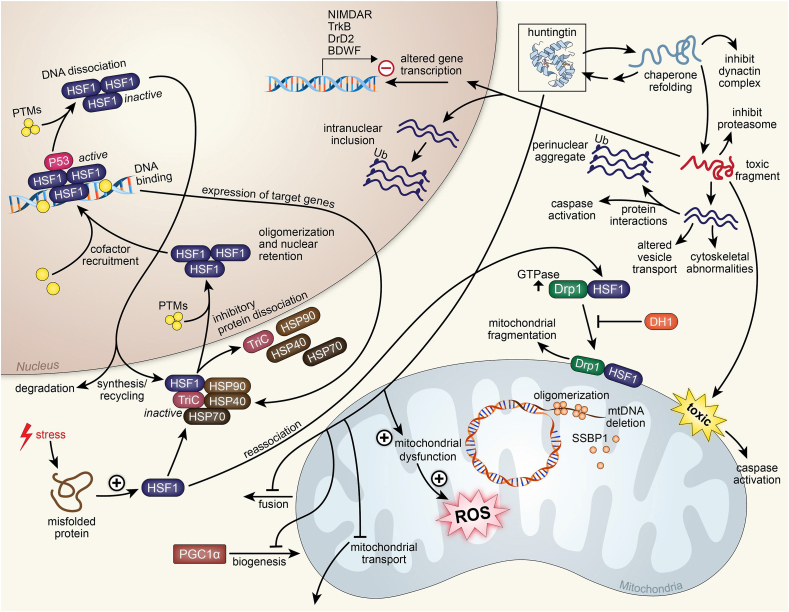
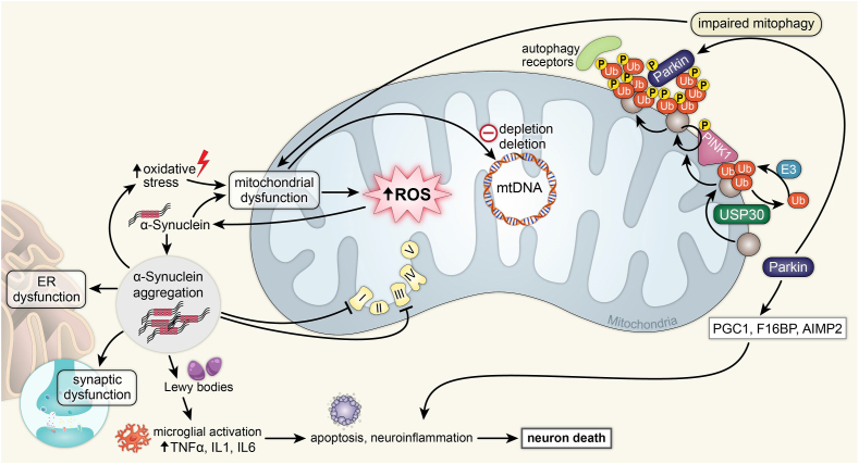
Recent advances in understanding the role of mitochondrial dysfunction in neurodegenerative diseases have expanded the opportunities for neurotherapeutics targeting mitochondria to alleviate symptoms and slow disease progression. In this review, we offer a historical account of advances in mitochondrial biology and neurodegenerative disease. Additionally, we summarize current knowledge of the normal physiology of mitochondria and the pathogenesis of mitochondrial dysfunction, the role of mitochondrial dysfunction in neurodegenerative disease, current therapeutics and recent therapeutic advances, as well as future directions for neurotherapeutics targeting mitochondrial function. A focus is placed on reactive oxygen species and their role in the disruption of telomeres and their effects on the epigenome. The effects of mitochondrial dysfunction in the etiology and progression of Alzheimer's disease, amyotrophic lateral sclerosis, Parkinson's disease, and Huntington's disease are discussed in depth. Current clinical trials for mitochondria-targeting neurotherapeutics are discussed.

Keywords: Aging; Bioenergetics; Mitochondria; Neurodegeneration; Reactive oxygen species (ROS).


Conflict of interest statement

Declaration of competing interest The authors declare that they have no known competing financial interests or personal relationships that could have appeared to influence the work reported in this paper.

Figures

Image 1
Graphical abstract
Fig. 1
Fig. 1
Timeline. Above is a historical timeline outlining seminal discoveries linking mitochondria to neurodegenerative disorders.
Fig. 2
Fig. 2
Alzheimer's disease. Above is a figure illustrating mitochondrial affecting pathways in Alzheimer's disease associated with Aβ, tau, Drp1, SIRT proteins, and Nrf2. Aβ and AβAD work synergistically to increase mitochondrial ROS, inhibit the CAC, and inhibit complexes 3 and 4 of the ETC. Aβ inhibitors inhibit Aβ and inhibit this synergistic activity. NADH and DH inhibit Aβ and AβAD synergy as well. Tau protein increases mitochondrial ROS and inhibits complexes 1, 4, and 5 of the ETC. Tau also increases the activity of VDAC, leading to the loss of the mitochondrial membrane potential. Tau inhibitors inhibit the tau protein. Drp1 works synergistically with GTPase to increase mitochondrial fission. Drp1 and tau inhibitors inhibit this activity. SIRT4 inhibits CAC. SIRT5 inhibits PDH while SIRT3 activates PDH. ROS activates the Keap1, Nrf2 complex. Nrf2 dissociates and enters the nucleus, where it increases transcription for antioxidants to inhibit ROS. SOD, CAT, mGSH, and empagliflozin also inhibit ROS.
Fig. 3
Fig. 3
Amyotrophic lateral sclerosis. Above is a figure illustrating mitochondrial affecting pathways in ALS associated with CHCHD2, CHCHD10, C9orf72, TDP-43, and SOD1. Superoxide activates the SOD1 protein. SOD1, with hydrogen peroxide and a CHK2 ATM complex, is phosphorylated, which allows it to enter the nucleus through SOD1 nuclear translocation. In the nucleus, it functions as a transcription factor on SBM to transcript antioxidants proteins, DNA repair proteins, and proteins for the DNA replication stress response. Pathogenic SOD1 creating mutant RNA will produce no protein. Tofersen inhibits this process. Pathological TDP-43 leads to increased TDP-43 cleavage, increased phosphorylation of TDP-43, decreased solubility, and increased TDP-43 ubiquitination. Pathogenic (G4C2)n inhibits the expression of normal C9orf72, leading to the loss of function of normal C9orf72 protein. (G4C2)n C9orf72 RNA is transcribed, leading to DPRs that inhibit the proteasome and lead to mitochondrial dysfunction. DPRs also lead to DNA damage. The (G4C2)n C9orf72 RNA can also create a complex with TDP and FUS, leading to RNA dysfunction. With hypoxic stress, CHCHD2 will enter the nucleus and increase COX4I2 and CHCHD2. Under ER stress, CHCHD2 will enter the nucleus and increase ATSF. CHCHD2 works with AbI2k to phosphorylate a CHCHD2, CHCHD10, Cyt c, and MICS1 complex in the ETC. Oligomerization of CHCHD2 leads to inhibition of the Bcl-xL, Bax, and Bax complex, leading to MOMP and apoptosis. Aggregates of CHCHD2 and CHCHd10 in the mitochondria lead to mitochondrial dysfunction. A CHCHD2 and CHCHD10 complex activate a TOM complex, creating disulfide bonds with MIA40. A complex of CHCHD2, CHCHD10, and p32 transfers the p32 to YME1L. The YME1L p32 complex cleaves L-OPA1 to become S-OPA1 with OMA1. OMA1 is inhibited by the CHCHD2, CHCHD10, and p32 complex, decreasing mitochondrial fusion.
Fig. 4
Fig. 4
Huntington's disease. Above is a figure illustrating mitochondrial affecting pathways in Huntington's disease associated with huntingtin and HSF1. Huntingtin in the mitochondria leads to mitochondrial dysfunction, leading to the production of ROS. Huntingtin also inhibits mitochondrial transport, inhibits PGC1α mitochondrial biogenesis, and inhibits mitochondrial fusion. With chaperone refolding, huntingtin can inhibit the dynactin complex. This refold can also become a toxic fragment that can inhibit the proteasome, and is also toxic to the mitochondria, leading to caspase activation. The toxic fragment can enter the mitochondria and negatively alter gene transcription, specifically affecting genes NIMDAR, TrkB, DrD2, and BDWF. In the nucleus, the toxic fragment can further associate with more fragments and create an intranuclear inclusion. The association of multiple toxic fragments can lead to cytoskeletal abnormalities and altered vesicle transport. These fragments can interact with proteins and lead to caspase activation. The association of more toxic fragments creates a perinuclear aggregate. Stress in the cell causing a misfolded protein causes the increase of HSF1. HSF1 can reassociate with Drp1 and in high GTPase conditions associate with mitochondria and lead to fragmentation. HSF1 with DRP1 allows mtDNA deletion by SSBP1. DH1 inhibits this process. HSF1 can form an inactive complex with proteins TriC, HSP70, HSP40, and HSP90 that allows it to travel into the nucleus where it dissociates with that complex and oligomerizes with other HSF1 proteins and is modified with PTMs for nuclear retention. With cofactors and P53, the HSF1 oligomer actively binds to DNA and expresses target genes TriC, HSP70, HSP40, and HSP90. Inhibitory PTMs allow the HSF1 oligomer to dissociate from DNA where it is either degraded in the cytoplasm or recycled for further synthesis of target genes.
Fig. 5
Fig. 5
Parkinson's disease. Above is a figure illustrating mitochondrial affecting pathways in Parkinson's associated with α-synuclein and Parkin. Increased oxidative stress leads to mitochondrial dysfunction, which leads to increased mitochondrial ROS and mitochondrial DNA depletion and deletion. Mitochondrial ROS activate α-synuclein, leading to α-synuclein aggregation in the cytoplasm. These aggregates cause ER dysfunction, synaptic dysfunction, inhibition of ETC complexes 1 and 3, and the formation of Lewy bodies. Lewy bodies cause microglial activation by increasing TNFα, IL1, and IL6, which leads to apoptosis, neuroinflammation, and neuron death. Parkin leads to impaired mitophagy involving both Parkin and PINK1, leading to mitochondrial dysfunction. Parkin also leads to the increase of PGC1, F1GBP, and AIMP2, which leads to neuroinflammation and neuron death.

References

    1. Ballard J.W., Whitlock M.C. The incomplete natural history of mitochondria. Mol Ecol. 2004;13:729–744. - PubMed
    1. Di Donato S. Disorders related to mitochondrial membranes: pathology of the respiratory chain and neurodegeneration. J Inherit Metab Dis. 2000;23:247–263. - PubMed
    1. Klecker T., Westermann B. Pathways shaping the mitochondrial inner membrane. Open Biol. 2021;11 - PMC - PubMed
    1. Kramer P., Bressan P. Our (Mother's) mitochondria and our mind. Perspect Psychol Sci. 2018;13:88–100. - PMC - PubMed
    1. Frey T.G., Mannella C.A. The internal structure of mitochondria. Trends Biochem Sci. 2000;25:319–324. - PubMed

MeSH terms

Substances