. 2024 Jan 19;10(3):eadi7203.
doi: 10.1126/sciadv.adi7203. Epub 2024 Jan 19.

Microstructural and chemical features of impact melts on Ryugu particle surfaces: Records of interplanetary dust hit on asteroid Ryugu

Affiliations

Microstructural and chemical features of impact melts on Ryugu particle surfaces: Records of interplanetary dust hit on asteroid Ryugu

Megumi Matsumoto et al. Sci Adv. .

Abstract

The Hayabusa2 spacecraft delivered samples of the carbonaceous asteroid Ryugu to Earth. Some of the sample particles show evidence of micrometeoroid impacts, which occurred on the asteroid surface. Among those, particles A0067 and A0094 have flat surfaces on which a large number of microcraters and impact melt splashes are observed. Two impact melt splashes and one microcrater were analyzed to unveil the nature of the objects that impacted the asteroid surface. The melt splashes consist mainly of Mg-Fe-rich glassy silicates and Fe-Ni sulfides. The microcrater trapped an impact melt consisting mainly of Mg-Fe-rich glassy silicate, Fe-Ni sulfides, and minor silica-rich glass. These impact melts show a single compositional trend indicating mixing of Ryugu surface materials and impactors having chondritic chemical compositions. The relict impactor in one of the melt splashes shows mineralogical similarity with anhydrous chondritic interplanetary dust particles having a probable cometary origin. The chondritic micrometeoroids probably impacted the Ryugu surface during its residence in a near-Earth orbit.


Figures

Fig. 1.
Fig. 1.. Optical microscope and SEM images of the flat surfaces of A0067 and A0094 particles.
(A and B) Optical microscope images of the A0067 and the A0094 flat surfaces (indicated by black dotted lines). The particles are held by indium. (C) Secondary electron (SE) image of wrinkled phyllosilicate layer covering the A0067 flat surface. (D) SE image of the A0094 flat surface showing vesicular structure. (E) SE image of A0067-crater#1. (F) Back scattered electron (BSE) image of A0067-melt#1 consisting of a silicate glass main body and an attached Fe-rich droplet. Splashes of glassy material spreading radially from A0067-melt#1 are indicated by a white arrow. (G) BSE image of the Fe-rich droplet splashed onto the silicate glass main body. (H) BSE image of A0094-melt#1 consisting of two glassy silicate droplets attached to each other (I and II parts). A0094-melt#1 containing abundant Fe-Ni sulfides (bright grains). Cracked-shell–shaped melt splashes occurring nearby A0094-melt#1 are indicated by a white arrow.
Fig. 2.
Fig. 2.. XnCT images of A0067-melt#1.
(A and B) Absorption and (C) phase shift XnCT images of A0067-melt#1 taken at 7, 7.35, and 8 keV, respectively. (D) Color-merged image of the three types of images (red: LAC, 0 to 1500 cm−1 at 7.35 keV; green: RID, 0 × 10−6 to 20 × 10−6 at 8 keV; blue: LAC, 0 to 1000 cm−1 at 7 keV). A0067-melt#1 contains a large void and is attached to the saponite-rich layer (Sap-layer) covering the A0067 main body (E) Magnified image of A0067-melt#1 in another slice of the color merged XnCT image. The slice was subjected to TEM analysis (see Fig. 3). A0067-melt#1 consisting mainly of glassy silicate and an Fe-rich droplet with a mottled texture. A few cone-shaped pores are recognized at the boundary between A0067-melt#1 and the saponite-rich layer. Stripe noise and unevenness in contrast of the glassy silicate are lighting artifacts. Mgt, magnetite; Dol, dolomite; Pt*, Pt deposition formed during the FIB sample processing.
Fig. 3.
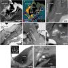
Fig. 3.. TEM and STEM images of A0067-melt#1.
(A and B) High-angle annular dark field (HAADF) STEM image and a combined elemental map for Fe (red), Mg (green), Si (blue), and S (yellow) of A0067-melt#1. A0067-melt#1 is attached to the saponite-rich layer covering the A0067 main body. A thin Si-rich layer is observed on the surface of the saponite-rich layer. (C) Bright-field (BF) TEM image of the silicate glass in A0067-melt#1. Inset is the SAED pattern of the silicate glass obtained from the circled area. (D) HAADF-STEM image of the Fe-rich spherules in the silicate glass. The spherules have a core-mantle structure with Fe-Ni metal core and Fe-Ni sulfide mantle.
Fig. 4.
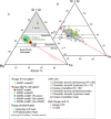
Fig. 4.. (Si + Al)–Mg–Fe atomic % ternary diagram of compositions of the melt splashes.
(A) Major element compositions of the melt splashes studied and (B) those of IDPs. The compositions are plotted along with an extension of a line combining the bulk CI chondrite composition (25) and Fe vertex (CI-Fe line). The compositions of the saponite-rich layer are also plotted. The compositions of IDPs are from (41), Ryugu’s bulk composition is from (18), and the representative composition of the phyllosilicates in the Ryugu major lithology is from (19). The compositional field of Ryugu phyllosilicates (774 analyses) (19) is shown as a green colored oval, while that of bulk ordinary and carbonaceous chondrites (OC and CC, respectively) is indicated by a black open oval. The hatched area indicates the two liquid field in the MgO-FeO-SiO2 system (58).
Fig. 5.
Fig. 5.. TEM and STEM images of an iron-rich opaque droplet in A0067-melt#1.
(A and B) HAADF-STEM image and a combined elemental map for Fe (magenta), Ni (green), and S (blue) of the Fe-rich droplet. (C) BF-TEM image of an α-(Fe-Ni) crystal. (D) SAED pattern obtained from the α-(Fe-Ni) in (C). (E) BF-TEM image of the rectangular E area in (A). SAED pattern obtained from the circled area is shown in (F). Narrow gaps parallel to the (001) troilite plane are recognized in the troilite matrix. (G and H) BF-TEM image and a combined elemental map for Fe (magenta), Ni (green), and S (blue) in the rectangular G area in (A). SAED pattern obtained from the circled area is shown in (I). The SAED pattern indicates that pentlandite has a specific crystallographic relationship with the troilite: (001)Tro//(111¯)Pen and (010)Tro//(101)Pen.
Fig. 6.
Fig. 6.. XnCT images of A0094-melt#1.
(A and B) Absorption and (C) phase shift XnCT images of A0094-melt#1 taken at 7, 7.35, and 8 keV, respectively. (D) Color merged image of the three types of images (red: LAC, 0 to 1500 cm−1 at 7.35 keV; green: RID, 0 × 10−6 to 20 × 10−6 at 8 keV; blue: LAC, 0 to 1000 cm−1 at 7 keV). A0094-melt#1 contains numerous voids and is attached directly to the A0094 main body. A0094-melt#1 consists mainly of glassy silicate and shows a patchy structure with Fe-rich and Fe-poor regions (the bright and the dark regions in the XnCT images). The boundary between the two regions is unclear. The I and the II parts of A0094-melt#1 (compare Figs. 1H and 6B) largely consist of the Fe-poor and the Fe-rich regions, respectively. TEM analysis was performed on the slice shown in this figure (see Fig. 7).
Fig. 7.
Fig. 7.. TEM and STEM images of A0094-melt#1.
(A to C) HAADF-STEM image and combined elemental maps for Fe (red), Mg (green), Si (blue), and S (yellow) and for S (magenta), Fe (green), and Mg (blue) of A0094-melt#1 attached onto the A0094 main body, respectively. Some large Fe-Ni sulfide grains formed on the upper surface of A0094-melt#1 [indicated by red arrows in (C). A0094-melt#1 is separated into Fe-rich and the Fe-poor regions. Two Fe-rich regions (#1 and #2) are recognized in the TEM section. A thin Si-rich layer is observed on the surface of the A0094 main body. The Fe-Ni sulfides in A0094-melt#1 show higher Fe/S atomic ratios and appear redder in (B) compared to those in the A0094 main body. (D) BF-TEM image of voids and Fe-Ni sulfide spherules in A0094-melt#1. (E) BF-TEM image of the Fe-rich region with irregular-shaped Fe-Ni sulfides (indicated by white dotted lines). (F to H) BF-TEM images of olivine#1 and #2 grains in the Fe-rich region. The insets are SAED patterns obtained from the circled areas in the olivines. Olivine#1 in (F and G) contains abundant voids and Fe-Ni sulfide inclusions. Olivine#2 in (H) consists of domains elongated along the crystallographic b axis. One of these domains is indicated by a black dotted line. (I) BF-TEM image of carbonaceous aggregates (CA) consisting of spongy carbonaceous material, irregular-shaped Fe-Ni sulfides, and Mg-rich amorphous silicates. The inset is a SAED pattern obtained from the carbonaceous material dominant portion (circled area).
Fig. 8.
Fig. 8.. XnCT images of A0067-crater#1.
(A and B) Absorption and (C) phase shift XnCT images of A0067-crater#1 taken at 7, 7.35, and 8 keV, respectively. (D) Color merged image of the three types of images (red: LAC, 0 to 1500 cm−1 at 7.35 keV; green: RID, 0 × 10−6 to 20 × 10−6 at 8 keV; blue: LAC, 0 to 1000 cm−1 at 7 keV). A0067-crater#1 intrudes into the saponite-rich-layer and shows a tapered structure. A mixture of glassy silicate and Fe-Ni sulfide is trapped on the crater wall. The A0067 main body under the saponite-rich layer contains plaquette magnetite (Mgt) and Fe-Ni sulfides embedded in the phyllosilicate matrix.
Fig. 9.
Fig. 9.. TEM and STEM images of A0067-crater#1.
(A and B) HAADF-STEM image and a combined elemental map for Fe (red), Mg (green), Si (blue), and S (yellow) of A0067-crater#1. A0067-crater#1 intrudes into the saponite-rich layer. The crater wall is indicated by white dotted lines in (A). A0067-crater#1 contains crater-filling material consisting of stacked glassy silicate and Fe-Ni sulfide layers. The glassy silicate is separated into Mg-Fe–rich and Si-rich parts. The Fe-Ni sulfides in A0067-crater#1 show higher Fe/S atomic ratios and are redder in (B) compared to those in the A0067 main body. (C) BF-TEM image of the boundary between the crater-filling material and the saponite-rich layer. The inset is an enlargement of the boxed area. The phyllosilicates constituting the saponite-rich layer are well crystallized. (D to F) BF-TEM image of the glassy silicates. The Si-rich glass occurs selectively along with the crater wall (indicated by a white dotted line). Small Fe-Ni spherules and voids are present both in the Mg-Fe–rich and Si-rich glasses. Small carbonaceous aggregates (CA) occur adjacent to the troilite grains composing the Fe-Ni sulfide layers. (G) BF-TEM image of a large troilite grain in the Fe-Ni sulfide layer. The troilite grain contains a small Fe-Ni metal spherule. The inset is a SAED pattern obtained from the circled area. (H) BF-TEM image of Fe oxide occurring at the boundary between troilite and the saponite-rich layer. Amorphous material with a saponite-like composition (amo-Sap) occurs around the boundary. Pt*, Pt deposition formed in the FIB sample processing.

Similar articles

  • Noble gases and nitrogen in samples of asteroid Ryugu record its volatile sources and recent surface evolution.
    Okazaki R, Marty B, Busemann H, Hashizume K, Gilmour JD, Meshik A, Yada T, Kitajima F, Broadley MW, Byrne D, Füri E, Riebe MEI, Krietsch D, Maden C, Ishida A, Clay P, Crowther SA, Fawcett L, Lawton T, Pravdivtseva O, Miura YN, Park J, Bajo KI, Takano Y, Yamada K, Kawagucci S, Matsui Y, Yamamoto M, Righter K, Sakai S, Iwata N, Shirai N, Sekimoto S, Inagaki M, Ebihara M, Yokochi R, Nishiizumi K, Nagao K, Lee JI, Kano A, Caffee MW, Uemura R, Nakamura T, Naraoka H, Noguchi T, Yabuta H, Yurimoto H, Tachibana S, Sawada H, Sakamoto K, Abe M, Arakawa M, Fujii A, Hayakawa M, Hirata N, Hirata N, Honda R, Honda C, Hosoda S, Iijima YI, Ikeda H, Ishiguro M, Ishihara Y, Iwata T, Kawahara K, Kikuchi S, Kitazato K, Matsumoto K, Matsuoka M, Michikami T, Mimasu Y, Miura A, Morota T, Nakazawa S, Namiki N, Noda H, Noguchi R, Ogawa N, Ogawa K, Okada T, Okamoto C, Ono G, Ozaki M, Saiki T, Sakatani N, Senshu H, Shimaki Y, Shirai K, Sugita S, Takei Y, Takeuchi H, Tanaka S, Tatsumi E, Terui F, Tsukizaki R, Wada K, Yamada M, Yamada T, Yamamoto Y, Yano H, Yokota Y, Yoshihara K, Yoshikawa M, Yoshikawa K, Furuya S, Hatakeda K, Hayashi T, Hitomi Y, Kumagai K, Miyazaki A, Nakato A, Nishimura M, Soejima H, Iwama… See abstract for full author list ➔ Okazaki R, et al. Science. 2023 Feb 24;379(6634):eabo0431. doi: 10.1126/science.abo0431. Epub 2023 Feb 24. Science. 2023. PMID: 36264828
  • Formation and evolution of carbonaceous asteroid Ryugu: Direct evidence from returned samples.
    Nakamura T, Matsumoto M, Amano K, Enokido Y, Zolensky ME, Mikouchi T, Genda H, Tanaka S, Zolotov MY, Kurosawa K, Wakita S, Hyodo R, Nagano H, Nakashima D, Takahashi Y, Fujioka Y, Kikuiri M, Kagawa E, Matsuoka M, Brearley AJ, Tsuchiyama A, Uesugi M, Matsuno J, Kimura Y, Sato M, Milliken RE, Tatsumi E, Sugita S, Hiroi T, Kitazato K, Brownlee D, Joswiak DJ, Takahashi M, Ninomiya K, Takahashi T, Osawa T, Terada K, Brenker FE, Tkalcec BJ, Vincze L, Brunetto R, Aléon-Toppani A, Chan QHS, Roskosz M, Viennet JC, Beck P, Alp EE, Michikami T, Nagaashi Y, Tsuji T, Ino Y, Martinez J, Han J, Dolocan A, Bodnar RJ, Tanaka M, Yoshida H, Sugiyama K, King AJ, Fukushi K, Suga H, Yamashita S, Kawai T, Inoue K, Nakato A, Noguchi T, Vilas F, Hendrix AR, Jaramillo-Correa C, Domingue DL, Dominguez G, Gainsforth Z, Engrand C, Duprat J, Russell SS, Bonato E, Ma C, Kawamoto T, Wada T, Watanabe S, Endo R, Enju S, Riu L, Rubino S, Tack P, Takeshita S, Takeichi Y, Takeuchi A, Takigawa A, Takir D, Tanigaki T, Taniguchi A, Tsukamoto K, Yagi T, Yamada S, Yamamoto K, Yamashita Y, Yasutake M, Uesugi K, Umegaki I, Chiu I, Ishizaki T, Okumura S, Palomba E, Pilorget C, Potin SM, Alasli A, Anada S, Araki Y, Sakatani N,… See abstract for full author list ➔ Nakamura T, et al. Science. 2023 Feb 24;379(6634):eabn8671. doi: 10.1126/science.abn8671. Epub 2023 Feb 24. Science. 2023. PMID: 36137011
  • The Albedo of Ryugu: Evidence for a High Organic Abundance, as Inferred from the Hayabusa2 Touchdown Maneuver.
    Potiszil C, Tanaka R, Kobayashi K, Kunihiro T, Nakamura E. Potiszil C, et al. Astrobiology. 2020 Jul;20(7):916-921. doi: 10.1089/ast.2019.2198. Epub 2020 Jun 15. Astrobiology. 2020. PMID: 32543220 Free PMC article.
  • Organic Matter in the Asteroid Ryugu: What We Know So Far.
    Potiszil C, Yamanaka M, Sakaguchi C, Ota T, Kitagawa H, Kunihiro T, Tanaka R, Kobayashi K, Nakamura E. Potiszil C, et al. Life (Basel). 2023 Jun 26;13(7):1448. doi: 10.3390/life13071448. Life (Basel). 2023. PMID: 37511823 Free PMC article. Review.
  • Cometary dust: the diversity of primitive refractory grains.
    Wooden DH, Ishii HA, Zolensky ME. Wooden DH, et al. Philos Trans A Math Phys Eng Sci. 2017 Jul 13;375(2097):20160260. doi: 10.1098/rsta.2016.0260. Philos Trans A Math Phys Eng Sci. 2017. PMID: 28554979 Free PMC article. Review.

Cited by

  • Sulfide minerals bear witness to impacts across the solar system.
    Thompson MS, Davidson J, Schrader DL, Zega TJ. Thompson MS, et al. Nat Commun. 2025 Jul 1;16(1):5975. doi: 10.1038/s41467-025-61201-6. Nat Commun. 2025. PMID: 40592914 Free PMC article.
  • Nonmagnetic framboid and associated iron nanoparticles with a space-weathered feature from asteroid Ryugu.
    Kimura Y, Kato T, Anada S, Yoshida R, Yamamoto K, Tanigaki T, Akashi T, Kasai H, Kurosawa K, Nakamura T, Noguchi T, Sato M, Matsumoto T, Morita T, Kikuiri M, Amano K, Kagawa E, Yada T, Nishimura M, Nakato A, Miyazaki A, Yogata K, Abe M, Okada T, Usui T, Yoshikawa M, Saiki T, Tanaka S, Terui F, Nakazawa S, Yurimoto H, Okazaki R, Yabuta H, Naraoka H, Sakamoto K, Watanabe SI, Tsuda Y, Tachibana S. Kimura Y, et al. Nat Commun. 2024 Apr 29;15(1):3493. doi: 10.1038/s41467-024-47798-0. Nat Commun. 2024. PMID: 38684653 Free PMC article.

References

    1. Keller L. P., McKay D. S., Discovery of vapor deposits in the lunar regolith. Science 261, 1305–1307 (1993). - PubMed
    1. Noguchi T., Nakamura T., Kimura M., Zokensky M. E., Tanaka M., Hashimoto H., Konno M., Nakato A., Ogami T., Fujimura A., Abe M., Yada T., Mukai T., Ueno M., Okada T., Shirai K., Ishibashi Y., Okazaki R., Incipient space weathering observed on the surface of Itokawa dust particles. Science 333, 1121–1125 (2011). - PubMed
    1. Noguchi T., Kimura M., Hashimoto T., Konno M., Nakamura T., Zolensky M. E., Okazaki R., Tanaka M., Tsuchiyama A., Nakato A., Ogami T., Ishida H., Sagae R., Tsujimori S., Matsumoto T., Matsuno J., Fujiwara A., Abe M., Yada T., Mukai T., Ueno M., Okada T., Shirai K., Ishibashi Y., Space weathered rims found on the surfaces of the Itokawa dust particles. Meteorit. Planet. Sci. 49, 188–214 (2014).
    1. Noble S. K., Keller L. P., Pieters C. M., Evidence of space weathering in regolith breccias I: Lunar regolith breccias. Meteorit. Planet. Sci. 40, 397–408 (2005).
    1. Noble S. K., Keller L. P., Pieters C. M., Evidence of space weathering in regolith breccias II: Asteroidal regolith breccias. Meteorit. Planet. Sci. 45, 2007–2015 (2011).