. 2024 Jan 19;10(3):eadk6524.
doi: 10.1126/sciadv.adk6524. Epub 2024 Jan 19.

MCJ: A mitochondrial target for cardiac intervention in pulmonary hypertension

Affiliations

MCJ: A mitochondrial target for cardiac intervention in pulmonary hypertension

Ayelén M Santamans et al. Sci Adv. .

Abstract

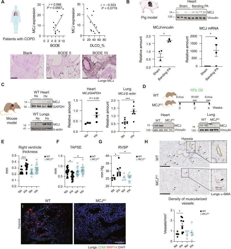
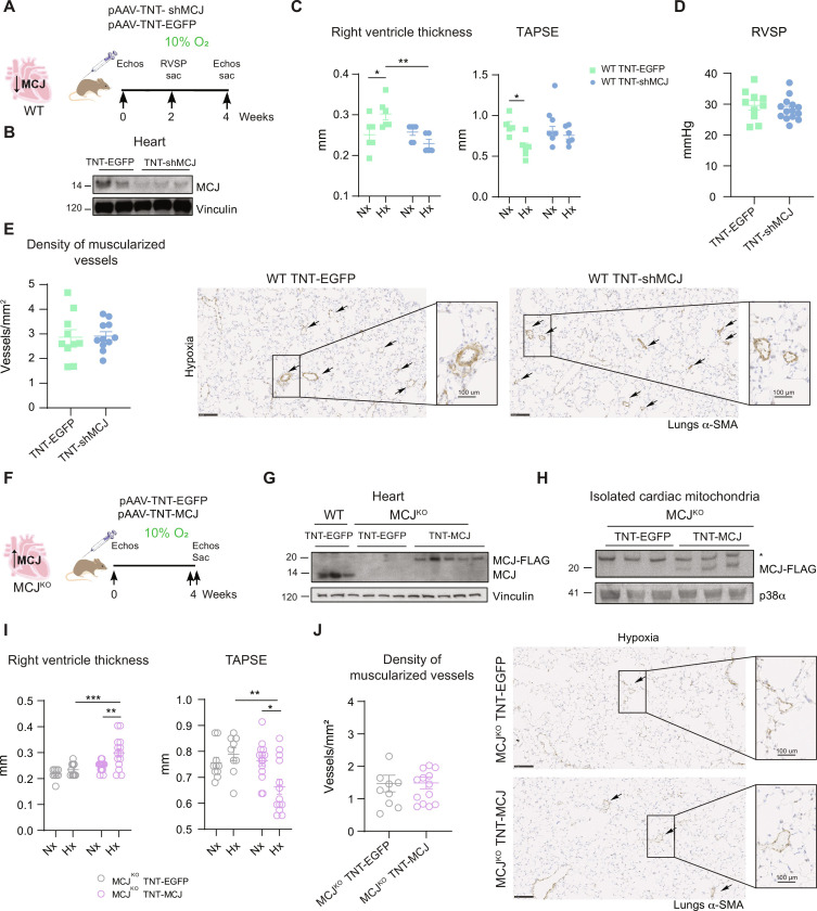
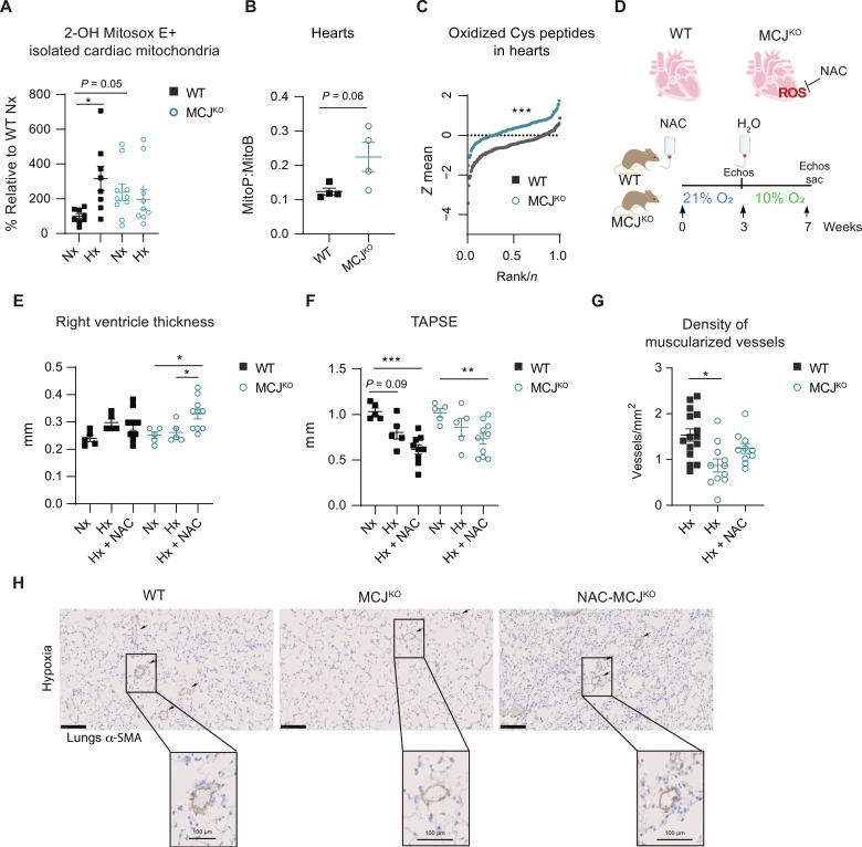
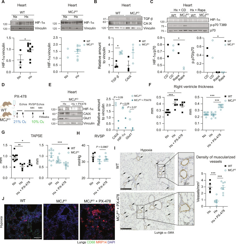
Pulmonary hypertension (PH) can affect both pulmonary arterial tree and cardiac function, often leading to right heart failure and death. Despite the urgency, the lack of understanding has limited the development of effective cardiac therapeutic strategies. Our research reveals that MCJ modulates mitochondrial response to chronic hypoxia. MCJ levels elevate under hypoxic conditions, as in lungs of patients affected by COPD, mice exposed to hypoxia, and myocardium from pigs subjected to right ventricular (RV) overload. The absence of MCJ preserves RV function, safeguarding against both cardiac and lung remodeling induced by chronic hypoxia. Cardiac-specific silencing is enough to protect against cardiac dysfunction despite the adverse pulmonary remodeling. Mechanistically, the absence of MCJ triggers a protective preconditioning state mediated by the ROS/mTOR/HIF-1α axis. As a result, it preserves RV systolic function following hypoxia exposure. These discoveries provide a potential avenue to alleviate chronic hypoxia-induced PH, highlighting MCJ as a promising target against this condition.


Figures

Fig. 1.
Fig. 1.. MCJ deficiency protects against hypoxia-induced pulmonary hypertension.
(A) Relation between MCJ lung expression in patients with COPD and the BODE index (body mass index, airflow obstruction, dyspnea, and exercise capacity) and the diffusing capacity of the lung for carbon monoxide (DLCO) (n = 11). Representative MCJ immunostaining in lung samples from patients with COPD with 5 and 10 points on the BODE index. Blank section incubated only with secondary antibody. Scale bars, 250 μm. (B) Swine model of chronic RV overload. MCJ protein and RNA levels were assayed by immunoblot and reverse transcription quantitative polymerase chain reaction in cardiac lysates from Yucatan pigs with surgical restrictive banding of the main PA (n = 4). (C) Representative immunoblot of cardiac and lung MCJ expression in wild-type (WT) mice maintained in normoxia (Nx; 21% O2) or exposed to chronic hypoxia (Hx; 28 days at 10% O2) (n = 3 to 8). GAPDH, glyceraldehyde-3-phosphate dehydrogenase. (D) Experimental scheme for (E) to (I): protocol for the analysis of lung and cardiac function in WT and MCJKO mice exposed to Nx or Hx. MCJ deletion in cardiac and lung lysates from MCJKO mice. (E and F) Echocardiography of RV hypertrophy (RV thickness) and function [tricuspid annular plane systolic excursion (TAPSE)] (n = 18 to 20, N = 3). (G) Right ventricular systolic pressure (RVSP) in WT and MCJKO mice (n = 5 to 10, N = 2). (H) Muscularized vessel density in WT and MCJKO lungs (n = 4 to 9, N = 2). Representative images show α–smooth muscle actin (α-SMA) immunostaining, amplification of a remodeled vessel in hypoxia. Scale bars, 250 μm, 100 μm (vessel). (I) Macrophages (CD68) and neutrophils (MRP14) infiltration in hypoxic mouse lungs. Nuclei stained with 4′,6-diamidino-2-phenylindole (DAPI). Scale bar, 100 μm. n, number of biological samples; N, number of times that the experiment was repeated. All data are presented as means ± SEM. Statistical comparison by Pearson correlation analysis (A), two-tailed Student’s t test [(B) and (C)], or two-way analysis of variance (ANOVA) with Tukey posttest [(E) to (H)]: nonsignificant (NS), *P < 0.05 and ***P < 0.001. Panels (A) to (D) prepared using modified figures from Servier Medical Art (https://smart.servier.com/), licensed under a Creative Commons Attribution 3.0 unported license.
Fig. 2.
Fig. 2.. Modulation of cardiac-specific MCJ expression is sufficient to determine the cardiac fate.
(A) Experimental scheme for (B) to (E). WT mice were intravenously injected at postnatal day 1 with pAAV-TnT-shMCJ adenovirus to silence specifically MCJ expression in cardiomyocyte (WT TNT-shMCJ) or with control AAV-TNT-EGFP-Luc (WT TNT-EGFP). Adult mice were maintained in normoxia (Nx; 21% O2) or exposed to chronic hypoxia (Hx; 10% O2). (B) Immunoblot showing MCJ deletion in cardiac lysates from WT TNT-shMCJ mice. (C) Echocardiography of RV hypertrophy (RV thickness) and function (TAPSE) (n = 5 to 6, N = 1). (D) RVSP in hypoxic mice (n = 10 to 14, N = 2). (E) Muscularized vessel density in hypoxic lungs of mice (n = 5 to 10, N = 2). Representative immunohistochemistry of α-SMA in hypoxic lungs, amplification of remodeled vessel. Scale bars, 250 μm, 100 μm (vessel). (F) Experimental scheme for (G) to (J). MCJKO mice were intravenously injected at 4 weeks with pAAV-TnT-MCJ adenovirus to achieve cardiomyocyte-specific overexpression of MCJ (MCJKO TNT-MCJ) or with control AAV-TNT-EGFP-Luc (MCJKO TNT-EGFP). Adult mice were maintained in normoxia (Nx; 21% O2) or exposed to chronic hypoxia (Hx; 10% O2). (G) MCJ overexpression in cardiac lysates from MCJKO TNT-MCJ mice. (H) MCJ overexpression in cardiac isolated mitochondria from MCJKO TNT-MCJ mice. Nonspecific band with an asterisk. (I) Echocardiography of RV hypertrophy (RV thickness) and function (TAPSE) (n = 9 to 14, N = 1). (J) Muscularized vessel density in hypoxic lungs with a representative immunohistochemistry of α-SMA, amplification of remodeled vessels (n = 8 to 14, N = 1). Scale bars, 250 μm, 100 μm (vessel). n, number of biological samples; N, number of times that the experiment was repeated. All data are presented as means ± SEM. Statistical comparison by two-tailed Student’s t test [(D), (E), and (J)] or two-way ANOVA with Tukey posttest [(C) and (I)]: NS, *P < 0.05, **P < 0.01, ***P < 0.001. Panels (A) and (F) prepared using modified figures from Servier Medical Art (https://smart.servier.com/), licensed under a Creative Commons Attribution 3.0 unported license.
Fig. 3.
Fig. 3.. Lack of MCJ leads to elevated baseline levels of ROS and impaired burst in response to hypoxia in cardiac tissue.
(A) High-performance liquid chromatography (HPLC)–based quantification of 2-OH–mito-E+ in isolated cardiac mitochondria from normoxic (Nx; 21% O2) or hypoxic (Hx; 7 days at 10% O2) WT and MCJKO mice (n = 8 to 10, N = 2). (B) Ex vivo measurement of mitochondrial H2O2 with the ratiometric mass spectrometry MitoB probe which reacts with H2O2 forming MitoP. Mice were injected with MitoB and euthanized after 30 min (n = 4, N = 1). (C) Thiol redox proteomics using the FASILOX technique in cardiac tissue from normoxic (Nx; 21% O2) WT and MCJKO mice (n = 4, N = 1). Plot showing the distributions of standardized log2-abundance ratios of oxidized Cys peptides. (D) Experimental protocol for (E) to (H). MCJKO mice received N-acetylcysteine (NAC) in drinking water for 2 weeks. After withdrawal of the treatment, they were exposed to chronic hypoxia (Hx; 10% O2). (E and F) Echocardiography assessment of RV hypertrophy (RV thickness) and function (TAPSE) in Nx or Hx mice pretreated or not with NAC (n = 5 to 10, N = 2). (G) Muscularized vessel density in hypoxic lungs of WT, MCJKO, and MCJKO mice treated with NAC (n = 10 to 15, N = 2). Data for hypoxic WT and MCJKO mice from Fig. 1H were added to increase the power of the statistical test. (H) Representative images show α-SMA immunostaining, with amplification of a remodeled vessel in hypoxia. Scale bars, 250 μm, 100 μm (vessel). n, number of biological samples; N, number of times that the experiment was repeated. All data are presented as means ± SEM. Statistical comparison by one-way ANOVA with Dunnet’s posttests compared to the control WT Nx (A), two-tailed Student’s t test (B), Kolmogorov-Smirnov test (C), or one-way ANOVA with Tukey’s posttest [(E) to (G)] [comparisons performed within the same genotype for (E) and (F)]: NS, *P < 0.05, **P < 0.01, and ***P < 0.001. Panel (D) prepared using modified figures from Servier Medical Art (https://smart.servier.com/), licensed under a Creative Commons Attribution 3.0 unported license.
Fig. 4.
Fig. 4.. MCJKO mice have higher baseline mTOR activation due to increased ROS production.
(A) Representative immunoblots of mTOR pathway activation in heart lysates from normoxic WT and MCJKO mice (n = 4 to 11). (B) Experimental protocol to assess the effect of ROS scavenging in MCJKO mice. Mice were intraperitoneally injected during four consecutive days with the ROS scavenger NAC and euthanized at day 5. Representative immunoblots of mTOR pathway activation in normoxic MCJKO mice injected with NAC or vehicle (control) (n = 2 to 5). (C) Experimental protocol for (D) to (F). WT and MCJKO mice were fed a control diet or a diet supplemented with rapamycin (mTOR inhibitor) for 1 week and then exposed to chronic (Hx; 10% O2). Diets were maintained until euthanasia. (D and E) Echocardiography assessment of RV hypertrophy (RV thickness) and function (TAPSE) in Nx or Hx mice fed with the control or rapamycin diet (n = 8 to 10, N = 2). (F) Muscularized vessel density in normoxic and hypoxic WT, MCJKO, and MCJKO lungs treated with rapamycin (n = 3 to 5, N = 2). Representative images show α-SMA immunostaining, with amplification of a remodeled vessel in hypoxia. Scale bars, 250 μm, 100 μm (vessel). n, number of biological samples; N, number of times that the experiment was repeated. All data are presented as means ± SEM. Statistical comparison by two-tailed Student’s t test [(A) and (C)] or one-way ANOVA with Tukey’s posttest [(E) to (G)]; NS, *P < 0.05, **P < 0.01, and ***P < 0.001. Panels (B) and (C) prepared using modified figures from Servier Medical Art (https://smart.servier.com/), licensed under a Creative Commons Attribution 3.0 unported license.
Fig. 5.
Fig. 5.. MCJKO mice presented elevated HIF-1α levels, and its inhibition leads to the development of PH.
(A) Representative immunoblot analysis of HIF-1α levels in cardiac lysates from mice maintained in normoxia (Nx; 21% O2) or exposed to chronic hypoxia (Hx; 28 days at 10% O2). WT and MCJKO samples were loaded and ran in the same gel (n = 2 to 10). (B) Representative immunoblot of HIF-1α downstream targets: transforming growth factor–β (TGF-β) and carbonic anhydrase IX (CA IX) in cardiac lysates from normoxic mice (n = 3 to 4). (C) mTOR pathway activation and HIF-1α expression in hypoxic WT and MCJKO mice fed with control or rapamycin diet. (D) Experimental protocol for (E) to (J). WT and MCJKO mice received the HIF-1 α inhibitor PX-478 in drinking water for 1 week and were then exposed to hypoxia (10% O2) for 15 or 28 days. PX-478 treatment was maintained until euthanasia. (E) Immunoblots shows PX-478 efficiency in hypoxic MCJKO mice. (F and G) Echocardiography of RV hypertrophy (RV thickness) and function (TAPSE) in mice maintained in Nx or exposed to Hx with or without PX-478 treatment (n = 5 to 10, N = 2). (H) RVSP in WT and MCJKO mice exposed to hypoxia with or without PX-478 (n = 5 to 7, N = 2). (I) Muscularized vessel density in hypoxic lungs of WT and MCJKO mice (n = 5 to 7, N = 2). Representative images show α-SMA immunostaining in hypoxic WT and MCJKO lungs, amplification of a remodeled vessel. Scale bars, 250 μm, 100 μm (vessel). (J) Macrophages (CD68) and neutrophils (MRP14) infiltration in hypoxic lungs. Nuclei stained with DAPI. Scale bar, 100 μm. n, number of biological samples; N, number of times that the experiment was repeated. All data are presented as means ± SEM. Statistical comparison by one-way ANOVA with Tukey test [(F) to (I)]: NS, *P < 0.05, **P < 0.01, and ***P < 0.001. Panel (D) prepared using modified figures from Servier Medical Art (https://smart.servier.com/), licensed under a Creative Commons Attribution 3.0 unported license.
Fig. 6.
Fig. 6.. Cardiac-specific modulation of MCJ is enough to rescue the cardiac function in mice with substantial lung remodeling.
Lack of MCJ results in cardiac activation of the ROS/mTOR/HIF-1α pathway acting as a preconditioning for posterior hypoxic insults preventing the development of PH. In addition, specific silencing of MCJ in cardiomyocytes is enough to protect from RV dysfunction. Figure prepared using modified figures from Servier Medical Art (https://smart.servier.com/), licensed under a Creative Commons Attribution 3.0 unported license.

References

    1. Arias-Stella J., Saldana M., The terminal portion of the pulmonary arterial tree in people native to high altitudes. Circulation 28, 915–925 (1963). - PubMed
    1. Zhao L., Chronic hypoxia-induced pulmonary hypertension in rat: The best animal model for studying pulmonary vasoconstriction and vascular medial hypertrophy. Drug Discov. Today Dis. Model. 7, 83–88 (2010).
    1. Nathan S. D., Barbera J. A., Gaine S. P., Harari S., Martinez F. J., Olschewski H., Olsson K. M., Peacock A. J., Pepke-Zaba J., Provencher S., Weissmann N., Seeger W., Pulmonary hypertension in chronic lung disease and hypoxia. Eur. Respir. J. 53, 1801914 (2019). - PMC - PubMed
    1. Humbert M., Morrell N. W., Archer S. L., Stenmark K. R., MacLean M. R., Lang I. M., Christman B. W., Weir E. K., Eickelberg O., Voelkel N. F., Rabinovitch M., Cellular and molecular pathobiology of pulmonary arterial hypertension. J. Am. Coll. Cardiol. 43, 13S–24S (2004). - PubMed
    1. Humbert M., Guignabert C., Bonnet S., Dorfmüller P., Klinger J. R., Nicolls M. R., Olschewski A. J., Pullamsetti S. S., Schermuly R. T., Stenmark K. R., Rabinovitch M., Pathology and pathobiology of pulmonary hypertension: State of the art and research perspectives. Eur. Respir. J. 53, 1801887 (2019). - PMC - PubMed

Publication types

Substances