

Comparison of disc position stability and condylar bone remodelling between two open disc repositioning surgeries: a retrospective single-centre cohort study
- PMID: 38241415
- PMCID: PMC11020046
- DOI: 10.1097/JS9.0000000000001129
Comparison of disc position stability and condylar bone remodelling between two open disc repositioning surgeries: a retrospective single-centre cohort study
Abstract
Background and objective: Open suturing (OSu) and mini-screw anchor (MsA) are two commonly used open disc repositioning surgeries for anterior disc displacement (ADD) of the temporomandibular joint (TMJ). This study assesses the differences in disc position stability (DPS) and condylar bone remodelling (CBR) between these two surgical procedures in a single centre.
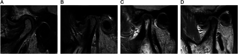
Methods: A retrospective cohort study using MRI scans (pre-operation, 1 week and 12 months post-operation) of all patients who had open TMJ disc repositioning surgery from January 2016 to June 2021 at one centre through two surgical techniques (OSu and MsA) was performed. The predictor variable was technique (OSu and MsA). Outcome variables were DPS and CBR. During follow-up, DPS was rated as good, acceptable and poor, and CBR was graded as improved, unchanged, and degenerated. Multivariate analysis was used to compare the DPS and CBR at 12 months after adjusting five factors including age, sex, Wilkes stage, preoperative bone status (normal, mild/moderate abnormal) and the degree of disc repositioning (normal, overcorrected, and posteriorly repositioned). Relative risk (RR) for DPS and CBR was calculated by multivariate logistic regression.
Results: Three hundred eighty-five patients with 583 joints were included in the study. MRIs at 12 months showed that 514 joints (93.5%) had good DPS, and 344 joints (62.5%) had improved CBR. Multivariate analysis revealed that OSu had higher DPS (RR=2.95; 95% CI, 1.27-6.85) and better CBR (RR=1.58; 95% CI, 1.02-2.46) than MsA. Among the factors affecting DPS, females had better results than males (RR=2.63; 95% CI, 1.11-6.26) and overcorrected or posteriorly repositioned discs were more stable than normally repositioned discs (RR=5.84; 95% CI, 2.58-13.20). The improvement in CBR decreased with age increasing (RR=0.91; 95% CI, 0.89-0.93). Preoperative mild/moderate abnormal bone status had a higher probability of improved CBR compared to normal preoperative bone status (RR=2.60; 95% CI, 1.76-3.83).
Conclusion: OSu had better DPS and CBR than MsA. Sex and the degree of disc repositioning impacted DPS, while age and preoperative bone status affected CBR.
Copyright © 2024 The Author(s). Published by Wolters Kluwer Health, Inc.
Conflict of interest statement
None of the authors have any relevant financial relationship(s) with a commercial interest.
Sponsorships or competing interests that may be relevant to content are disclosed at the end of this article.
Figures


Similar articles
-
Effect of sagittal position of the articular disc on condylar bone remodeling after disc repositioning surgeries in adolescents: A retrospective cohort study.J Craniomaxillofac Surg. 2025 Sep;53(9):1626-1637. doi: 10.1016/j.jcms.2025.07.001. Epub 2025 Jul 16. J Craniomaxillofac Surg. 2025. PMID: 40675877
-
Predicting anterior repositioning splint efficacy in disc displacement with reduction using MRI-based texture and quantitative analysis: a retrospective study.Clin Oral Investig. 2025 Aug 9;29(9):402. doi: 10.1007/s00784-025-06481-4. Clin Oral Investig. 2025. PMID: 40782193
-
Three-dimensional retrospective comparison of disc repositioning, condylar remodelling, and mandibular symmetry following traditional versus digital articulation techniques in patients with temporomandibular disorders.BMC Oral Health. 2025 Aug 31;25(1):1389. doi: 10.1186/s12903-025-06722-8. BMC Oral Health. 2025. PMID: 40887635 Free PMC article.
-
Clinical outcomes of the discopexy using suture anchors for repositioning disc displacement in temporomandibular joints: Systematic review and meta-analysis.J Craniomaxillofac Surg. 2023 Jul-Aug;51(7-8):475-484. doi: 10.1016/j.jcms.2023.06.007. Epub 2023 Jun 27. J Craniomaxillofac Surg. 2023. PMID: 37517977
-
Sertindole for schizophrenia.Cochrane Database Syst Rev. 2005 Jul 20;2005(3):CD001715. doi: 10.1002/14651858.CD001715.pub2. Cochrane Database Syst Rev. 2005. PMID: 16034864 Free PMC article.
References
-
- List T, Jensen RH. Temporomandibular disorders: OLd ideas and new concepts. Cephalalgia 2017;37:692–704. - PubMed
-
- Lamot U, Strojan P, Surlan Popovic K. Magnetic resonance imaging of temporomandibular joint dysfunction-correlation with clinical symptoms, age, and gender. Oral Surg Oral Med Oral Pathol Oral Radiol 2013;116:258–263. - PubMed
-
- Moncada G, Cortes D, Millas R, et al. . Relationship between disk position and degenerative bone changes in temporomandibular joints of young subjects with TMD. An MRI study. J Clin Pediatr Dent 2014;38:269–276. - PubMed
-
- Dias IM, Cordeiro PC, Devito KL, et al. . Evaluation of temporomandibular joint disc displacement as a risk factor for osteoarthrosis. Int J Oral Maxillofac Surg 2016;45:313–317. - PubMed
Publication types
MeSH terms
LinkOut - more resources
Full Text Sources
Miscellaneous
