. 2024 Apr:70:103006.
doi: 10.1016/j.redox.2023.103006. Epub 2023 Dec 23.

Therapeutic effects of long-term HBOT on Alzheimer's disease neuropathologies and cognitive impairment in APPswe/PS1dE9 mice

Affiliations

Therapeutic effects of long-term HBOT on Alzheimer's disease neuropathologies and cognitive impairment in APPswe/PS1dE9 mice

Cui Yang et al. Redox Biol. 2024 Apr.

Abstract

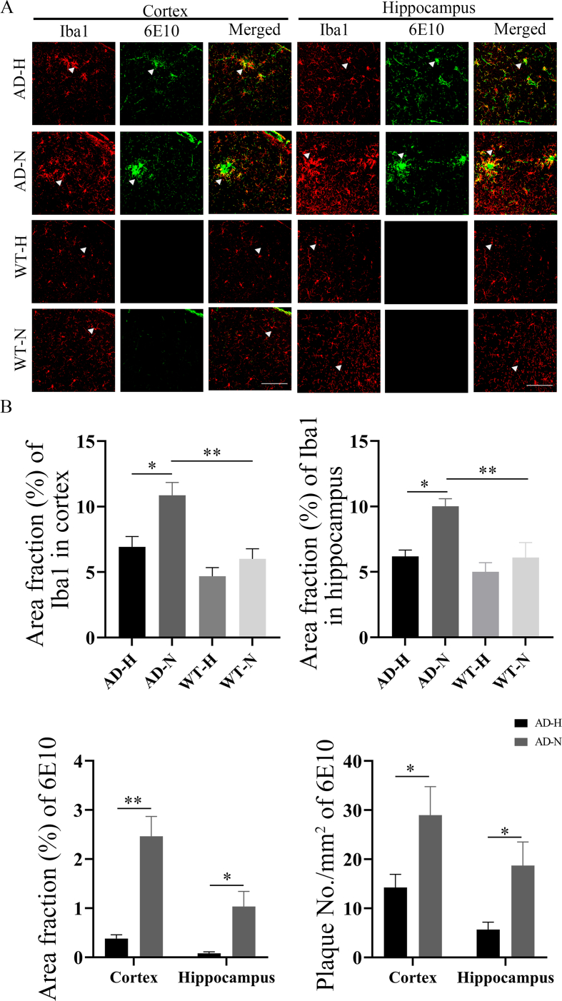
Alzheimer's disease (AD) is the most common neurodegenerative disorder with the pathological hallmarks of amyloid beta (Aβ) plaques and neurofibrillary tangles (NFTs) in the brain. Although there is a hope that anti-amyloid monoclonal antibodies may emerge as a new therapy for AD, the high cost and side effect is a big concern. Non-drug therapy is attracting more attention and may provide a better resolution for the treatment of AD. Given the fact that hypoxia contributes to the pathogenesis of AD, hyperbaric oxygen therapy (HBOT) may be an effective intervention that can alleviate hypoxia and improve AD. However, it remains unclear whether long-term HBOT intervention in the early stage of AD can slow AD progression and ultimately prevent cognitive impairment in this disease. In this study we applied consecutive 3-month HBOT interventions on 3-month-old APPswe/PS1dE9 AD mice which represent the early stage of AD. When the APPswe/PS1dE9 mice at 9-month-old which represent the disease stage we measured cognitive function, 24-h blood oxygen saturation, Aβ and tau pathologies, vascular structure and function, and neuroinflammation in APPswe/PS1dE9 mice. Our results showed that long-term HBOT can attenuate the impairments in cognitive function observed in 9-month-old APPswe/PS1dE9 mice. Most importantly, HBOT effectively reduced the progression of Aβ plaques deposition, hyperphosphorylated tau protein aggregation, and neuronal and synaptic degeneration in the AD mice. Further, long-term HBOT was able to enhance blood oxygen saturation level. Besides, long-term HBOT can improve vascular structure and function, and reduce neuroinflammation in AD mice. This study is the first to demonstrate that long-term HBOT intervention in the early stage of AD can attenuate cognitive impairment and AD-like pathologies. Overall, these findings highlight the potential of long-term HBOT as a disease-modifying approach for AD treatment.

Keywords: Alzheimer's disease; Amyloid plaques deposition; Blood oxygen saturation; Long-term hyperbaric oxygen therapy; Neuroinflammatory.


Conflict of interest statement

Declaration of competing interest Authors declare that there are no conflicts of interest.

Figures

Fig. 1
Fig. 1
HBOT improves Cognitive function. (A) The escape latency of mice during the training period after HBOT. (B) The number of times mice passed through the targeted position in quadrant after HBOT. (C) The percentage of swimming time in the target platform quadrant of AD and WT mice in the total swimming time during Morris water maze test period. (D) The active escape count in shuttle box test of mice in each group. (E) The passive escape count in shuttle box test of mice in each group. (F) The number of rearing in open field test of mice in each group. (G) Total distance of mice in open field test. In (A), *represents AD-H vs. AD-N; # represents AD-N vs.WT-N. Data were the mean ± SEM values, with 6 mice per group.*p < 0.05, **p < 0.01, ***p < 0.001, ****p < 0.0001, by two-way ANOVA with Tukey's multiple comparisons test.
Fig. 2
Fig. 2
HBOT alleviates Aβ pathology in the brain of AD mice. (A) Representative images 6E10 immunohistochemical staining in cortex and hippocampus in AD-H and AD-N mice. Scale bar = 500 μm. (B) Comparison of the area fraction and density of 6E10-positive Aβ plaques in the cortex and hippocampus between AD-H and AD-N mice. (C) Representative images of Congo red in cortex and hippocampus in AD-H and AD-N mice. (D) Comparison of the area fraction and density of Congo red in the cortex and hippocampus between AD-H and AD-N mice. (E) Comparison of Aβ40 and Aβ42 levels measured with ELISA of cortex and hippocampus between AD-H and AD-N mice. Data were the mean ± SEM values, with 6 mice per group.*p < 0.05, **p < 0.01, ***p < 0.001, ****p < 0.0001, by unpaired t-test. (For interpretation of the references to colour in this figure legend, the reader is referred to the Web version of this article.)
Fig. 3
Fig. 3
Immunofluorescent staining of p-Tau Thr231 in the cortex and hippocampus of AD and WT mice after HBOT. (A) The level of p-Tau Thr231 positive neurons in the cortex and hippocampus of AD and WT mice after HBOT. scale bar = 100 μm. (B) Quantitative statistical analysis showed that fluorescence area of p-Tau Thr231 staining in the cortex and hippocampus of AD mice decreased after HBOT. Data were the mean ± SEM values, with 6 mice per group.*p < 0.05, **p < 0.01, ***p < 0.001, ****p < 0.0001, by two-way ANOVA with Tukey's multiple comparisons test.
Fig. 4
Fig. 4
NeuN + Map2 and SYP staining in the brain of AD and WT mice after HBOT. (A) The level of NeuN+Map2 and SYP in hippocampus of AD mice increased after HBOT. scale bar = 100 μm. (B) Quantitative statistical analysis showed that the fluorescence area of NeuN+map2 and SYP staining in the hippocampus of AD increased after HBOT. Data were the mean ± SEM values, with 6 mice per group. *p < 0.05, **p < 0.01, ***p < 0.001, ****p < 0.0001, by two-way ANOVA with Tukey's multiple comparisons test.
Fig. 5
Fig. 5
HBOT improves blood oxygen saturation and ameliorates oxygen saturation fluctuations. (A) Changes of 24 h blood oxygen saturation in AD-H, AD-N, WT-H and WT-N mice. (B) The level of oxygen saturation of AD and WT mice after HBOT. (C) The level of oxygen saturation fluctuations of AD and WT mice after HBOT. Data were the mean ± SEM values, with 6 mice per group. *p < 0.05, **p < 0.01, ***p < 0.001, ****p < 0.0001, by two-way ANOVA with Tukey's multiple comparisons test.
Fig. 6
Fig. 6
HBOT attenuates cerebrovascular hypoperfusion and microvascular injury in the AD mice. (A) Cerebral perfusion images at age of 9 months in the control and HBOT groups (1 session and 90 sessions). (B) Histograms comparing the cerebral blood perfusion. (C) Representative images of 6E10+1A4 in AD-H and AD-N mice (scale bar = 100 μm). (D) Histograms comparing number of 6E10+1A4 in the AD-H and AD-N. Data were the mean ± SEM values, with 6 mice per group.*p < 0.05, **p < 0.01, ***p < 0.001, ****p < 0.0001, by unpaired t-test.
Fig. 7
Fig. 7
Immunoflourescence staining of microglia cells in the cortex and hippocampus of AD and WT mice after HBOT. (A) Iba1 staining showed a reduced in microglia in the cortex and hippocampus of AD mice after HBOT, and 6E10 staining showed a significant decrease in Aβ plaques deposition in the cortex and hippocampus of AD mice after HBOT (scale bar = 100 μm). (B) Statistical analysis showed that the staining positive area of microglia in the cortex and hippocampus of AD mice after HBOT was decreased, and the Aβ plaques area fraction and density in the cortex and hippocampus of AD mice after HBOT was decreased. Data were the mean ± SEM values, with 6 mice per group.*p < 0.05, **p < 0.01, ***p < 0.001, ****p < 0.0001, by unpaired t-test and two-way ANOVA with Tukey's multiple comparisons test.
Fig. 8
Fig. 8
Immunoflourescence staining of astrocytes in the cortex and hippocampus of AD and WT mice after HBOT. (A) GFAP staining showed a reduced in astrocytes in the cortex and hippocampus of AD mice after HBOT, and 6E10 staining showed a decrease in Aβ plaques deposition in the cortex and hippocampus of AD mice after HBOT (scale bar = 100 μm). (B) Statistical analysis showed that the staining positive area of astrocytes in the cortex and hippocampus of AD mice after HBOT was significantly decreased, and the Aβ plaques area fraction and density in the cortex of AD mice after HBOT was significantly decreased. Data were the mean ± SEM values, with 6 mice per group.*p < 0.05, **p < 0.01, ***p < 0.001, ****p < 0.0001, by unpaired t-test and two-way ANOVA with Tukey's multiple comparisons test.
Fig. 9
Fig. 9
Levels of inflammatory cytokines in the cortex and hippocampus of AD and WT mice after HBOT. (A)–(D) The expressions of pro-inflammatory cytokines IL-1β, IL-6, TNF-α and IFN-γ in the cortex and hippocampus of AD mice were decreased after HBOT treatment. Data were the mean ± SEM values, with 6 mice per group.*p < 0.05, **p < 0.01, ***p < 0.001, ****p < 0.0001, by two-way ANOVA with Tukey's multiple comparisons test.
figs1
figs1
figs2
figs2
figs3
figs3

Similar articles

Cited by

References

    1. Reitz C., Brayne C., Mayeux R. Epidemiology of alzheimer disease. Nat. Rev. Neurol. 2011;7(3):137–152. doi: 10.1038/nrneurol.2011.2. - DOI - PMC - PubMed
    1. Tang M.Y., Gorin F.A., Lein P.J. Review of evidence implicating the plasminogen activator system in blood-brain barrier dysfunction associated with Alzheimer's disease. Ageing Neurodegener Dis. 2022;2 doi: 10.20517/and.2022.05. - DOI - PMC - PubMed
    1. Liu H., Qiu H., Yang J., Ni J., Le W. Chronic hypoxia facilitates Alzheimer's disease through demethylation of gamma-secretase by downregulating DNA methyltransferase 3b. Alzheimers Dement. 2016;12(2):130–143. doi: 10.1016/j.jalz.2015.05.019. - DOI - PubMed
    1. Cummings J. Anti-amyloid monoclonal antibodies are transformative treatments that redefine alzheimer's disease therapeutics. Drugs. 2023;83(7):569–576. doi: 10.1007/s40265-023-01858-9. - DOI - PMC - PubMed
    1. Jutkowitz E., Pizzi L.T., Shewmaker P., Alarid-Escudero F., Epstein-Lubow G., Prioli K.M., Gaugler J.E., Gitlin L.N. Cost effectiveness of non-drug interventions that reduce nursing home admissions for people living with dementia. Alzheimers Dement. 2023;19(9):3867–3893. doi: 10.1002/alz.12964. - DOI - PMC - PubMed

MeSH terms

Substances