. 2024 Jun 3;147(6):2023-2037.
doi: 10.1093/brain/awae020.

Neurodevelopmental and synaptic defects in DNAJC6 parkinsonism, amenable to gene therapy

Affiliations

Neurodevelopmental and synaptic defects in DNAJC6 parkinsonism, amenable to gene therapy

Lucia Abela et al. Brain. .

Abstract

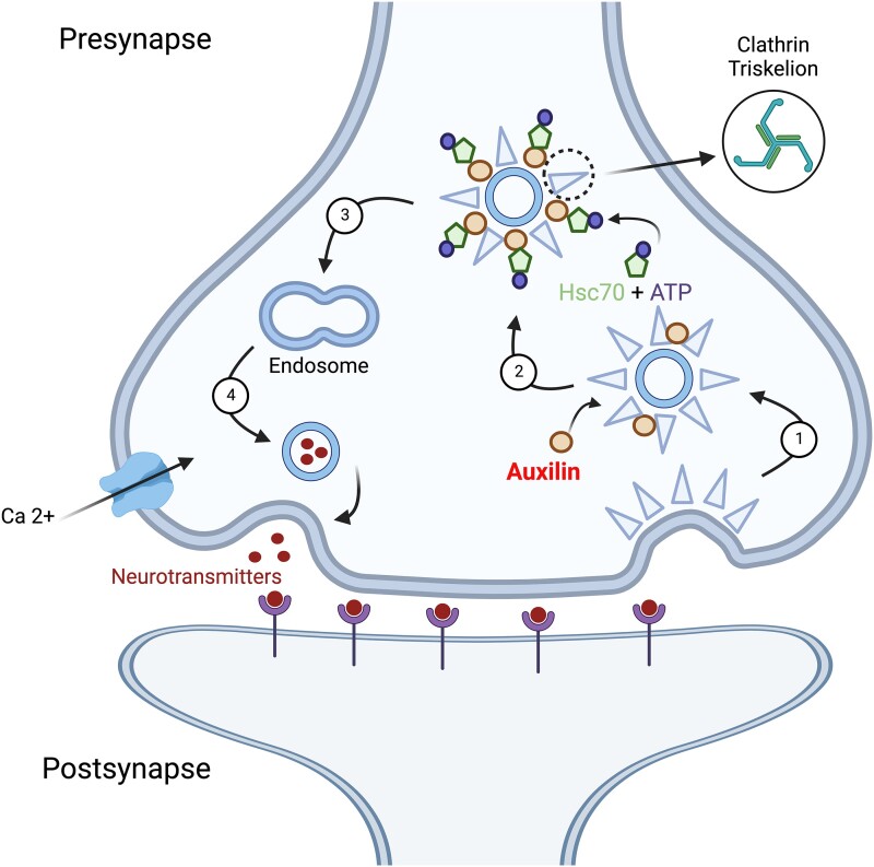
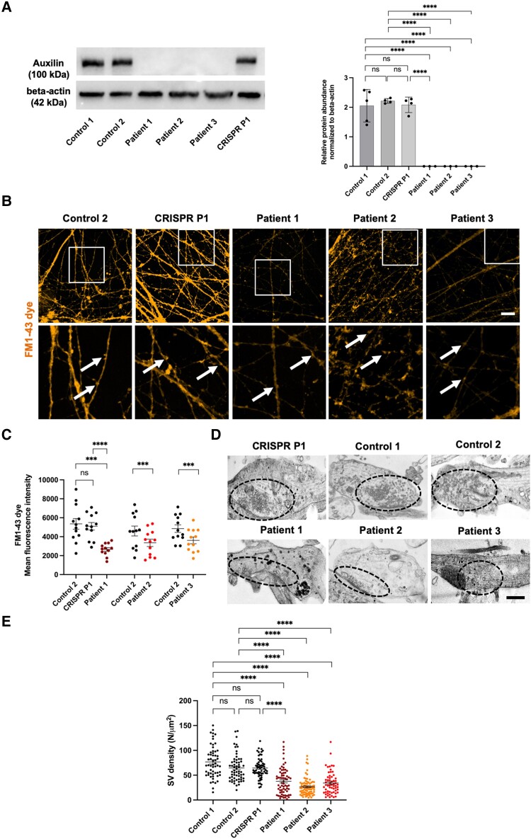
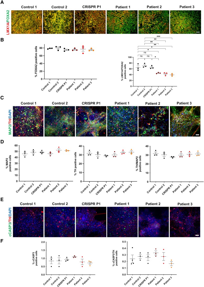
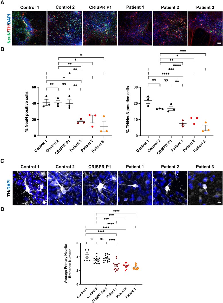
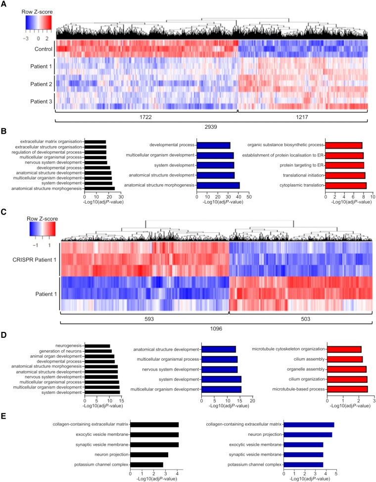
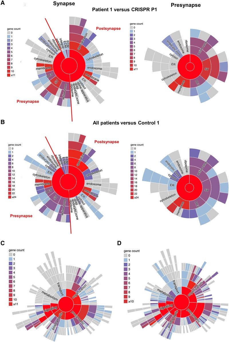
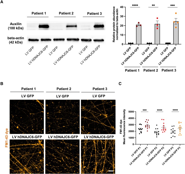
DNAJC6 encodes auxilin, a co-chaperone protein involved in clathrin-mediated endocytosis (CME) at the presynaptic terminal. Biallelic mutations in DNAJC6 cause a complex, early-onset neurodegenerative disorder characterized by rapidly progressive parkinsonism-dystonia in childhood. The disease is commonly associated with additional neurodevelopmental, neurological and neuropsychiatric features. Currently, there are no disease-modifying treatments for this condition, resulting in significant morbidity and risk of premature mortality. To investigate the underlying disease mechanisms in childhood-onset DNAJC6 parkinsonism, we generated induced pluripotent stem cells (iPSC) from three patients harbouring pathogenic loss-of-function DNAJC6 mutations and subsequently developed a midbrain dopaminergic neuronal model of disease. When compared to age-matched and CRISPR-corrected isogenic controls, the neuronal cell model revealed disease-specific auxilin deficiency as well as disturbance of synaptic vesicle recycling and homeostasis. We also observed neurodevelopmental dysregulation affecting ventral midbrain patterning and neuronal maturation. To explore the feasibility of a viral vector-mediated gene therapy approach, iPSC-derived neuronal cultures were treated with lentiviral DNAJC6 gene transfer, which restored auxilin expression and rescued CME. Our patient-derived neuronal model provides deeper insights into the molecular mechanisms of auxilin deficiency as well as a robust platform for the development of targeted precision therapy approaches.

Keywords: CME; DNAJC6; auxilin; gene therapy; neurodevelopmental; parkinsonism.


Conflict of interest statement

The authors report no competing interests.

Figures

Figure 1
Figure 1
Schematic representation of CME. Clathrin-mediated endocytosis with (1) nucleation of clathrin-coated vesicles and clathrin coat assembly; (2) uncoating and release of cargo; (3) endolysosomal recycling of synaptic vesicles (SV); and (4) synaptic vesicle exocytosis. CME = clathrin-mediated endocytosis. Created with BioRender.com.
Figure 2
Figure 2
Patient-derived neurons show auxilin deficiency and disturbance of CME and synaptic vesicle homeostasis. (A) Immunoblot analysis for auxilin protein in control, patient and CRISPR Patient P1-derived neurons at Day 65 of differentiation. Quantification of protein abundance relative to the loading control (β-actin) (auxilin n = 5, 4, 4, 3, 3, 3). (B) Representative images of FM1–43 dye uptake assay for control, patient and CRISPR Patient P1-derived neurons at Day 65 of differentiation. Arrows in the high magnification images (bottom row) show representative synaptic boutons. Scale bar = 20 μm. (C) Quantification of FM1–43 mean fluorescence intensity in patients relative to the control and Patient 1 versus CRISPR Patient P1, respectively. (D) Electron microscopy analysis of presynaptic terminals in control, patient and CRISPR Patient P1-derived neuronal cultures at Day 65 of differentiation. Scale bar = 500 nm. Dotted circles indicate area with synaptic vesicles. (E) Quantification of synaptic vesicle (SV) density in the presynaptic terminal on electron microscopy images. CME = clathrin-mediated endocytosis; ns = not significant.
Figure 3
Figure 3
Patient-derived neurons show alterations in ventral midbrain patterning without neurodegeneration. (A) Representative immunofluorescence images for LMX1A and FOXA2 in control, patient and CRISPR Patient P1-derived neural progenitors at Day 11 of differentiation. Scale bar = 100 μm. (B) Quantification of total number of FOXA2-positive cells and LMX1A/FOXA2 double-positive cells at Day 11 (n = 3 for all). (C) Representative immunofluorescence images for MAP2 and TH in control, patient and CRISPR Patient P1-derived midbrain dopaminergic (mDA) neurons at Day 65 of differentiation. Scale bar = 150 μm. (D) Quantification of total number of MAP2 and TH positive cells, and TH/MAP2 double-positive cells at Day 65 of differentiation (n = 3 for all). (E) Representative immunofluorescence images for cCASP3 in control, patient and CRISPR Patient P1-derived mDA neurons at Day 65 of differentiation. Scale bar = 150 μm. (F) Quantification of total numbers of cCASP3-positive cells and cCASP3/TH double-positive cells at Day 65 of differentiation (n = 3 for all). ns = not significant.
Figure 4
Figure 4
Patient-derived neurons show defects in neuronal maturation. (A) Representative immunofluorescence images for NeuN and TH in control, patient and CRISPR Patient P1-derived midbrain dopaminergic (mDA) neurons. Scale bar = 150 μm. (B) Quantification of NeuN positive and TH/NeuN double-positive cells in control, patient and CRISPR Patient P1-derived mDA neurons at Day 65 of differentiation (n = 3 for all). (C) Representative immunofluorescence images for control, patient and CRISPR Patient P1-derived mDA neuron branching. Scale bar = 20 μm. (D) Quantification of average primary neurite branching in control, patient-and CRISPR Patient P1-derived mDA neurons at Day 65 of differentiation (n = 4 for all). ns = not significant.
Figure 5
Figure 5
Bulk RNA sequencing analysis highlights developmental and synaptic vesicle defects in patient-derived neurons. (A) Heat map showing hierarchical clustering of protein-coding differentially expressed genes (DEGs) in all patients compared to Control 1 (n = 3). (B) Gene Ontology (GO) terms enrichment for biological process of total (black), under-expressed (blue) and over-expressed (red) protein-coding DEGs in patients compared to control. The top 10 or five categories are shown. (C) Heat map showing hierarchical clustering of protein-coding DEGs in Patient 1 compared to CRISPR Patient P1 (n = 3). (D) GO terms enrichment for biological process of total (black), under-expressed (blue) and over-expressed (red) protein-coding DEGs in Patient 1 compared to CRISPR Patient P1. The top 10 or five categories are shown. (E) GO terms enrichment for cellular component of total (black) and under-expressed (blue) protein-coding DEGs in Patient 1 compared to CRISPR Patient P1.
Figure 6
Figure 6
Synaptic location enrichment analysis reveals structural abnormalities in patient-derived neurons. (A) Pie charts showing synaptic location mapping of under-expressed protein-coding differentially expressed genes (DEGs) in Patient 1 compared to CRISPR Patient P1. Left: general synapse; right: presynapse zoom-in. (B) Pie charts showing synaptic location mapping of under-expressed protein-coding DEGs in all patients compared to Control 1. Left: general synapse; right: presynapse zoom-in. (C) Pie charts showing synaptic function mapping of under-expressed protein-coding DEGs in Patient 1 compared to CRISPR Patient P1. (D) Pie charts showing synaptic function mapping of under-expressed protein-coding DEGs in all patients compared to Control 1.
Figure 7
Figure 7
Lentiviral DNAJC6 gene transfer restores auxilin and improves CME. (A) Left: Representative immunoblot images for auxilin and loading control (β-actin) in patient-derived midbrain dopaminergic (mDA) neurons transfected with LV GFP (lentivirus encoding green fluorescent protein) and LV DNAJC6-GFP (lentivirus encoding DNAJC6-green fluorescent protein) at Day 65 of maturation. Right: Quantification relative to the loading control (β-actin) (n = 3). (B) Representative immunofluorescence images for FM1–43 dye uptake assay in patient-derived mDA neurons transfected with LV GFP and LV DNAJC6-GFP at Day 65 of maturation. Scale bar = 20 μm. (C) Quantification of FM1–43 mean fluorescence intensity in patient-derived mDA neurons transfected with LV GFP and LV DNAJC6-GFP.

Similar articles

  • DNAJC6 Mutations Disrupt Dopamine Homeostasis in Juvenile Parkinsonism-Dystonia.
    Ng J, Cortès-Saladelafont E, Abela L, Termsarasab P, Mankad K, Sudhakar S, Gorman KM, Heales SJR, Pope S, Biassoni L, Csányi B, Cain J, Rakshi K, Coutts H, Jayawant S, Jefferson R, Hughes D, García-Cazorla À, Grozeva D, Raymond FL, Pérez-Dueñas B, De Goede C, Pearson TS, Meyer E, Kurian MA. Ng J, et al. Mov Disord. 2020 Aug;35(8):1357-1368. doi: 10.1002/mds.28063. Epub 2020 May 30. Mov Disord. 2020. PMID: 32472658 Free PMC article.
  • A deleterious mutation in DNAJC6 encoding the neuronal-specific clathrin-uncoating co-chaperone auxilin, is associated with juvenile parkinsonism.
    Edvardson S, Cinnamon Y, Ta-Shma A, Shaag A, Yim YI, Zenvirt S, Jalas C, Lesage S, Brice A, Taraboulos A, Kaestner KH, Greene LE, Elpeleg O. Edvardson S, et al. PLoS One. 2012;7(5):e36458. doi: 10.1371/journal.pone.0036458. Epub 2012 May 1. PLoS One. 2012. PMID: 22563501 Free PMC article.
  • DNAJC6 Mutations Associated With Early-Onset Parkinson's Disease.
    Olgiati S, Quadri M, Fang M, Rood JP, Saute JA, Chien HF, Bouwkamp CG, Graafland J, Minneboo M, Breedveld GJ, Zhang J; International Parkinsonism Genetics Network; Verheijen FW, Boon AJ, Kievit AJ, Jardim LB, Mandemakers W, Barbosa ER, Rieder CR, Leenders KL, Wang J, Bonifati V. Olgiati S, et al. Ann Neurol. 2016 Feb;79(2):244-56. doi: 10.1002/ana.24553. Epub 2016 Jan 14. Ann Neurol. 2016. PMID: 26528954
  • DNAJC6 Parkinson Disease.
    Kurian MA, Abela L. Kurian MA, et al. 2021 May 13. In: Adam MP, Feldman J, Mirzaa GM, Pagon RA, Wallace SE, Amemiya A, editors. GeneReviews® [Internet]. Seattle (WA): University of Washington, Seattle; 1993–2025. 2021 May 13. In: Adam MP, Feldman J, Mirzaa GM, Pagon RA, Wallace SE, Amemiya A, editors. GeneReviews® [Internet]. Seattle (WA): University of Washington, Seattle; 1993–2025. PMID: 33983693 Free Books & Documents. Review.
  • DNAJC proteins and pathways to parkinsonism.
    Roosen DA, Blauwendraat C, Cookson MR, Lewis PA. Roosen DA, et al. FEBS J. 2019 Aug;286(16):3080-3094. doi: 10.1111/febs.14936. Epub 2019 Jun 20. FEBS J. 2019. PMID: 31120186 Free PMC article. Review.

Cited by

References

    1. McMahon HT, Boucrot E. Molecular mechanism and physiological functions of clathrin-mediated endocytosis. Nat Rev Mol Cell Biol. 2011;12:517–533. - PubMed
    1. Edvardson S, Cinnamon Y, Ta-Shma A, et al. . A deleterious mutation in DNAJC6 encoding the neuronal-specific clathrin-uncoating co-chaperone auxilin, is associated with juvenile parkinsonism. PLoS One. 2012;7:e36458. - PMC - PubMed
    1. Köroĝlu Ç, Baysal L, Cetinkaya M, Karasoy H, Tolun A. DNAJC6 is responsible for juvenile parkinsonism with phenotypic variability. Park Relat Disord. 2013;19:320–324. - PubMed
    1. Olgiati S, Quadri M, Fang M, et al. . DNAJC6 mutations associated with early-onset Parkinson’s disease. Ann Neurol. 2015;79:244–256. - PubMed
    1. Elsayed LEO, Drouet V, Usenko T, et al. . A novel nonsense mutation in DNAJC6 expands the phenotype of autosomal-recessive juvenile-onset Parkinson’s disease. Ann Neurol. 2016;79:335–337. - PubMed

Publication types