Review
. 2024 Aug;51(10):2931-2940.
doi: 10.1007/s00259-024-06602-3. Epub 2024 Jan 20.

Current clinical applications of Cerenkov luminescence for intraoperative molecular imaging

Affiliations
Review

Current clinical applications of Cerenkov luminescence for intraoperative molecular imaging

Natalie Boykoff et al. Eur J Nucl Med Mol Imaging. 2024 Aug.

Erratum in

Abstract

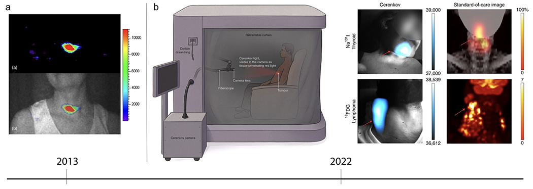
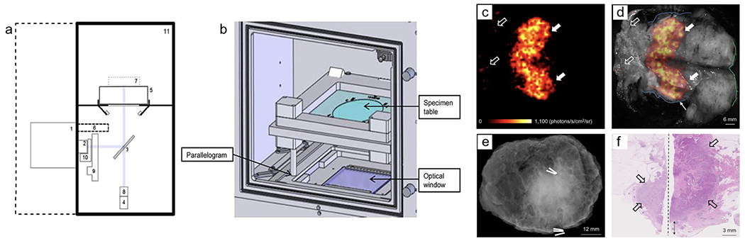
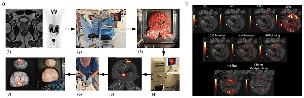
Background: Cerenkov luminescence imaging (CLI) is a new emerging technology that can be used for optical imaging of approved radiotracers, both in a preclinical, and even more recently, in a clinical context with rapid imaging times, low costs, and detection in real-time (Grootendorst et al. Clin Transl Imaging 4(5):353-66, 2016); Wang et al. Photonics 9(6):390, 2022). This brief review provides an overview of clinical applications of CLI with a focus on intraoperative margin assessment (IMA) to address shortcomings and provide insight for future work in this application.

Methods: A literature review was performed using PubMed using the search words Cerenkov luminescence imaging (CLI), intraoperative margin assessment (IMA), and image-guided surgery. Articles were selected based on title, abstract, content, and application.

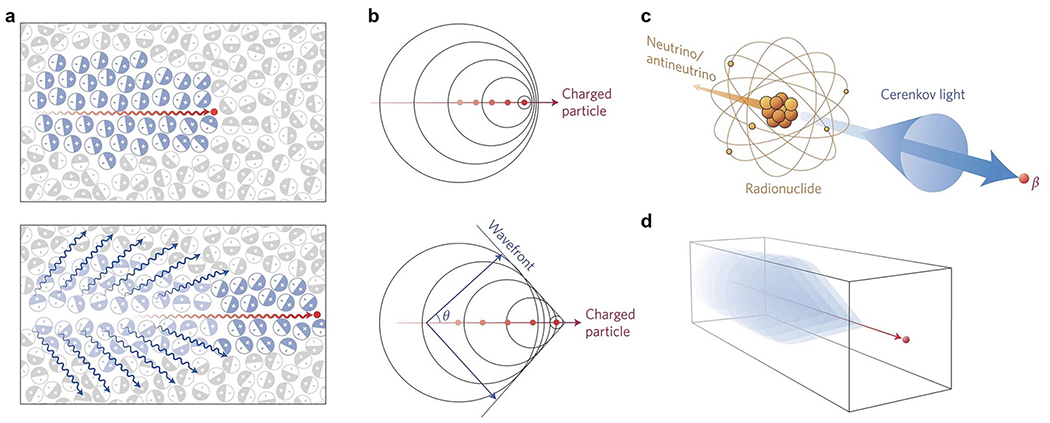
Results: Original research was summarized to examine advantages and limitations of CLI compared to other modalities for IMA. The characteristics of Cerenkov luminescence (CL) are defined, and results from relevant clinical trials are discussed. Prospects of ongoing clinical trials are reviewed, along with technological advancements related to CLI.

Conclusion: CLI is a proven method for molecular imaging and shows feasibility for determining intraoperative margins if future work involves establishing quantitative approaches for attenuation and scattering, depth analysis, and radiation safety for CLI at a larger scale.

Keywords: Cerenkov luminescence imaging; Image-guided surgery; Intraoperative margin assessment; Molecular imaging.


Conflict of interest statement

Competing Interests

The authors have no relevant financial or non-financial interests to disclose.

Figures

Fig. 1
Fig. 1
Cerenkov luminescence phenomenon. a Top: Charged particle (red line) traveling faster than the velocity of light in a particular dielectric medium, i.e. water or tissue, asymmetrically polarizing the medium to create a dipole electric field. Bottom: As particle moves through medium, the molecules return to ground state, emitting blue-weighted light in the forward direction (blue lines). b A photonic wavefront is formed from constructive interference of the coherent waves produced in a forward angle θ. c Radionuclides that emit β-particles in a dielectric medium with energies greater than the Cerenkov threshold produce CL. d A medium with a negative refractive index shows the CL cone is pointing in the reverse direction. Reproduced with permission from [20], © 2017 Springer Nature Limited
Fig. 2
Fig. 2
Clinical Cerenkov fiberscope imaging of cancer patients. a (top) Early CLI of a patient’s thyroid. (bottom) CL and gray scale overlay confirms CL is coming from the thyroid gland. Reproduced with permission from [28], © 2013 SPIE. b (left) After radiotracer injection, the patient sits in the lightproof enclosure for up to 15 min of imaging time. The Cerenkov camera is placed outside the enclosure and connected to relay optics, fiberscope, and a f-0.95 lens. Most of the CL detected is red-weighted, since the blue-weighted wavelengths are attenuated and scattered through the patients’ tissue. (right) CL image produced using the clinical Cerenkov setup vs. a standard-of-care PET image of (top) thyroid cancer using [131I]-sodium iodide and (bottom) lymphoma using [18F]-FDG. Reproduced with permission from [30], © 2022 Springer Nature Limited
Fig. 3
Fig. 3
Investigational intraoperative CLI imaging of WLE specimen. a Schematic diagram of Lightpath® (Lightpoint Medical Ltd. UK) imaging system: (1) EMCCD camera, (2) f-0.95 lens, (3) Hinged reflex mirror, (4) complementary metal oxide semiconductor reference camera, (5) specimen table, (6) lead radiation shielding for EMCCD camera, (7) focal zone, (8) fixed lens for reference camera, (9) filter wheel, (10) LED RGB light array, (11) specimen chamber. b Specimen Chamber: Table can be positioned using a parallelogram to center specimen within the optical window. c Cerenkov image of WLE specimen from patient with grade 3, estrogen receptor-negative/human epidermal growth factor receptor 2-negative, NST carcinoma, mean radiance of 871±131 photons/s/cm2/sr and mean TBR is 3.22 (white arrows); phosphorescent signal was detected from pathology ink (open arrows). d Gray-scale image overlaid with Cerenkov image; both surgeons measured the posterior margin to be 2 mm (blue outline). e Specimen radiography image. f Histopathology image of primary tumor confirmed to have a posterior margin of 3 mm (double arrow). This research was originally published in J Nuclear Med.[33] Grootendorst, M. R. et al. J Nucl Med. 2017;58(6):891-898. © 2017 SNMMI
Fig. 4
Fig. 4
Workflow and settings used for intraoperative CLI of prostate tumor. a (1) Preoperative 68Ga-PSMA PET-CT and MRI scan. (2) 68Ga-PSMA is administered via i.v. injection during surgery. Once prostate is removed using the da Vinci® surgical system, the specimen is rinsed with NaCl solution. (3) Prostate is placed on specimen table. (4) Lightpath® images all six sides. (5) Unfiltered Cerenkov image produced. (6) Specimen is inked and cleaved ~1 cm from the apex. (7) White-light (right) and Cerenkov (left) image of cleaved prostate. b, Top row: 8 × 8 binning and 550 nm filter used for images of varying exposures of 30 s (TBR 1.10), 60 s (TBR 1.18), 150 s (TBR 1.85), 300 s (TBR 1.98). Center row: 150 s exposure and 550 nm filter used for images of varying pixel binning of 2 × 2 (TBR 1.06), 4 × 4 (TBR 1.26), 8 × 8 (TBR 1.85). Bottom row: 150 s exposure and 8 × 8 binning used for images without filter and with 550 nm filter. This research was originally published in Eur. J. Nucl. Med. Mol. Imaging. [4] Olde Heuvel, J. et al. EJNMMI. 2020;47(11):2624-2632. © 2020 Springer Science + Business Media

Similar articles

Cited by

References

    1. Grootendorst MR, et al. , Cerenkov luminescence imaging (CLI) for image-guided cancer surgery. Clinical and Translational Imaging, 2016. 4(5): p. 353–366. - PMC - PubMed
    1. Wang X, et al. , Cherenkov Luminescence in Tumor Diagnosis and Treatment: A Review. Photonics, 2022. 9(6): p. 390.
    1. Rosenthal EL, et al. , The Status of Contemporary Image-Guided Modalities in Oncologic Surgery. Annals of Surgery, 2015. 261(1): p. 46–55. - PMC - PubMed
    1. Wilson BC and Eu D, Optical spectroscopy and imaging in surgical management of cancer patients. Translational Biophotonics, 2022. 4(3).
    1. Olde Heuvel J, et al. , 68Ga-PSMA Cerenkov luminescence imaging in primary prostate cancer: first-in-man series. European Journal of Nuclear Medicine and Molecular Imaging, 2020. 47(11): p. 2624–2632. - PMC - PubMed

LinkOut - more resources