Review
. 2024 Jun;244(6):887-899.
doi: 10.1111/joa.14009. Epub 2024 Jan 19.

Functional anatomy of the orbit in strabismus surgery: Connective tissues, pulleys, and the modern surgical implications of the "arc of contact" paradigm

Affiliations
Review

Functional anatomy of the orbit in strabismus surgery: Connective tissues, pulleys, and the modern surgical implications of the "arc of contact" paradigm

Paulo Freitas-da-Costa et al. J Anat. 2024 Jun.

Abstract

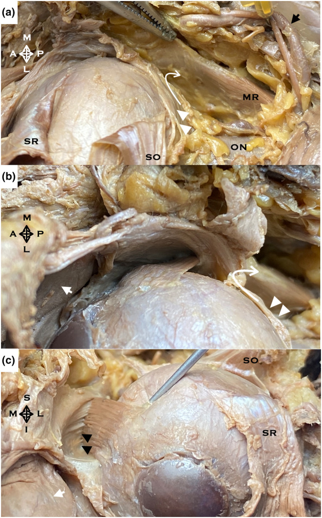
Oculomotricity is a multidimensional domain characterised by a delicate interplay of anatomical structures and physiological processes. This manuscript meticulously dissects the nuances of this interplay, bringing to the fore the integral role of the extraocular muscles (EOMs) and their intricate relationship with the myriad orbital connective tissues as it harmoniously orchestrates binocular movements, ensuring synchronised and fluid visual tracking. Historically, the peripheral oculomotor apparatus was conceptualised as a rudimentary system predominantly driven by neural directives. While widely accepted, this perspective offered a limited view of the complexities inherent in ocular movement mechanics. The twentieth century heralded a paradigm shift in this understanding. With advances in anatomical research and imaging techniques, a much clearer picture of the gross anatomy of the EOMs emerged. This clarity challenged traditional viewpoints, suggesting that the inherent biomechanical properties of the EOMs, coupled with their associated tissue pulleys, play a pivotal role in dictating eye movement dynamics. Central to this revised understanding is the "arc of contact" paradigm. This concept delves deep into the mechanics of eye rotation, elucidating the significance of the point of contact between the EOMs and the eyeball. The arc of contact is not just a static anatomical feature; its length and orientation play a crucial role in determining the effective torque generated by a muscle, thereby influencing the amplitude and direction of eye rotation. The dynamic nature of this arc, influenced by the position and tension of the muscle pulleys, offers a more comprehensive model for understanding ocular kinematics. Previously overlooked in traditional models, muscle pulleys have now emerged as central players in the biomechanics of eye movement. These anatomical structures, formed by dense connective tissues, guide the paths of the EOMs, ensuring that their pulling angles remain optimal across a range of gaze directions. The non-linear paths resulting from these pulleys provide a more dynamic and intricate understanding of eye movement, challenging two-dimensional, linear models of orbital anatomy. The implications of these revelations extend beyond mere theoretical knowledge. The insights garnered from this research promise transformative potential in the realm of strabismus surgery. Recognising the pivotal role of muscle pulleys and the "arc of contact" paradigm allows for more precise surgical interventions, ensuring better post-operative outcomes and minimising the risk of complications. Surgical procedures that previously relied on basic mechanical principles now stand to benefit from a more nuanced understanding of the underlying anatomical and physiological dynamics. In conclusion, this manuscript serves as a testament to the ever-evolving nature of scientific knowledge. Challenging established norms and introducing fresh perspectives pave the way for more effective and informed clinical interventions in strabismus surgery.

Keywords: anatomy; connective tissue; eye movements; oculomotor muscles; orbit; rectus muscle pulleys; regional; strabismus.


Conflict of interest statement

The authors declare that they have no conflict of interest.

Figures

FIGURE 1
FIGURE 1
Anatomical dissection of an orbit of a formalin‐fixed cadaver in a superior approach. The extraconal adipose tissue was removed. (a, b) left orbit, same dissection, different steps, superior aspect. (a) After periorbita opening. Adipose tissue forms the filling of the muscular cone in the mid‐to‐deep orbit. (b) The intraconal adipose was removed, and the LR‐SR/LPS band (white arrow) of connective tissue in the anterior orbit was exposed. (c) Right orbit, superior aspect. After opening and excising the LR‐SR/LPS band, the Tenon's capsule is exposed and pulled here with forceps. A, anterior; Black arrowhead, periorbita; Black star, residual lacrimal gland tissue; P, posterior; L, lateral; LPS levator palpebrae superioris; LR lateral rectus; M, medial; SR superior rectus; White arrowhead, lateral check ligament or lateral capsulopalpebral fascia.
FIGURE 2
FIGURE 2
Orbital anatomical dissection in different formalin‐fixed cadavers. Exposure after muscle individualisation, cleaning and careful dissection of all surrounding connective and adipose tissue. (a) Left orbit, superolateral aspect. The superior oblique tendon rolls into a cylinder as it transits the trochlea; (b) Left orbit, anterosuperior aspect. Insertion of the superior oblique muscle tendon into the superolateral aspect of the globe after opening and dissection of Tenon's capsule (white arrowheads); (c) Left orbit, anterior aspect. Inferior oblique muscle origin and trajectory; (d) Right orbit, lateral aspect. Insertion of the inferior oblique (IO) muscle into the globe in the posterolateral and inferior quadrant. A, anterior; I, inferior; L, lateral; LG, lacrimal gland; LPS, levator palpebrae superioris; LR, lateral rectus; M, medial; P, posterior; S, superior; SR, superior rectus.
FIGURE 3
FIGURE 3
Relation of the orbital axis (equivalent to the superior rectus muscle plane) and the superior oblique muscle plane with the y‐axis.
FIGURE 4
FIGURE 4
Orbit diagram. Representation of the pulley concept and its influence on the rectus muscles' non‐linear path and their tendon's insertion angle. (a) Lateral rectus muscle with loss of tangency created by the pulley. (b) Posterior fixations suture of the medial rectus—the concept of cancelling the arc of contact with reduced lever arm length, compared to what happens in a recession (c). At the same time, there is a restriction in the longitudinal movement of the pulley with the inability of the tendon to telescope on it. (d) The traditional concept of a linear path of the lateral rectus muscle with the definition of the tangential point (TP) and arc of contact (curved arrow). OS left eye, OD right eye.
FIGURE 5
FIGURE 5
The right orbit of a formalin‐fixed cadaver in a superior approach dissection. (a, b) Superior aspect. (c) Anterior aspect. The medial rectus muscle (MR) courses anteriorly through lobules of orbital fat until it enters the connective tissue pulley that ensheaths it as it penetrates posterior Tenon's fascia (white arrowhead). Curved White Arrow, the constitution of the muscular fascia by the reflection of the Tenon's capsule on the muscular belly; Black arrowhead, MR insertion on its pulley; White arrow, superior tarsus; SR superior rectus tendon anteriorly reflected, SO tendon of the superior oblique muscle reflected posterolaterally, ON optic nerve; Black arrow, ophthalmic artery and nasociliary nerve retracted with a needle. A, anterior; I, inferior; L, lateral; M, medial; P, posterior; S, superior.
FIGURE 6
FIGURE 6
(a–c) Surgical and sequential exposure of a lateral rectus (LR) muscle (view of the surgeon on the patient's head). (d–f) The left orbit of a formalin‐fixed cadaver in a superior approach dissection exposes the trajectory of the LR muscle in its relationship with Tenon's capsule, its pulley and interdependence with neighbouring structures. The figure seeks to establish a correlation between vertically arranged images: (a–d, b–e and c–f). (a) Following the engagement of the LR muscle on a surgical hook, the white anterior pulley slings are visible just posterior to the tendon insertion (black arrowheads) and easily displaced with forceps. (d) Note its arrangement concerning the globe's curvature towards the superior rectus muscle insertion (black arrowheads), bending or with the convexity towards the orbital wall (black curved arrow 1). (b) After blunt dissection of the anterior slings of the pulley from the most anterior part of the tendon, the LR‐SR band (black arrows) becomes apparent more posteriorly at the level of the equator (this position is more posterior in the case of the lateral rectus relative to the medial rectus). (e, f) More robust, dense and elastic (black arrows), practically inseparable from the muscular belly with forceps (black curved arrow 2). (c) In the same position, on the external surface of the muscle, we observed a dense connective tissue strongly adherent to the muscle, corresponding to its insertion of the pulley (black asterisk). (f) When dissecting the orbit, its relationship with the corresponding check ligament (black asterisk) and its adhesion to the lateral wall of the orbit and anteriorly in continuity with the tarsus and the lateral palpebral ligament (white asterisk) are observed. The posterior end of this sleeve is shaped into slings that bend inwards towards the centre of the orbit (schematically represented by the curved arrow 3). A, anterior; I, inferior; L, lateral; M, medial; ON, optic nerve; P, posterior; SO, superior oblique; SR, superior rectus; S, superior; White arrowheads, Tenon's capsule.
FIGURE 7
FIGURE 7
Medial rectus muscle force vectors on the globe after a recession with the inclusion of the pulley complex.

Similar articles

Cited by

References

    1. Beisner, D.H. (1971) Reduction of ocular torque by medial rectus recession. Archives of Ophthalmology, 85, 13–17. - PubMed
    1. Boeder, P. (1962) Co‐operative action of extra‐ocular muscles. The British Journal of Ophthalmology, 46, 397–403. - PMC - PubMed
    1. Chaudhuri, Z. & Demer, J.L. (2013) Sagging eye syndrome: connective tissue involution as a cause of horizontal and vertical strabismus in older patients. JAMA Ophthalmology, 131, 619–625. - PMC - PubMed
    1. Clark, R.A. & Demer, J.L. (2002a) Effect of aging on human rectus extraocular muscle paths demonstrated by magnetic resonance imaging. American Journal of Ophthalmology, 134, 872–878. - PubMed
    1. Clark, R.A. & Demer, J.L. (2002b) Rectus extraocular muscle pulley displacement after surgical transposition and posterior fixation for treatment of paralytic strabismus. American Journal of Ophthalmology, 133, 119–128. - PubMed

MeSH terms